Chapter 11: Cell Communication. Why do cells need to signal?
advertisement
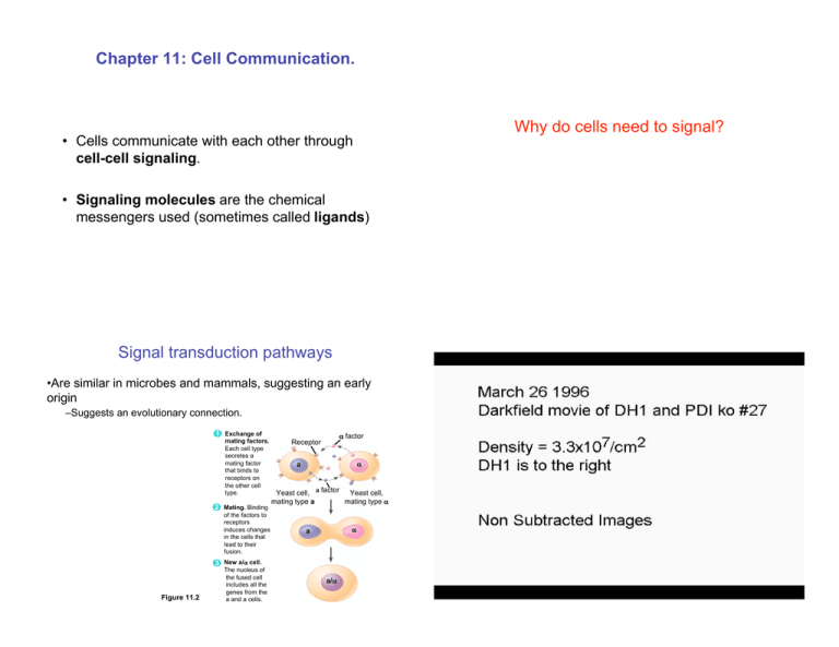
Chapter 11: Cell Communication. Why do cells need to signal? • Cells communicate with each other through cell-cell signaling. • Signaling molecules are the chemical messengers used (sometimes called ligands) Signal transduction pathways •Are similar in microbes and mammals, suggesting an early origin –Suggests an evolutionary connection. 1 Exchange of mating factors. Each cell type secretes a mating factor that binds to receptors on the other cell type. 2 Mating. Binding of the factors to receptors induces changes in the cells that lead to their fusion. ! factor Receptor a ! Yeast cell, a factor Yeast cell, mating type ! mating type a a ! 3 New a/! cell. Figure 11.2 The nucleus of the fused cell includes all the genes from the a and a cells. a/! Signaling in multicellular organisms • Can be both Local or Long-Distance • Local Signaling – Paracrine: The signaling molecule is released and diffuses to the neighboring cells. This is local Signaling. – Synaptic Signaling. Nerve cells signal across a synapse. Long Distance Signaling •Endocrine: Signal is released into a carrier system, such as blood, which carries the molecules to the target cells, which can be far away. – Examples of this are hormones. Long-distance signaling Endocrine cell Blood vessel Local signaling Target cell Electrical signal along nerve cell triggers release of neurotransmitter Neurotransmitter diffuses across synapse Secretory vesicle Local regulator diffuses through extracellular fluid Figure 11.5 A B (a) Paracrine signaling. A secreting cell acts on nearby target cells by discharging molecules of a local regulator (a growth factor, for example) into the extracellular fluid. Hormone travels in bloodstream to target cells Target cell Target cell is stimulated (b) Synaptic signaling. A nerve cell releases neurotransmitter molecules into a synapse, stimulating the target cell. Figure 11.5 (c) Hormonal signaling. Specialized endocrine cells secrete hormones into body fluids, often the blood. Hormones may reach virtually all C body cells. The signal-transduction pathway • For most signals the signaling molecule does not enter the cell • – The signal is relayed through the membrane, from the outside to the inside. • – Reception – Transduction – Response • How is this done? • EXTRACELLULAR FLUID The signaling molecule (ligand) binds to a membrane receptor on the outside of the cell. • This receptor spans the membrane and has both an extracellular and a cytosolic domain. • The cytosolic domain changes shape when bound to ligand. The response of the cell to a signaling molecule is mediated through a signal-transduction pathway. This occurs through 3 steps. 1 Reception Plasma membrane CYTOPLASM 2 Transduction 3 Response Receptor Activation of cellular response Relay molecules in a signal transduction pathway – The ligand is not always a diffusible molecule Signal molecule Figure 11.6 G-Protein linked receptors Receptors • There are three major types of membrane receptors. – G-Protein linked receptors. Figure 11.7a – Tyrosine-Kinase Receptors – Ligand-gated Ion channels. Focus on the G-Protein linked receptors Know the other pathways! • • • Are connected to a G-protein that is activated when a ligand binds to the receptor. The activation triggers the displacement of the GDP by GTP. This activated G-protein then goes on to activate other proteins. Example: Slime mold cAMP receptor system. Tyrosine-Kinase Receptors. • • • • Ligand-gated Ion channels Form dimers. Phosphorylate the tyrosine amino acids on the other receptor This activates the receptor dimer Example: Growth factor receptor. Signal Signal-binding sitea molecule !Helix in the Membrane Tyr Tyr Tyr Tyrosines Signal molecule • Binding of ligand opens up an ion channel. • The opening of the channel leads to a net flow of ions into or out of the cell. Gate closed Ligand-gated ion channel receptor Ions Plasma Membrane Gate open Tyr Tyr Tyr Tyr Tyr Tyr Tyr Tyr Tyr Tyr Tyr Tyr Receptor tyrosine kinase proteins (inactive monomers) CYTOPLASM Signal molecule (ligand) Tyr Tyr Tyr • This triggers an intracellular response. Na+, Ca2+ Dimer Cellular response Activated relay proteins Tyr Tyr Tyr Figure 11.7 Tyr Tyr Tyr 6 ATP Activated tyrosinekinase regions (unphosphorylated dimer) 6 ADP P Tyr P Tyr P Tyr P Tyr P Tyr P Tyr Tyr P Tyr P Tyr P Fully activated receptor tyrosine-kinase (phosphorylated dimer) Tyr P Tyr P Tyr P Cellular response 1 Cellular response 2 Gate close Inactive relay proteins Figure 11.7 Intracellular Receptors. • • Small non polar molecules, Hormones, can travel through the membrane where they can bind to an internal receptor protein Usually activates a transcription factor. Hormone EXTRACELLULAR (testosterone) FLUID 1 The steroid hormone testosterone passes through the plasma membrane. Plasma membrane Receptor protein Hormonereceptor complex Transduction The signal is relayed via cascades of molecular interactions in the cell. • • • • Amplification of the signal can be achieved by phosphorylation cascades and second messengers Adding or removing phosphate groups regulates protein activity. Kinases use ATP to phosphorylate molecules. Phosphatases remove phosphate groups from molecules. Signal molecule 2 Testosterone binds to a receptor protein in the cytoplasm, activating it. e Figure 11.9 ad 5 The mRNA is translated into a specific protein. sc CYTOPLASM New protein ca stimulates the transcription of the gene into mRNA. io n NUCLEUS la t 4 The bound protein PP o ry mRNA Active P protein kinase 2 Inactive protein kinaseATP Active P ADP 3 protein kinase Pi PP 3 Inactive protein ATP ADP P Active protein P i PP Pi ph Figure 11.6 receptor complex enters the nucleus and binds to specific genes. os Active protein kinase 1 Inactive protein kinaseATP ADP 2 Ph Inactive protein kinase 1 3 The hormone- DNA Activated relay molecule Receptor Cellular response Second messengers Nuclear response to a signal Ca2+ Regulate genes by activating transcription factors that turn genes on or off – cAMP and or other small molecules (IP3, DAG) 1 A signal molecule binds 2 Phospholipase C cleaves a to a receptor, leading to plasma membrane phospholipid activation of phospholipase C. called PIP2 into DAG and IP3. EXTRACELLULAR FLUID 3 DAG functions as a second messenger in other pathways. Growth factor Signal molecule (first messenger) Reception Receptor G protein DAG Phosphorylation cascade GTP Figure 11.13 G-protein-linked receptor Phospholipase C PIP2 Transduction IP3 (second messenger) CYTOPLASM IP3-gated calcium channel Endoplasmic reticulum (ER) Various proteins activated Ca2+ Ca2+ Inactive transcription Active factor transcription factor P Cellular response DNA (second messenger) 4 IP3 quickly diffuses through the cytosol and binds to an IP3– gated calcium channel in the ER membrane, causing it to open. 5 Calcium ions flow out of the ER (down their concentration gradient), raising the Ca2+ level in the cytosol. Gene 6 The calcium ions activate the next protein in one or more signaling pathways. Figure 11.14 The signaling is very specific • The same signaling molecule can lead to different responses in different types of cells. Signal molecule EXTRACELLULAR FLUID G protein DAG Relay molecules GTP Response Response 2 G-protein-linked receptor Phospholipase C PIP2 IP3 (second messenger) 3 IP3-gated calcium channel Endoplasmic reticulum (ER) Activation or inhibition Response 4 Figure 11.17 Signal molecule (first messenger) Receptor Response 1 mRNA NUCLEUS Signal Transduction • Receptors only bind to a specific ligand. – Example: Epinephrine – In liver it triggers breakdown of glycogen – In the heart it increase the heart beat. Response Ca2+ Ca2+ (second messenger) Response 5 Various proteins activated Cellular response Summary of signaling. • Via membrane proteins. – G-Protein linked receptors. Many different G-proteins – Tyrosine-Kinase Receptors. – Ligand-gated Ion channels. • Directly Intracellular Receptors – Non-polar molecules that bind to receptors inside cells. Usually activate transcription factors. • Both can lead to amplification of signal. – Branching: Binding of signaling molecule can trigger several different responses inside cell. – Combination of two different signals. Chapter 12 The Cell Cycle. Every living organism must be able to reproduce in order to survive. • Reproduction occurs by Cell Division. – It is part of the Cell Cycle. – It results in genetically identical daughter cells • The Cell Cycle is the foundation of life. • Unicellular organisms – Reproduce by cell division • Multicellular organisms depend on cell division for – Development from a fertilized cell – Growth – Repair Genome Brief review • Eukaryotic cell division consists of – Mitosis, the division of the nucleus – Cytokinesis, the division of the cytoplasm • In meiosis – Sex cells are produced after a reduction in chromosome number • The cells genetic information is called its genome. • The genome contains the recipe for running and building the cell. • The genome contains all the chromosomes. • The chromosome is made up of a very long linear DNA strand containing up to thousands of genes, each gene specifying a specific protein. • This strand is associated with various proteins that maintain the structure and control the activity of genes. • This DNA protein complex is called chromatin. – A cell can have 3 m long DNA strands even though it is only 10 !m long. The Cell Cycle is divided into Two Phases Interphase and Mitosis (Mitotic phase) • • • • • • Interphase is split into 3 phases G1, S, G2 After G2 the cell enters mitosis. • DNA is Duplicated in S phase! Not in mitosis. After the S phase each chromosome has an identical copy, the pair are called sister chromatids. They are joined at the Centeromere, a waist like region near the center of the chromosome. Kinetochores, where the microtubules attach, are formed here as well. INTERPHASE S (DNA synthesis) Chromosome duplication (including DNA synthesis) Once duplicated, a chromosome consists of two sister chromatids connected at the centromere. Each chromatid contains a copy of the DNA molecule. Centromere C M y to ito k i si n e s si s G1 0.5 !m A eukaryotic cell has multiple chromosomes, one of which is represented here. Before duplication, each chromosome has a single DNA molecule. G2 MI (M TOT ) P IC HA SE Figure 12.5 Separation of sister chromatids Mechanical processes separate the sister chromatids into two chromosomes and distribute them to two daughter cells. Figure 12.4 Sister chromatids Centromeres Sister chromatids Mitosis is split into five subphases It is made up of mitosis and cytokinesis • Prophase – Two centerosomes already formed begin to move towards opposite poles of the cell. Microtubules grow from the centerosomes. Chromosomes begin to condense and sister chromatid pairs become visible. • Prometaphase – Nuclear envelope begins to break down. – Each of the two sister chromatids has now a structure called Kinetochore located at the centeromer region. Microtubules begin to attach to the kinetochores. • G2 OF PROMETAPHASE PROPHASE INTERPHASE Centrosomes Aster Fragments Early mitotic Kinetochore (with centriole pairs) Chromatin of nuclear Centromere (duplicated) spindle Nonkinetochore envelope microtubules Figure 12.6 Nucleolus Nuclear Plasma envelopemembrane Chromosome, consisting of two sister chromatids Kinetochore microtubule Metaphase – Centerosomes are at opposite poles. Chromosomes convene in the center between the two centerosomes . • METAPHASE Anaphase – The two sister chromatids are separated as the microtubules shorten. • ANAPHASE Metaphase plate TELOPHASE AND CYTOKINESIS Cleavage furrow Telophase – Two daughter nuclei form and the nuclear envelope arises from the fragments of the parents nuclear envelope. The chromosomes become less tightly coiled. Figure 12.6 Spindle Centrosome at Daughter one spindle polechromosomes Nuclear envelope forming Nucleolus forming Animal Mitosis (Bio Flix) • Every Eukaryotic cell has a characteristic number of chromosomes. – Diploid cells have pairs of homologous chromosomes. – Haploid cells have single chromosomes. The only haploid cells in humans are the gametes. Plant and animal Cytokinesis • Bacteria divide by binary fission Cytokinesis, the division of the cytoplasm usually finishes at the end of telophase. •In animal cells • –Cytokinesis occurs by a process known as cleavage, forming a cleavage furrow In plant cells, during cytokinesis – A cell plate forms Origin of replication E. coli cell Chromosome replication 1 begins. Soon thereafter, one copy of the origin moves rapidly toward the other end of the cell. 2 Replication continues. One copy of the origin is now at each end of the cell. Cleavage furrow 100 !m Vesicles forming cell plate Wall of 1 !m patent cell Cell plateNew cell wall 3 Replication finishes. The plasma membrane grows inward, and new cell wall is deposited. Figure 12.11 4 Two daughter cells result. Contractile ring of microfilaments Figure 12.9 A Daughter cells (a) Cleavage of an animal cell (SEM) Daughter cells (b) Cell plate formation in a plant cell (SEM) Figure 12.9 B Cell wall Two copies of origin Origin Plasma Membrane Bacterial Chromosome Origin How many chromatids does a human cell have after undergoing S-phase? Can the repeated cycle of cell divisions continue unchecked? A) 46 B) 19 C) 23 D) 32 E) 92 Cell check points • The cell cycle is regulated by a molecular control system • There are 3 major checkpoints during the cell cycle. (a) Fluctuation of MPF activity and cyclin concentration during the cell cycle M G1 S G2 M G1 S G2 M MPF activity Cyclin Time (b) Molecular mechanisms that help regulate the cell cycle 1 Synthesis of cyclin begins in late S phase and continues through G2. Because cyclin is protected from degradation during this stage, it accumulates. G1 checkpoint M Figure 12.14 G2 Cdk Cyclin is degraded M checkpoint G2 checkpoint 4 During anaphase, the cyclin component Figure 12.17 A, B of MPF is degraded, terminating the M phase. The cell enters the G1 phase. 2 Degraded Cyclin M G1 G S the cell favor degradation of cyclin, and the Cdk component of MPF is recycled. S Control system 1 5 During G1, conditions in G • – The G1 checkpoint goes into S or into G0 – The G2 checkpoint – And the Mitotic checkpoint. Why are checkpoints necessary? Cell cycle clock – Cyclins proteins whose concentrations cycles during the cell cycle – Cyclin-dependent-Kinases (CDK’s) that are activated when bound to the cyclins. – MPF maturation (mitotic) promotion factor at the G2 checkpoint, a cdkcyclin complex. G2 Cdk checkpoint MPF Cyclin 2 Accumulated cyclin molecules combine with recycled Cdk molecules, producing enough molecules of MPF to pass the G2 checkpoint and initiate the events of mitosis. 3 MPF promotes mitosis by phosphorylating various proteins. MPF‘s activity peaks during metaphase. • • • Growth factors PDGF ( Platelet derived growth factor) for fibroblasts affect the G1 checkpoint. Density dependence and anchorage dependence inhibition. • Growth factors – PDGF ( Platelet derived growth factor) for fibroblasts affect the G1 checkpoint. • – Can lower density of growth factors. – There is also contact inhibition. (a) Normal mammalian cells. The availability of nutrients, growth factors, and a substratum for attachment limits cell density to a single layer. Control of Cell growth Density dependence and anchorage dependence inhibition. – Can lower density of growth factors. – There is also contact inhibition. Cells anchor to dish surface and divide (anchorage dependence). When cells have formed a complete single layer, they stop dividing (density-dependent inhibition). (a) Normal mammalian cells. The availability of nutrients, growth factors, and a substratum for attachment limits cell density to a single layer. If some cells are scraped away, the remaining cells divide to fill the gap and then stop (density-dependent inhibition). Figure 12.18 A Cancer • Cancer cells – – – – – Exhibit neither density-dependent inhibition nor anchorage dependence Have escaped from cell cycle control Make excessive amounts of GF Have lost the ability to be controlled by GF. GF signaling pathway abnormal. Lymph vessel Tumor Glandular tissue Figure 12.20 Blood vessel Cancer cell Metastatic Tumor When cells have formed a complete single layer, they stop dividing (density-dependent inhibition). If some cells are scraped away, the remaining cells divide to fill the gap and then stop (density-dependent inhibition). Figure 12.19 A 25 !m Cells anchor to dish surface and divide (anchorage dependence). 25 !m
