The nucleus subglomerulosus of the trout visual systems: A DiI study
advertisement
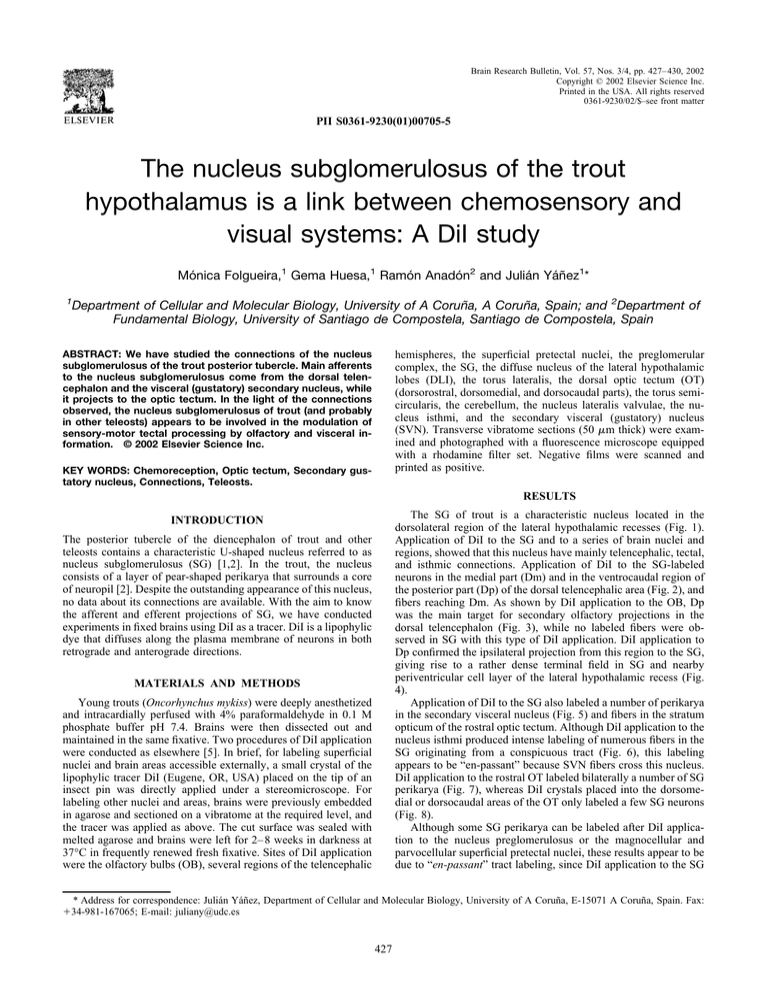
Brain Research Bulletin, Vol. 57, Nos. 3/4, pp. 427– 430, 2002 Copyright © 2002 Elsevier Science Inc. Printed in the USA. All rights reserved 0361-9230/02/$–see front matter PII S0361-9230(01)00705-5 The nucleus subglomerulosus of the trout hypothalamus is a link between chemosensory and visual systems: A DiI study Mónica Folgueira,1 Gema Huesa,1 Ramón Anadón2 and Julián Yáñez1* 1 Department of Cellular and Molecular Biology, University of A Coruña, A Coruña, Spain; and 2Department of Fundamental Biology, University of Santiago de Compostela, Santiago de Compostela, Spain ABSTRACT: We have studied the connections of the nucleus subglomerulosus of the trout posterior tubercle. Main afferents to the nucleus subglomerulosus come from the dorsal telencephalon and the visceral (gustatory) secondary nucleus, while it projects to the optic tectum. In the light of the connections observed, the nucleus subglomerulosus of trout (and probably in other teleosts) appears to be involved in the modulation of sensory-motor tectal processing by olfactory and visceral information. © 2002 Elsevier Science Inc. hemispheres, the superficial pretectal nuclei, the preglomerular complex, the SG, the diffuse nucleus of the lateral hypothalamic lobes (DLI), the torus lateralis, the dorsal optic tectum (OT) (dorsorostral, dorsomedial, and dorsocaudal parts), the torus semicircularis, the cerebellum, the nucleus lateralis valvulae, the nucleus isthmi, and the secondary visceral (gustatory) nucleus (SVN). Transverse vibratome sections (50 m thick) were examined and photographed with a fluorescence microscope equipped with a rhodamine filter set. Negative films were scanned and printed as positive. KEY WORDS: Chemoreception, Optic tectum, Secondary gustatory nucleus, Connections, Teleosts. RESULTS The SG of trout is a characteristic nucleus located in the dorsolateral region of the lateral hypothalamic recesses (Fig. 1). Application of DiI to the SG and to a series of brain nuclei and regions, showed that this nucleus have mainly telencephalic, tectal, and isthmic connections. Application of DiI to the SG-labeled neurons in the medial part (Dm) and in the ventrocaudal region of the posterior part (Dp) of the dorsal telencephalic area (Fig. 2), and fibers reaching Dm. As shown by DiI application to the OB, Dp was the main target for secondary olfactory projections in the dorsal telencephalon (Fig. 3), while no labeled fibers were observed in SG with this type of DiI application. DiI application to Dp confirmed the ipsilateral projection from this region to the SG, giving rise to a rather dense terminal field in SG and nearby periventricular cell layer of the lateral hypothalamic recess (Fig. 4). Application of DiI to the SG also labeled a number of perikarya in the secondary visceral nucleus (Fig. 5) and fibers in the stratum opticum of the rostral optic tectum. Although DiI application to the nucleus isthmi produced intense labeling of numerous fibers in the SG originating from a conspicuous tract (Fig. 6), this labeling appears to be “en-passant” because SVN fibers cross this nucleus. DiI application to the rostral OT labeled bilaterally a number of SG perikarya (Fig. 7), whereas DiI crystals placed into the dorsomedial or dorsocaudal areas of the OT only labeled a few SG neurons (Fig. 8). Although some SG perikarya can be labeled after DiI application to the nucleus preglomerulosus or the magnocellular and parvocellular superficial pretectal nuclei, these results appear to be due to “en-passant” tract labeling, since DiI application to the SG INTRODUCTION The posterior tubercle of the diencephalon of trout and other teleosts contains a characteristic U-shaped nucleus referred to as nucleus subglomerulosus (SG) [1,2]. In the trout, the nucleus consists of a layer of pear-shaped perikarya that surrounds a core of neuropil [2]. Despite the outstanding appearance of this nucleus, no data about its connections are available. With the aim to know the afferent and efferent projections of SG, we have conducted experiments in fixed brains using DiI as a tracer. DiI is a lipophylic dye that diffuses along the plasma membrane of neurons in both retrograde and anterograde directions. MATERIALS AND METHODS Young trouts (Oncorhynchus mykiss) were deeply anesthetized and intracardially perfused with 4% paraformaldehyde in 0.1 M phosphate buffer pH 7.4. Brains were then dissected out and maintained in the same fixative. Two procedures of DiI application were conducted as elsewhere [5]. In brief, for labeling superficial nuclei and brain areas accessible externally, a small crystal of the lipophylic tracer DiI (Eugene, OR, USA) placed on the tip of an insect pin was directly applied under a stereomicroscope. For labeling other nuclei and areas, brains were previously embedded in agarose and sectioned on a vibratome at the required level, and the tracer was applied as above. The cut surface was sealed with melted agarose and brains were left for 2– 8 weeks in darkness at 37°C in frequently renewed fresh fixative. Sites of DiI application were the olfactory bulbs (OB), several regions of the telencephalic * Address for correspondence: Julián Yáñez, Department of Cellular and Molecular Biology, University of A Coruña, E-15071 A Coruña, Spain. Fax: ⫹34-981-167065; E-mail: juliany@udc.es 427 428 FIG. 1. Nissl-stained transverse section through the trout hypothalamus showing the rostral part of the nucleus subglomerulosus (SG). Scale bar: 70 m. See List of Abbreviations. FIG. 2. Transverse section through the caudal telencephalon showing retrograde labeled cells (arrowheads) in the ventrocaudal region of Dp after DiI application to the nucleus subglomerulosus. Scale bar: 100 m. See List of Abbreviations. FIG. 3. Transverse section through the caudal telencephalon showing intensely labeled secondary olfactory projections in Dp after DiI application in the olfactory bulb. Scale bar: 100 m. See List of Abbreviations. FOLGUEIRA ET AL. FIG. 4. Detail of a transverse section through the nucleus subglomerulosus (SG) to show anterograde varicose fibers (arrows) after DiI application to the Dp. Scale bar: 100 m. See List of Abbreviations. FIG. 5. Transverse section through the isthmus showing a dense group of retrograde labeled cells (arrowhead) in the secondary visceral nucleus (SVN) and their axons (arrows) crossing the nucleus isthmi (NI) after DiI application to the SG. Scale bar: 100 m. See List of Abbreviations. FIG. 6. Transverse section through the hypothalamus showing anterogradely labeled fibres (arrow) in the SG after DiI application to the isthmus. Scale bar: 100 m. See List of Abbreviations. CONNECTIONS OF THE NUCLEUS SUBGLOMERULOSUS FIGS. 7 and 8. Transverse section through the SG after DiI application to the rostro- (7) and caudo-dorsal (8) region of the optic tectum (OT) showing retrogradely labeled perikarya (arrowheads). Scale bar: 60 m. See List of Abbreviations. 429 appear to be mainly telencephalic (Dp and Dm) and isthmal (SVN). The afferent neurons located in Dp appear to overlap the main terminal field of the secondary olfactory projections ([4], present results). These results suggest that Dp transmits olfactory information to the SG. Extratelencephalic projections originating from Dp were not reported previously in teleosts (see [3]). The SVN of trout projects mostly to the ipsilateral tertiary gustatory nucleus of the hypothalamus [5]. The present results indicate that the SG is also a specific target of the SVN, a connection that was not previously reported in teleosts. Therefore, both tertiary visceral (gustatory?) and olfactory projections appear to be the most significant afferents to the SG. The OT is the main visual center of teleosts [4]. Interestingly, it was the only target identified for the SG neurons. Our results also indicate that many more SG neurons were labeled when DiI was applied to the rostral OT than when it was applied to the caudal tectum, which indicates some topographical organization of the SG-tectal projection. Together, present experiments suggest that the SG of trout may have a role in the modulation of sensory-motor processing in the OT by both taste and olfactory inputs. ACKNOWLEDGEMENTS does not label terminal fields in these nuclei. DiI application to other structures mentioned above did not label neurons or fibers in the SG, with the exception of DLI. This work was supported by a grant from the Xunta de Galicia (PGIDT99BIO20002) and the Science and Technology Ministry (BXX2000-0453-CO2-01 and O2). DISCUSSION REFERENCES The present study demonstrates for the first time the connections of the SG. The afferents of the SG (summarized in Fig. 9) 1. Gómez Segade, P.; Anadón, R. Specialization in the diencephalon of advanced teleosts. J. Morphol. 197:71–104; 1988. FIG. 9. Schematic drawings of transverse sections (A–E) of the trout brain (levels are indicated in the inset) showing the main connections of the SG. See List of Abbreviations. 430 FOLGUEIRA ET AL. 2. Holmgren, N. Zur Anatomie und Histologie des Vorder- und Zwischenhirns der Knochenfischen hauptsächlich nach Untersuchungen an Osmerus eperlanus. Acta Zool. (Stockh.) 1:137–153; 1920. 3. Matz, S. P. Connections of the olfactory bulb in the chinook salmon (Oncorhynchus tshawytscha). Brain Behav. Evol. 46:108 –120; 1995. 4. Meek, J.; Nieuwenhuys, R. Holosteans and teleosts. In: Nieuwenhuys, R.; ten Donkelaar, H. J.; Nicholson, C., eds. The central nervous system of vertebrates, vol. 2. Berlin: Springer Verlag; 1998:759 –937. 5. Pérez, S. E.; Yáñez, J.; Marı́n, O.; Anadón, R.; González, A.; Rodrı́guez-Moldes, I. Distribution of choline acetyltransferase (ChAT) immunoreactivity in the brain of the adult trout, and tract-tracing observations on the connections of the nuclei of the isthmus. J. Comp. Neurol. 428:450 – 474; 2000. LIST OF ABBREVIATIONS CB cerebellum DLI diffuse nucleus of the lateral hypothalamic lobes Dm medial part of the dorsal telencephalic area Dp ENT H HL LR NI OB OC OT PG PON PSP SG SVN T TS VC posterior part of the dorsal telencephalic area entopeduncular nucleus habenula lateral hypothalamic lobes lateral hypothalamic recess nucleus isthmi olfactory bulb optic chiasm optic tectum nucleus preglomerulosus preoptic nucleus superficial pretectal nucleus, parvocellular part nucleus subglomerulosus secondary visceral nucleus telencephalon torus semicircularis valvula cerebelli

