A bioinformatic assay for BRIEF COMMUNICATIONS
advertisement

BRIEF COMMUNICATIONS
© 2011 Nature America, Inc. All rights reserved.
A bioinformatic assay for
pluripotency in human cells
Franz-Josef Müller1,11, Bernhard M Schuldt2,11,
Roy Williams3, Dylan Mason4, Gulsah Altun5,
Eirini P Papapetrou6, Sandra Danner7,
Johanna E Goldmann5,8, Arne Herbst1, Nils O Schmidt9,
Josef B Aldenhoff1, Louise C Laurent5,10 &
Jeanne F Loring5
Pluripotent stem cells (PSCs) are defined by their potential
to generate all cell types of an organism. The standard assay
for pluripotency of mouse PSCs is cell transmission through
the germline, but for human PSCs researchers depend on
indirect methods such as differentiation into teratomas in
immunodeficient mice. Here we report PluriTest, a robust openaccess bioinformatic assay of pluripotency in human cells based
on their gene expression profiles.
The current standard for demonstrating that human stem cells are
pluripotent is based on their ability to generate a complex variety
of tissues in tumors developed in immunodeficient mice. This
teratoma assay is widely considered to be the most reliable and
informative assay for pluripotency in human cells1 and its use has
increased substantially after the report of induction of pluripotency in somatic cells2. However, the generation of teratomas is
technically challenging, resource-intensive, primarily qualitative
and difficult to standardize, and as we have previously argued,
may have limited value as a criterion for pluripotency3. With the
rapid increase in generation of pluripotent human cells, especially
induced pluripotent stem cell (iPSC) lines, there is a need for a
cost-effective, animal-free alternative to the teratoma assay for
assessing pluripotency in human cells4. The low cost and accessibility of microarray-based gene expression datasets makes transcription profiling an attractive alternative. We hypothesized that
machine-learning methods that are capable of delineating stem
cell phenotypes5 based on microarray data could also predict the
presence or absence of pluripotent features for unknown samples
of cells.
We considerably expanded the gene expression database that
we previously used for defining stem cell phenotypes5 to a much
larger dataset we term ‘stem cell matrix 2’ (SCM2). The SCM2
database contains ~450 genome-wide transcriptional profiles
from diverse stem cell preparations from multiple laboratories,
differentiated cell types, and developing and adult human tissues
(Supplementary Table 1). The SCM2 dataset contains expression profiles from 223 human embryonic stem cell (hESC) and
41 iPSC lines. We analyzed the samples for the SCM2 dataset in
a highly quality controlled pipeline, using Illumina microarrays.
After appropriate transformation and normalization, we used nonnegative matrix factorization (NMF) for dimension reduction and
to identify unexpected patterns engrained in the datasets6. NMF
is a systematic, unbiased approach to identify multigene features,
frequently termed ‘metagenes’ in gene-expression studies7, which
can be used to characterize stem cell phenotypes3.
We then assessed pluripotency of an unknown, potentially
pluripotent sample by comparison of a ‘query gene expression
profile’ from the sample to data models derived from the SCM2
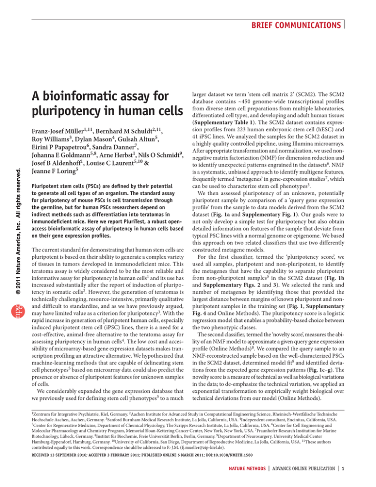
dataset (Fig. 1a and Supplementary Fig. 1). Our goals were to
not only develop a simple test for pluripotency but also obtain
detailed information on features of the sample that deviate from
typical PSC lines with a normal genome or epigenome. We based
this approach on two related classifiers that use two differently
constructed metagene models.
For the first classifier, termed the ‘pluripotency score’, we
used all samples, pluripotent and non-pluripotent, to identify
the metagenes that have the capability to separate pluripotent
from non-pluripotent samples5 in the SCM2 dataset (Fig. 1b
and Supplementary Figs. 2 and 3). We selected the rank and
number of metagenes by identifying those that provided the
largest distance between margins of known pluripotent and nonpluripotent samples in the training set (Fig. 1, Supplementary
Fig. 4 and Online Methods). The pluripotency score is a logistic
regression model that enables a probability-based choice between
the two phenotypic classes.
The second classifier, termed the ‘novelty score’, measures the ability of an NMF model to approximate a given query gene expression
profile (Online Methods)8. We compared the query sample to an
NMF-reconstructed sample based on the well-characterized PSCs
in the SCM2 dataset, determined model fit8 and identified deviations from the expected gene expression patterns (Fig. 1c–g). The
novelty score is a measure of technical as well as biological variations
in the data; to de-emphasize the technical variation, we applied an
exponential transformation to empirically weight biological over
technical deviations from our model (Online Methods).
1Zentrum für Integrative Psychiatrie, Kiel, Germany. 2Aachen Institute for Advanced Study in Computational Engineering Science, Rheinisch-Westfälische Technische
Hochschule Aachen, Aachen, Germany. 3Sanford Burnham Medical Research Institute, La Jolla, California, USA. 4Independent consultant, Encinitas, California, USA.
5Center for Regenerative Medicine, Department of Chemical Physiology, The Scripps Research Institute, La Jolla, California, USA. 6Center for Cell Engineering and
Molecular Pharmacology and Chemistry Program, Memorial Sloan-Kettering Cancer Center, New York, New York, USA. 7Fraunhofer Research Institution for Marine
Biotechnology, Lübeck, Germany. 8Institut für Biochemie, Freie Universität Berlin, Berlin, Germany. 9Department of Neurosurgery, University Medical Center
Hamburg-Eppendorf, Hamburg, Germany. 10University of California, San Diego, Department of Reproductive Medicine, La Jolla, California, USA. 11These authors
contributed equally to this work. Correspondence should be addressed to F.-J.M. (fj.mueller@zip-kiel.de).
RECEIVED 13 SEPTEMBER 2010; ACCEPTED 3 FEBRUARY 2011; PUBLISHED ONLINE 6 MARCH 2011; DOI:10.1038/NMETH.1580
NATURE METHODS | ADVANCE ONLINE PUBLICATION | 1
BRIEF COMMUNICATIONS
Offline
SCM2
computation
Model generation
phenotypic
with NMF
sample information
and microarray data
Raw microarray
data from stem
cell samples
NMF
model
database
Pluripotency score
and novelty score
Report
Pluripotency score selected
for maximal class separation
0
–50
–100
0
–20
–40
–60
f
50
20
0
–50
0.31
0.32
0.33
Novelty score
0.34
0.35
1.0
1.5 2.0 2.5 3.0
1.67
Novelty score
3.5
Good
g
40
20
0
–20
–40
–60
1.5 2.0 2.5 3.0
Novelty score
3.5
Bad
Model fit (simulated)
0
–20
–40
–60
–80
–100
–120
–100
0.30
–100
1.0
–80
–100
–80
–50
Somatic cells, cancer tissue, cell lines
and in vitro preparations differentiated
from pluripotent stem cells
Pluripotency score
0
Pluripotency score
Pluripotency score
© 2011 Nature America, Inc. All rights reserved.
e
50
0
100
200
300
400
Number of samples
Human pluripotent stem cell preparations
Germ cell tumor cells and tissues
Parthenogenic pluripotent stem cells
d
50
Pluripotency score
Microarray data
upload
http://www.pluritest.org/
c
50
Pluripotency score
Online
computation
User
Component
microarray
projection
database
b
High
Researcher
output
Pluripotency score
PluriTest
Pluripotency signature (simulated)
Researcher
input
Low
a
1.0
1.5
2.0
2.5
Novelty score
3.0
2.8
3.0
3.2
3.4
3.6
Novelty score
Figure 1 | A multidimensional data model for assessing PSCs. (a) Schematic for PluriTest. (b,c) Assessment of pluripotent and somatic cell samples in
the training dataset with the pluripotency score only (b) and with both PluriTest scores (c). (d–g) PluriTest classifiers tested on datasets generated
using four different microarray platforms: Illumina WG6v1 (d, 177 samples)5, HT12v3 (e, 498 samples), HT12v4 (f, 38 samples) and Affymetrix U133A (g,
5,372 samples)10. Samples for these datasets were independently generated (e,f) and/or curated from published stu dies (d,e,g). In e, the lines in the
plot indicate empirically determined thresholds for defining normal pluripotent lines.
The combination of the pluripotency score and the novelty
score enables open-ended assessment of pluripotent features in
a query sample when that sample is a new kind of PSC. The first
classifier reports to what extent a query sample contains a pluripotent signature, and the second classifier reports how much of
the signal measured in a query sample can be explained by the
normal PSC lines contained in the SCM2 dataset (Supplementary
Note 1 and Supplementary Fig. 1). To test the two-classifier
approach, we analyzed germ cell tumor cell lines. These cells
are pluripotent and resemble normal PSCs but have genetic and
epigenetic abnormalities 9. These cells had high pluripotency
scores, as expected, but the novelty score indicated that they
deviate from the normal PSCs in the SCM2 dataset (Fig. 1 and
Supplementary Fig. 2).
We tested the combined classification approach and communication framework, which we termed PluriTest (http://www.
pluritest.org/), using several independently generated test datasets containing pluripotent and non-pluripotent samples: Illumina
WG6v1 (ref. 5), HT12v3 and HT12v4 datasets (Fig. 1d–f) generated in-house on our own microarray scanner and datasets that had
been generated in six different core facilities (Online Methods and
Supplementary Table 1). We also used PluriTest to examine data
from a recently published human transcriptome atlas10 based on
Affymetrix U133A arrays (Fig. 1g).
PluriTest predicted pluripotency with excellent sensitivity and
specificity. We set thresholds that separated pluripotent from nonpluripotent samples in HT12v3 test datasets with 98% sensitivity and 100% specificity, and also distinguished germ cell tumor
cell lines and parthenogenetic stem cell lines from the bulk of
2 | ADVANCE ONLINE PUBLICATION | NATURE METHODS
PSCs (Fig. 1d–f and Supplementary Fig. 2). A few pluripotent
samples had unusually high novelty scores (Fig. 1e), indicating that these test samples should be additionally evaluated for
epigenetic or genetic abnormalities or unwanted differentiation
(Supplementary Fig. 1). For the most informative analysis, the
query sample should be analyzed on the same platform as the
training dataset (Illumina HT12), but acceptable results can be
obtained with data from other platforms (Fig. 1f, Supplementary
Fig. 3 and Supplementary Note 2).
We examined the performance of PluriTest on hESC lines
(SIVF014, SIVF011, SIVF042, F4.2 and WA01) and human iPSC
lines (HDF51IPS12 and HDF51IPS1), which were part of the
training dataset; these lines grouped together and were separated from somatic samples (Fig. 2a). PluriTest also separated
fully and partially reprogrammed iPSC lines (samples that were
not in the training datatset; Fig. 2b); partially reprogrammed
cell lines clustered with non-pluripotent cells. We then applied
PluriTest to samples from a neural differentiation time-course
experiment that also were not in the training dataset (Fig. 2c,d).
We differentiated WA09 cells into neural precursors and collected
three biological replicates on day 0, day 3, day 6 and day 14 after
neural induction. The novelty score changed after 3 d of differentiation, but the pluripotency score was still high at this time
point, whereas samples from later time points dropped out of
the pluripotency score space and had increasingly higher novelty
scores (Fig. 2c). In a mixing experiment in which we combined
RNA samples from different time points (day 0 and day 14) at
varying ratios, PluriTest could separate the differentially mixed
samples (Fig. 2d).
BRIEF COMMUNICATIONS
a
Scaled empirical density distribution
PSC
b
PSC
0
–50
–100
2
3
Novelty score
hESC
Fibroblasts
Heart muscle
–50
4
1
hiPSC
Astrocytes
Skeletal muscle
2
3
Novelty score
4
Fully reprogrammed hiPSC, low passage
Fully reprogrammed hiPSC, high passage
Partially reprogrammed hiPSC, low passage
Scaled empirical density distribution
PSC
d
Scaled empirical density distribution
Somatic cells
PSC
Pluripotency score
0
–50
–100
METHODS
Methods and any associated references are available in the online
version of the paper at http://www.nature.com/naturemethods/.
0
Note: Supplementary information is available on the Nature Methods website.
–50
–100
1
2
3
Novelty score
Day 0
Day 3
Day 6
Day 14
4
a few markers is no longer necessary. Using all of the expression
information available provides much higher discriminatory power
and the ability to identify deviations from known patterns that
may lead to additional insights into cellular phenotypes.
The PluriTest framework could be applied to any unbiased highcontent dataset, such as global DNA methylation analysis or RNA
sequencing data, provided that there is sufficient representation
of a defined target phenotype in the training dataset. Our results
suggest that it will be relatively straightforward to construct
similar models of developmental pathways such as differentiation
along the neural, endodermal or hematopoietic lineages. Such
databases will inform subsequent experiments and may be applicable as a rapid method to quality control PSC-derived preparations for experimental and preclinical investigations.
Somatic cells
50
50
Pluripotency score
0
–100
1
© 2011 Nature America, Inc. All rights reserved.
Somatic cells
50
Pluripotency score
Pluripotency score
50
c
Scaled empirical density distribution
Somatic cells
1
2
3
Novelty score
100%:0%
75%:25%
66%:33%
50%:50%
4
33%:66%
25%:75%
0%:100%
Figure 2 | Output of PluriTest. (a–c) PluriTest results for known pluripotent cells
and somatic cells and tissues (a), for fully and partially reprogrammed iPSC lines
(b) and for an hESC line (WA09) differentiated into neural precursors, at the
indicated time points (c). (d) PluriTest results for mixed samples of hESC and
hESC-derived neural precursor RNA (day 0 and day 14 from the data shown in c)
at the indicated ratios. hiPSC, human iPSC. The background encodes an empirical
density map indicating pluripotency and novelty as indicated by the color bar.
The PluriTest is contained in a single R/Bioconductor opensource, open-access workspace11 (Supplementary Data and
Supplementary Note 3) that also includes the SCM2 database–
derived NMF models. To enable easy access to PluriTest, we
programmed a rich internet application (RIA) using Microsoft
Silverlight 4 and C# (http://www.pluritest.org/). The RIA automatically performs all data extraction and preprocessing steps
after the upload of an unmodified microarray scanner output
file. All data and results are stored securely in a Microsoft structured query language (MS-SQL) database. We used the binary
microarray scanner output file (.idat file) as the most basic ‘stem
cell query term’. After upload, the results of our PSC-prediction
algorithm are reported back to the user via a web interface (Fig. 2
and Supplementary Fig. 5). PluriTest can run on every recent
(Mac OS 10.5 and Windows XP or later) operating system, and
requires internet access and a local installation of the Silverlight 4
plug-in. A typical online analysis with 12 samples takes less than
10 min including data upload (Supplementary Note 2).
Here we demonstrated the general feasibility of a web-based
prediction of stem cell properties12. PluriTest breaks from the
conventional marker-based approaches to assess pluripotency of
human cells, which typically assay a few markers by methods
such as quantitative real-time PCR. With the lowered cost of
whole-genome analysis, reduction of a gene expression profile to
ACKNOWLEDGMENTS
F.-J.M. is supported by an Else-Kröner Fresenius Stiftung fellowship. J.F.L. is
supported by grants from the California Institute for Regenerative Medicine
(RT1-01108, TR1-01250 and CL1-00502), the US National Institutes of Health
(R21 MH087925), the Bill and Melinda Gates Foundation, the Esther O’Keeffe
Foundation and the Millipore Foundation. B.M.S. is supported by Bayer Technology
Services GmbH and the Deutsche Forschungsgemeinschaft (GSC 111). L.C.L. is
supported by a US National Institutes of Health National Institute of Child Health
and Human Development K12 Career Development award. E.P.P. was supported by
New York State Stem Cell Science grant N08T-060. We thank C. Lynch and H. Tran
for preparing the samples and running the arrays; A. Schuppert, S. Peterson and
K. Nazor for comments, criticisms and reading the manuscript for clarity;
M. Sadelain (Memorial Sloan-Kettering Cancer Center) for providing samples
and data; K. Haden and I. Mikoulitch (Illumina) for help with handling Illumina
BeadArray data formats and letting us use the idat.reader.dll program module in
PluriTest RIA; and C. Becker, D. Barker and A. Fritz for helpful discussions.
AUTHOR CONTRIBUTIONS
F.-J.M. conceived and designed the study. F.-J.M. and B.M.S. developed the
PluriTest algorithm. F.-J.M., J.F.L., L.C.L. and J.B.A. oversaw the sample
collection, microarray analysis and coordinated biological and bioinformatic
experiments. R.W., D.M., B.M.S. and A.H. implemented the online bioinformatic
platform. R.W., D.M., F.-J.M., B.M.S. and G.A. provided bioinformatic analyses.
E.P.P., S.D., J.E.G. and N.O.S. prepared biological samples. F.-J.M., B.M.S. and
J.F.L. wrote the manuscript with input from all authors.
COMPETING FINANCIAL INTERESTS
The authors declare no competing financial interests.
Published online at http://www.nature.com/naturemethods/.
Reprints and permissions information is available online at http://npg.nature.
com/reprintsandpermissions/.
1.
2.
3.
4.
Daley, G.Q. et al. Cell Stem Cell 4, 200–201 (2009).
Takahashi, K. & Yamanaka, S. Cell 126, 663–676 (2006).
Müller, F.J. et al. Cell Stem Cell 6, 412–414 (2010).
Russell, W.M.S. & Burch, R.L. The Principles of Humane Experimental
Technique (Methuen, London, 1959).
5. Müller, F.J. et al. Nature 455, 401–405 (2008).
6. Lee, D.D. & Seung, H.S. Nature 401, 788–791 (1999).
7. Brunet, J.P. et al. Proc. Natl. Acad. Sci. USA 101, 4164–4169 (2004).
8. Tax, D.M.J. & Muller, K.-R. IEEE Proc. Pattern Recognition 3,
1051–4651/04 (2004).
9. Josephson, R. et al. Stem Cells 25, 437–446 (2007).
10. Lukk, M. et al. Nat. Biotechnol. 28, 322–324 (2010).
11. R Development Core Team. R: A Language and Environment for Statistical
Computing (R Foundation for Statistical Computing, Vienna, 2010).
12. Gray, J. in Data-Intensive Scientific Discovery (eds., Hey, T., Tansley, S. and
Tolle, K.) XVII–XXXI (Microsoft Research, Redmond, Washington, USA, 2009).
NATURE METHODS | ADVANCE ONLINE PUBLICATION | 3
© 2011 Nature America, Inc. All rights reserved.
ONLINE METHODS
Microarray analysis. Sample runs were analyzed in-house essentially as reported previously5, except that Illumina HT12 arrays
were used. We first filtered the probes that are present on both
Illumina HT12v3 and HT12v4 arrays to ensure identical results
when either of the two array versions were used with the PluriTest
application. We filtered for probes that were detected with a
P value of at least < 0.01 in at least ten samples of the SCM2 dataset.
After filtering, 22,135 probes were retained and raw probe expression values were transformed and normalized with the variance
stabilization transformation and robust spline normalization
functions as implemented in the lumi R/Bioconductor package13.
We normalized sample data to an in-house well-characterized
pluripotent target sample (WA09).
Sample collection, test and training datasets. We analyzed 468 human
samples for generating the PluriTest model. Of these, 204 were derived
from somatic cells and tissues, 264 were pluripotent samples (223 hESC
and 41 human iPSC; Fig. 1b,c). With these samples we trained both the
multiclass and one-class classifiers. For our test datasets we analyzed
samples in-house on Illumina HT12v3 (398 samples total; Fig. 1e) and
v4 arrays (39 samples total; Fig. 1f) but also combined these samples
with published datasets. J. Jeyakani (Genome Institute of Singapore),
A. Tarca (Wayne State University), Toshima Parris (University of
Gothenburg), M. Suarez-Farinas (Rockefeller University), S. Doulatov
(Ontario Cancer Institute, Toronto), K. Gandhi and D. Booth
(Westmead Millenium Institute, Sydney) shared the raw .idat files from
their published studies (National Center for Biotechnology Information
Gene Expression Omnibus (GEO) accession numbers GSE21973
(ref. 14), GSE204628 (ref. 15), GSE170489 (ref. 16), GSE2113510
(ref. 17) and GSE1868611 (ref. 18).
For the Illumina SCM1 dataset (GSE115081)3 we focused on
samples from our previous study that we analyzed on the WG6v1
platform (177 samples total; Fig. 1d).
For the Affymetrix U133A dataset (EM-Tab-6212; 5,372 samples total)10 we translated the gene identifiers from the HT12v3
PLATFORM to the respective gene array annotation with a mapping table provided by Illumina (http://www.switchtoi.com/
probemapping.ilmn, accessed 6 June 2010).
In the other cases (WG6v1 (GSE115081), Illumina WG6v3,
HT12v4), most probes targeting specific transcripts were identical
and matched based on their specific probe nucleotide universal
identifiers (NuIDs)13.
Details on all samples used for training and testing PluriTest
are available in Supplementary Table 1.
Partially reprogrammed cell preparations. Human dermal
fibroblasts (HDFs; Sciencell) were cultured in DMEM, 2 mM
GlutaMax, 10% FBS and 0.1 mM non-essential amino acids (Life
Technologies). HDFiPS cells were generated and maintained in
standard hESC medium containing DMEM/F12 supplemented
with 20% Knockout Serum Replacement (Life Technologies),
2 mM GlutaMax, 0.1 mM non-essential amino acids, 0.1 mM
2-mercaptoethanol and 12 ng ml−1 of bFGF (Stemgent). HDFiPS
cells were cultured on irradiated mouse embryonic fibroblasts
(MEFs) in hESC medium and mechanically passaged once a week.
The hESC medium was changed daily.
PLAT-A packaging cells were plated onto six-well plates coated
with poly(D-lysine) at a density of 1.5 × 10⁶ cells per well without
NATURE METHODS
antibiotics and incubated overnight. Cells were transfected with
4 Mg pMXs retroviral plasmids, which carry human POU5F1 (also
known as OCT4), SOX2, KLF4 or MYC (Addgene 17217, 17218,
17219 and 17220, respectively), using Lipofectamine 2000 (Life
Technologies) according to the manufacturer’s instructions. Viral
supernatants were collected at 48 h and 72 h after transfection,
and filtered through a 0.45-Mm pore size filter.
HDF cells were seeded onto a well of a six-well plate at a density of 1.5 × 10⁶ cells per well 1 d before transduction. Cells were
transduced (day 0) with equal volumes of fresh viral supernatants
from all four transfections on day 2 and day 3, supplemented with
6 Mg ml−1 of Polybrene (Sigma). On day 4, the transduced cells
were split onto MEFs at a density of 104 cells per well of a six-well
plate in hESC medium supplemented with 0.5 mM valproic acid
(VPA; Stemgent). Cells were fed every other day with VPAsupplemented hESC medium for 14 d before VPA was withdrawn.
The iPSC colonies were manually picked 3 weeks after transduction and transferred to MEF plates.
Twenty to thirty days after transduction the partially and fully
reprogrammed cells were identified based on morphology and
live staining with antibodies to TRA1-81 (1:200, R&D system,
MAB1435) and SSEA4 (1:100, Stemgent 09-0011) as described
previously19 (Supplementary Fig. 6). Colonies that stained positive for TRA1-81 and SSEA4 and had hESC-like morphology
(fully reprogrammed cells) were expanded on MEF feeders. Cells
were collected for microarray analysis at passage 4 and passage 57.
Colonies that showed no SSEA4 staining and very faint TRA1-81
staining (partially reprogrammed cells) were collected at passage 4.
Before the cells were collected for whole-genome transcription
microarrays they were again stained to confirm that the cells still
expressed the correct surface cell marker.
Neural differentiation. We used a standard protocol for generating neural precursors from hESCs. hESCs were grown on
Matrigel in StemPro medium (Life Technologies) until they were
30% confluent. We changed the medium then to DMEM/F12
(Life Technologies), 20% Knockout Serum Replacement with
5 mM dorsomorphin and 5 mM SB431542. Over the next 6 days,
cells differentiated along a neuroectodermal lineage; on day 6
(Fig. 2), the population was ~95% PAX6+, OTX2+ and NES+,
and POU5F1− and Tra1-81− as assessed with flow cytometry
(parallel cultures were analyzed by flow cytometry to estimate
percentages). The cells were then passaged with Accutase (Life
Technologies) onto a plate coated with Matrigel (BD Biosciences)
and cultured in DMEM/F12 supplemented with N2/B27 media
supplements (Life Technologies) and basic fibroblast growth
factor (bFGF) for 8 d; during this time the primordial neural
progenitor cells expanded and differentiated into more mature
neural cells that were PAX6+ and OTX2− (Fig. 2).
We profiled samples from this time course experiment in two
ways: biological replicates (3 replicates) were collected on day 0
(undifferentiated hESCs), day 3 (differentiating hESCs) and before
splitting the cells on day 6 (differentiating hESCs). Finally, three
more biological replicates were collected after an additional 8 d in
culture after the passage (day 14; neurally differentiated hESC).
In a second experiment we used the RNA obtained from the
day 0 and day 14 cultures and mixed pooled RNA from those
time points at seven ratios: 100% undifferentiated hESC RNA;
75% undifferentiated hESC RNA plus 25% neurally differentiated
doi:10.1038/nmeth.1580
© 2011 Nature America, Inc. All rights reserved.
RNA; 66% undifferentiated hESC RNA plus 33% neurally differentiated RNA; 50% undifferentiated hESC RNA plus 50% neurally
differentiated RNA; 33% undifferentiated hESC RNA plus 66%
neurally differentiated RNA; 25% undifferentiated hESC RNA
plus 75% neurally differentiated RNA; and 100% neurally differentiated RNA.
For each of the experiments shown in Figure 2 (different PSC
lines, partially and fully reprogrammed iPSC samples, neural differentiation and RNA mixing experiments), we ran 12 samples on
a single HT12v3 chip, which can be used to analyze 12 samples in
parallel to minimize batch effects.
With the variable p representing the probability of pluripotency
and the variable c denoting the logistic regression coefficients.
Next, this information was used to compare the quality of different choices of k and l. We defined a quality measure r based on
the margin between the pluripotent and non-pluripotent samples.
As we were interested in a model that generalized well to new
samples, logistic regression coefficients c < 0 were prohibited.
This prevents the classifier from using the absence of specific
non-pluripotent signatures, such as genes expressed specifically
in fibroblasts, which may lead to inferior generalizability of our
classifier and over-fitting to our training dataset. PSC is the set
of samples defined as pluripotent:
Model construction. We used a previously described dimension
reduction algorithm6 to compute NMFs.
Briefly, V is a data matrix from our microarray data. Each of
the m columns contains gene expression values of one sample.
Each of n rows in V contains the intensity values of a single gene
probe across all samples.
To allow comparison between different NMF factorizations we
scaled r by the range of s:
V y W r fH
r r (max(s) min(s))
The NMF algorithm approximates a non-negative matrix V by
the product of an n × r matrix W and an r × m matrix H with
non-negative values with the variable n representing the number
of rows, m the number of columns as above. r denotes the rank of
the NMF decomposition. The column vectors of W can be seen as
a basis that allows the approximation of V by linear combinations
of the basis vectors. The H matrix contains the coordinates of the
sample in the W basis6.
The columns in W are standardized to sum to 1. We used the
previously proposed procedure6 to minimize the Euclidian distance between V and as implemented in the NMF R/Bioconductor
package20. To compute the coordinates of a new sample in the
basis W we implemented a multiplicative update algorithm6 with
a fixed matrix W.
Hij Hij
(Wt V)ij
(Wt WH)ij
Wt denotes W transposed. The update process is iterated until
either convergence or a maximum of 2,000 cycles.
We constructed two classifiers based on two different data
subsets: a multiclass classifier based on all samples in the SCM2
dataset (tissues, somatic cells, PSCs and cells differentiated from
PSCs, and a one-class classifier based on all PSC samples).
Selection of rank k and maximum number of features l. We used
two different criteria to estimate the optimal number of factors
determined by NMF for each of two classifiers. For the two-class
classifier, we used NMF to find a low dimensional representation of all of our array data. Given a factorization of rank k we
decided the optimal number of features l < k to select for our
classifier (rows in the H matrix). We calculated the area under
the receiver operating characteristic (AUC) for each row of the
H matrix using the sample information (pluripotency experimentally demonstrated or not) provided in the annotation file. The
features hi were ordered by the AUC and used to train a logistic
regression model in R.
s log it( p) c0 c1h(1) "cl h(l )
doi:10.1038/nmeth.1580
¤ max(0, min(s PSC) max(s PSC)) , min(ci {1!l} ) 0³
r¥
0
, min(ci {1!ll} ) a 0´µ
¦
In a more general setting a more robust quality measure may be
required. We suggest using the other suitable quantiles instead the
maximum minus minimum quantiles used in this case.
To select the optimal k and l values, we randomly split the training
dataset (468 samples) in subtest and subtraining sets. NMF factorizations in the range from k = 2 to 25 were generated from the training set, with 8 random initializations for each k value. Classifiers
with l values in the range 1:4 were trained. Supplementary Figure 2
shows a plot of the mean r scaled by the range of s on the training
set for the training (50% of samples chosen randomly from the 468
training samples) and test data (the remaining training samples).
Classifiers with k-ranks lower than 10 achieved a good separation
on the training set but did not generalize well to the test dataset.
k-ranks of 13–17 resulted in classifiers that performed well on the
training data. We therefore choose k = 15 and l = 3, and recalculated
the classifier using the best out of 100 randomly initialized NMF
approximations on the whole training dataset (468 samples). We
tested the classifier on several independently generated datasets
(Fig. 1 and Supplementary Figs. 3 and 4).
We also derived a one-class novelty detection classifier on the
samples in the training dataset based on a factorization of only the
pluripotent samples in the SCM2 dataset, by using a previously
described consistency approach8 to limit the risk of over-fitting. We
chose a rejection rate of 5% in a fivefold cross-validation setting.
Well-characterized pluripotent samples in the SCM2 dataset were
randomly assigned to one of five groups. Four of the randomly
selected groups were used to train a NMF factorization, and the
cutoff on the reconstruction error was set to reject the top 5% of
samples with the biggest root mean squared error (RMSE).
The rejection of a sample can therefore be modeled as a binomial experiment. Given the number of test samples n we can compute the expectation and variance of the rejected samples based
on the n repeated binomial experiments8. The samples in the test
group were fitted to the Wmodel matrix and the number of rejected
samples was counted. This procedure was repeated for all five
groups. A classifier was considered consistent if the mean rejection rate did not exceed the 2 s.d. (S) bounds around the expected
rejection rate. Rank k = 12 was the highest NMF decomposition
that lead to a consistent classifier.
NATURE METHODS
© 2011 Nature America, Inc. All rights reserved.
For the novelty classifier, we gauged the ability of the one-class
NMF model to reconstruct a given query gene expression profile
by the Wmodel basis. We first considered RMSE as suitable measure for estimating model fit. We noticed that the RMSE detected
not only new biological features but also flagged some arrays
analyzed in other core facilities as diverging from the one class
classifier model; these particular samples were from the same PSC
lines that we had analyzed in-house that did not diverge substantially from our PSC model. On the basis of such observations,
we concluded that the RMSE as a novelty detection mechanism
was more sensitive to technical variation than the pluripotency
score. We observed in these cases that laboratory-specific variation changed most features on these arrays by a small distance,
but biological variation (such as that observed in germ cell tumor
cell lines) changed a restricted number of features in a sample by
a large distance.
We therefore generalized the RMSE score to the P-weighted
mean deviation (P-WMD) to empirically accommodate for technical variations across microarray core facilities. In the case P = 2
the P-WMD equals the RMSE and setting N = 1 the P-weighted
mean deviation is reduced to a one-dimensional p-norm.
We defined the P-WMD as a distance between the reconstructed
vector u and the measured vector v.
N
3 |u v |
P-WMD(u, v ) p i 1 i i
N
NATURE METHODS
p
where |ui − vi| denotes the component’s absolute distances for
N vector components and P the weighting exponent. As a result,
for P > 2, components < 1 are reduced and those larger >1 gain
more influence in P-WMD.
We determined that a P value in the range from 6 to 10 was optimal
to increase the weight of biological variation over the technically
induced deviations. Choosing P = 8 allowed us to reliably compare
samples from several different core facilities without calibration.
To enable a probability-based assessment of the output score by
PluriTest, we trained a logistical regression model for the novelty
score as implemented in R/Bioconductor11.
All model matrices and operations which are necessary
to use PluriTest on novel query samples are contained in an
R/Bioconductor workspace, which is available as Supplementary
Data, and used on a local R/Bioconductor instance.
All offline computations were performed on a Cray CX1 16core cluster with SUSE11 Enterprise and a custom compiled 64-bit
R/Bioconductor implementation.
13.
14.
15.
16.
17.
18.
19.
20.
Du, P., Kibbe, W.A. & Lin, S.M. Bioinformatics 24, 1547–1548 (2008).
Doulatov, S. et al. Nat. Immunol. 11, 585–593 (2010).
Parris, T.Z. et al. Clin. Cancer Res. 16, 3860–3874 (2010).
Gandhi, K.S. et al. Hum. Mol. Genet. 19, 2134–2143 (2010).
Kunarso, G. et al. Nat. Genet. 42, 631–634 (2010).
Fuentes-Duculan, J. et al. J. Invest. Dermatol. 130, 2412–2422 (2010).
Chan, E.M. et al. Nat. Biotechnol. 27, 1033–1037 (2009).
Gaujoux, R. & Seoighe, C. BMC Bioinformatics 11, 367 (2010).
doi:10.1038/nmeth.1580


