Physics Seminar Matthew Marone Advances in Electron Microscopy
Physics Seminar
Wednesday, February 19, 4:30 pm
Science & Engineering Building Auditorium
Matthew Marone
Department of Physics
Mercer University
Co-presenter Michael Boykin from Marine Reef Inc.
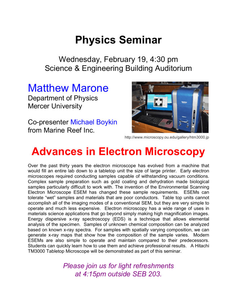
http://www.microscopy.ou.edu/gallery/htm3000.jp
Advances in Electron Microscopy
Over the past thirty years the electron microscope has evolved from a machine that would fill an entire lab down to a tabletop unit the size of large printer. Early electron microscopes required conducting samples capable of withstanding vacuum conditions.
Complex sample preparation such as gold coating and dehydration made biological samples particularly difficult to work with. The invention of the Environmental Scanning
Electron Microscope ESEM has changed these sample requirements. ESEMs can tolerate “wet” samples and materials that are poor conductors. Table top units cannot accomplish all of the imaging modes of a conventional SEM, but they are very simple to operate and much less expensive. Electron microscopy has a wide range of uses in materials science applications that go beyond simply making high magnification images.
Energy dispersive x-ray spectroscopy (EDS) is a technique that allows elemental analysis of the specimen. Samples of unknown chemical composition can be analyzed based on known x-ray spectra. For samples with spatially varying composition, we can generate x-ray maps that show how the composition of the sample varies. Modern
ESEMs are also simple to operate and maintain compared to their predecessors.
Students can quickly learn how to use them and achieve professional results. A Hitachi
TM3000 Tabletop Microscope will be demonstrated as part of this seminar.
Please join us for light refreshments at 4:15pm outside SEB 203.


