Resource
Integrated Omics Analysis of Pathogenic Host
Responses during Pandemic H1N1 Influenza Virus
Infection: The Crucial Role of Lipid Metabolism
Graphical Abstract
Authors
Jennifer Tisoncik-Go, David J. Gasper,
Jennifer E. Kyle, ..., M. Suresh,
Laurence Josset, Michael G. Katze
Correspondence
honey@uw.edu
In Brief
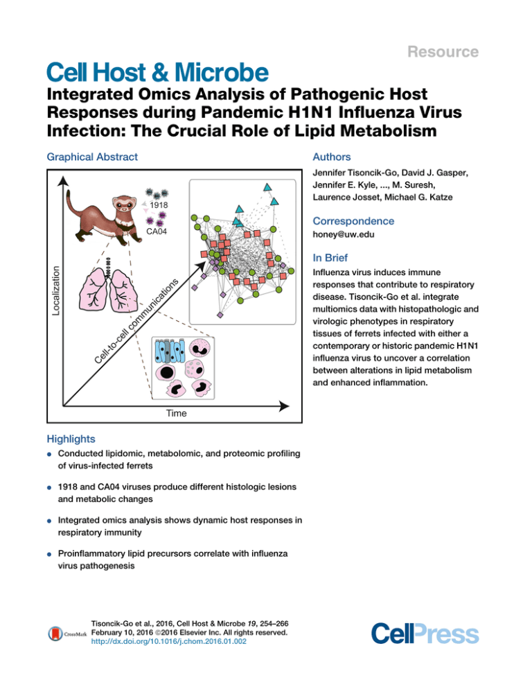
Influenza virus induces immune
responses that contribute to respiratory
disease. Tisoncik-Go et al. integrate
multiomics data with histopathologic and
virologic phenotypes in respiratory
tissues of ferrets infected with either a
contemporary or historic pandemic H1N1
influenza virus to uncover a correlation
between alterations in lipid metabolism
and enhanced inflammation.
Highlights
d
Conducted lipidomic, metabolomic, and proteomic profiling
of virus-infected ferrets
d
1918 and CA04 viruses produce different histologic lesions
and metabolic changes
d
Integrated omics analysis shows dynamic host responses in
respiratory immunity
d
Proinflammatory lipid precursors correlate with influenza
virus pathogenesis
Tisoncik-Go et al., 2016, Cell Host & Microbe 19, 254–266
February 10, 2016 ª2016 Elsevier Inc. All rights reserved.
http://dx.doi.org/10.1016/j.chom.2016.01.002
Cell Host & Microbe
Resource
Integrated Omics Analysis of Pathogenic Host
Responses during Pandemic H1N1 Influenza Virus
Infection: The Crucial Role of Lipid Metabolism
Jennifer Tisoncik-Go,1,11 David J. Gasper,3,11,12 Jennifer E. Kyle,4 Amie J. Eisfeld,3 Christian Selinger,1,13 Masato Hatta,3
Juliet Morrison,1 Marcus J. Korth,1 Erika M. Zink,4 Young-Mo Kim,4 Athena A. Schepmoes,4 Carrie D. Nicora,4
Samuel O. Purvine,5 Karl K. Weitz,4 Xinxia Peng,1 Richard R. Green,1 Susan C. Tilton,4,14 Bobbie-Jo Webb-Robertson,6
Katrina M. Waters,4 Thomas O. Metz,4 Richard D. Smith,4 Yoshihiro Kawaoka,3,7,8,9 M. Suresh,3 Laurence Josset,2
and Michael G. Katze1,10,*
1Department
of Microbiology, University of Washington, Seattle, WA 98195, USA
de Virologie, Centre de Biologie Est des Hospices Civils de Lyon, Université Claude Bernard Lyon 1, 69495 Lyon, France
3Department of Pathobiological Sciences, School of Veterinary Medicine, University of Wisconsin, Madison, WI 53706, USA
4Biological Sciences Division
5Environmental Molecular Sciences Laboratory
6Computational and Statistical Analytics Division
Pacific Northwest National Laboratory, Richland, WA 99354, USA
7Division of Virology, Department of Microbiology and Immunology, Institute of Medical Science
8Japan Department of Special Pathogens, International Research Center for Infectious Diseases, Institute of Medical Science
University of Tokyo, Minato-ku, Tokyo 108-8639, Japan
9Laboratory of Bioresponses Regulation, Department of Biological Responses, Institute for Virus Research, Kyoto University, Kyoto
606-8507, Japan
10Washington National Primate Research Center, Seattle, WA 98195, USA
11Co-first author
12Present address: Pacific Zoo & Wildlife Diagnostics, San Diego, CA 92130, USA
13Present address: Institute for Disease Modeling, Bellevue, WA 98004, USA
14Present address: Environmental and Molecular Toxicology; Oregon State University, Corvallis, OR 97331, USA
*Correspondence: honey@uw.edu
http://dx.doi.org/10.1016/j.chom.2016.01.002
2Laboratoire
SUMMARY
INTRODUCTION
Pandemic influenza viruses modulate proinflammatory responses that can lead to immunopathogenesis. We present an extensive and systematic profiling
of lipids, metabolites, and proteins in respiratory
compartments of ferrets infected with either 1918
or 2009 human pandemic H1N1 influenza viruses.
Integrative analysis of high-throughput omics data
with virologic and histopathologic data uncovered
relationships between host responses and phenotypic outcomes of viral infection. Proinflammatory
lipid precursors in the trachea following 1918 infection correlated with severe tracheal lesions. Using
an algorithm to infer cell quantity changes from
gene expression data, we found enrichment of
distinct T cell subpopulations in the trachea. There
was also a predicted increase in inflammatory monocytes in the lung of 1918 virus-infected animals that
was sustained throughout infection. This study presents a unique resource to the influenza research
community and demonstrates the utility of an integrative systems approach for characterization of
lipid metabolism alterations underlying respiratory
responses to viruses.
Highly pathogenic influenza viruses cause robust and sustained
proinflammatory responses that enhance immunopathology in
the lung. Significant progress has been made in elucidating
innate immune responses contributing to the pathogenesis of
these medically important viral pathogens, yet important questions remain. Chief among these is how alterations in the host
metabolic state during influenza virus infection impact respiratory disease severity and progression. The domestic ferret
(Mustela putorius furo) is highly utilized as a model of influenza
pathogenesis and transmission. Ferrets are naturally susceptible to human influenza virus and present clinical symptoms
akin to humans. Cellular sialyllactose receptors promoting viral
entry are similarly distributed within the respiratory tract between ferrets and humans. This was recently reinforced by
the discovery of a deletion in the ferret CMAH gene resulting
in the exclusive expression of N-acetylneuramininic acid
(Neu5Ac) on cell surfaces, as seen with humans (Ng et al.,
2014).
With the sequencing of the ferret genome (Peng et al., 2014),
it is now possible to perform systems-level analyses. We previously evaluated the global transcriptional response induced in
the trachea and lung of ferrets infected with either 1918 or
2009 human pandemic H1N1 influenza viruses and found an
enrichment of genes associated with lipid receptor signaling
in the trachea (Peng et al., 2014). Lipids are important
254 Cell Host & Microbe 19, 254–266, February 10, 2016 ª2016 Elsevier Inc. All rights reserved.
modulators of inflammation and their role in influenza virus
infection is the subject of increasing investigation. Tam et al.
recently found that the ratio of specific proinflammatory and
anti-inflammatory lipids may be markers of influenza virus pathogenicity in mice (Tam et al., 2013). In a separate study, treatment of ferrets with an agonist of sphingosine-1-phosphate
(S1P) 1 receptor was found to suppress cytokine and chemokine production during pandemic influenza H1N1 virus infection
(Teijaro et al., 2014). These studies indicate a role for lipid
signaling pathways in the regulation of inflammation during
influenza virus infection. Moreover, lipids and the pathways
that they regulate may provide a therapeutic strategy for
ameliorating disease by dampening inflammatory responses
attending viral infection.
Here, we present a systematic profiling of lipids, metabolites,
and proteins in upper and lower respiratory compartments of ferrets infected with either 1918 or 2009 human pandemic H1N1
influenza viruses. Through an integrative network analysis, we
identified relationships between groups of different molecular
species in the host response during influenza virus infection.
We also examined relationships in independent virus and tissue
networks that were correlated with viral replication and respiratory disease. We found groups of lipids and metabolites positively correlated with genes enriched for cell differentiation and
adhesion processes. We also found enrichment of T cell genes
late in infection that was enhanced in the trachea compared to
the lung and corroborated using Digital Cell Quantifier (DCQ), a
computational method to infer distinct immune cell subpopulations. Correlation analysis found tissue damage was associated
with phospholipids containing arachidonic acid (20:4) and DHA
(22:6) that may act as reservoirs for lipid mediators regulating
inflammatory responses to pandemic H1N1 influenza virus
infection.
RESULTS
Lipidomic and Metabolomic Analysis of Lung and
Trachea from Ferrets Infected with Pandemic H1N1
Influenza Viruses
Ferrets were inoculated intranasally with either influenza A/California/04/2009 (CA04) virus or influenza A/Brevig Mission/1/1918
(1918) virus. On days 1, 3, and 8 postinoculation (p.i.), lung and
trachea samples were collected to analyze lipids by LC-MS/
MS and metabolites by GC-MS. We identified 488 unique lipids
in the lung and 191 in the trachea. Fifteen different lipid subclasses were represented, with triacylglycerol (TG) and diacylglycerophosphocholine (PC) subclasses having the greatest
relative abundances in the two tissues (Figures 1A and 1B). There
was a larger relative percentage of diacylglycerophosphoglycerol (PG) in the lung compared to the trachea. We also identified
91 metabolites in the lung and 86 metabolites in the trachea.
Overall, there was a greater number of differentially abundant
(DA) metabolites with decreased abundance in the two tissues
for 1918 and CA04 infections (Figure 1C) that included essential
amino acids.
We next inferred a coexpression network based on log2FC
abundances for 50 DA lipids and 33 DA metabolites. The
network was composed of nodes, corresponding to lipids
and metabolites, and edges between nodes that show coabun-
dance patterns for 1918 and CA04 infections in the two tissues.
The nodes were organized into modules arbitrarily assigned
numbers (1–19) concatenated with the prefix ‘‘lm.’’ The modules represent groups of coexpressed molecular species that
often share similar biological functions. There were modules
that exhibited tissue-specific patterns in lipid and metabolite
abundance profiles, such as module lm18 containing ethanolamine, glyceric acid, and uracil (Figure 1D and see Table S1
available online). These metabolites showed increased abundance in the trachea compared to the lung and were coexpressed with sphingomyelin specie, SM(d18:1/24:1), and
PC(0:0/18:1) and PC(0:0/16:0) lipid species. Within this same
module, referred to as the ‘‘sphingomyelin module,’’ were two
unknown metabolites that may be functionally related to the
trachea metabolic response to influenza virus infection. This
network analysis identified groups of coexpressed lipids and
metabolites, known and unknown, capturing the dynamic
changes in the ferret lipidome and metabolome during
pandemic H1N1 influenza virus infection in ferrets.
Histological Differences in Severity, Distribution, and
Progression of Lung and Tracheal Lesions in CA04versus 1918-Infected Ferrets
To determine tissue-level responses, we performed a histopathologic assessment of paired tissue sections from each
ferret. No significant histologic findings were observed in the
lung on day 1 p.i. At day 3 p.i., multifocal submucosal gland necrosis was the most prominent and consistent lesion in both
the 1918 (Figures 2A and 2C) and CA04 groups (Figures 2B
and 2D). Compared to the 1918 group (Figure 2E), a severe
bronchiolitis was present in the CA04 group only (Figure 2F),
with necrotic debris, macrophages, eosinophils, and neutrophils obstructing the airways (see the inset in Figure 2F). Acute
alveolar damage and effusion were widespread in the
peripheral lungs of 1918-infected animals and generally not
associated with airways (Figure 2G). In CA04-infected animals,
alveolar damage tended to localize around affected bronchioles, which were lined by variably hypertrophic and hyperplastic type-II pneumocytes (see the left inset in Figure 2H).
Foamy macrophages were common in regions of effusion in
the CA04 group (see the right inset in Figure 2H), while neutrophils were frequent in lung infected with 1918. At day 8 p.i., the
bronchiolitis observed with CA04 infection was largely replaced
by mixed mesenchymal and epithelial proliferations (Figure S1).
These proliferations partially obstructed bronchioles (bronchiolitis obliterans) and widely extended into the adjacent parenchyma (organizing pneumonia), and have been previously
reported (Memoli et al., 2011). Though less severe compared
to CA04 infection, bronchiolitis in 1918-infected animals was
accompanied by neutrophilic and eosinophilic infiltrates in
areas with concurrent glandular necrosis.
In tracheal sections from mock-infected ferrets, there were
infrequent intraepithelial lymphocytes in most sections, and
sporadic individual or small clusters of intraepithelial eosinophils (Figure 3A). At day 1 p.i., there was extensive inflammatory infiltrate that was accompanied by epithelial changes in
1918-infected trachea (Figure 3B), whereas CA04 infection resulted in mildly increased submucosal lymphocytic infiltrate
(Figure 3C). Two of three ferrets in the 1918 group exhibited
Cell Host & Microbe 19, 254–266, February 10, 2016 ª2016 Elsevier Inc. All rights reserved. 255
A
D
Relative % lipid subclass in lung
100
triacylglycerols
diacylglycerols
monoacylglycerols
diacylglycerophosphoserines
monoacylglycerophosphoinositols
diacylglycerophosphoinositols
monoacylglycerophosphoglycerols
diacylglycerophosphoglycerols
1-(1Z-alkenyl),2-acylglycerophosphoethanolamines
monoacylglycerophosphoethanolamines
diacylglycerophosphoethanolamines
monoacylglycerophosphocholines
diacylglycerophosphocholines
sphingomyelins
ceramides
90
80
70
60
50
40
30
PC(18:0/18:1)
PE(P−16:0/22:6)
PC(18:1/20:4)
PC(16:0/22:6)
Unknown 20
D−mannitol
Unknown 08
Unknown 22
aminomalonic acid
PI(18:0/18:2)
Unknown 14
Unknown 02
carbonate ion
hypotaurine
myo−inositol
TG(16:0/16:0/18:1)
TG(16:0/16:1/18:1)
TG(16:1/18:2/18:2)
TG(16:0/16:1/18:2)
TG(16:1/16:1/18:1)
Unknown 09
TG(18:1/18:2/20:4)
TG(16:1/16:1/18:2)
PE(18:0/0:0)
adenosine
PE(P−18:0/20:4)
PE(P−18:0/18:1)
PE(18:0/18:2)
PE(16:0/18:2)
TG(16:0/18:0/18:0)
Unknown 24
Unknown 15
PE(18:1/20:4)
PG(16:0/18:1)
PE(18:0/18:1)
PE(18:0/22:4)
PE(18:0/22:6)
PE(P−16:0/20:4)
PC(0:0/18:0)
PC(18:0/20:3)
fructose
TG(18:1/18:1/22:4)
PC(16:0/18:2)
PC(16:0/16:0)
PC(18:0/20:4)
PC(16:0/18:1)
glycerol−3−phosphate
PE(P−18:0/22:4)
TG(18:1/18:1/20:4)
PE(P−18:0/18:2)
Unknown 13
Unknown 28
palmitic acid
Unknown 01
L−valine
urea
L−threonine
Cer(d18:1/16:0)
L−(+) lactic acid
PE(18:1/18:1)
PE(P−18:0/22:6)
PE(P−18:0/20:4)
Cer(d18:1/20:0)
TG(16:0/16:0/16:1)
TG(16:0/16:0/16:0)
PE(P−16:0/18:1)
Unknown 11
Unknown 12
Unknown 10
glyceric acid
Unknown 27
uracil
SM(d18:1/24:1)
PE(P−16:0/22:4)
ethanolamine
PC(0:0/18:1)
PC(0:0/16:0)
L−serine
PE(20:4/0:0)
Unknown 25
TG(18:1/18:1/20:1)
TG(16:0/18:2/18:2)
TG(16:1/18:1/18:2)
lm1
lm2
lm3
20
lm4
0
292
293
294
280
281
282
277
278
279
283
284
285
275
295
286
287
288
289
290
291
10
d1
d3
d8
d1
1918
d3
d8
lm5
d1
CA04
PBS
lm6
B
lm7
triacylglycerols
diacylglycerols
monoacylglycerols
diacylglycerophosphoserines
monoacylglycerophosphoinositols
diacylglycerophosphoinositols
monoacylglycerophosphoglycerols
diacylglycerophosphoglycerols
1-(1Z-alkenyl),2-acylglycerophosphoethanolamines
monoacylglycerophosphoethanolamines
diacylglycerophosphoethanolamines
monoacylglycerophosphocholines
diacylglycerophosphocholines
sphingomyelins
ceramides
90
80
70
60
50
40
30
lm8
lm9
lm10
lm11
lm12
lm13
20
lm14
10
0
292
293
294
280
281
282
277
278
279
283
284
285
275
295
286
287
288
289
290
291
Relative % lipid subclass in trachea
100
d1
d3
d8
d1
d3
d8
lm15
d1
1918
lm16
PBS
lm17
C
18
18
Increased
Decreased
No. of DA metabolites
16
16
lm18
14
14
12
12
lm19
10
10
88
d1 d3 d8 d1 d3 d8 d1 d3 d8 d1 d3 d8
66
1918
44
CA04
lung
1918
CA04
trachea
22
00
1
3
8
1
3
8
1
d1
d3
d8
d1
d3
d8
d1
lung
trachea
1918
3
d3
lung
8
1
3
8
d8
d1
d3
d8
−2
0
2
log2FC abun
trachea
CA04
Figure 1. Lipidomic and Metabolomic Analysis of Ferret Lung and Trachea Infected with 1918 and CA04 Viruses
(A) Lipid subclasses identified in the lung.
(B) Lipid subclasses identified in the trachea. Stacked bar graphs represent the relative percentages of lipid subclasses from individual animals. The lipid subclass
annotations are according to the LIPID MAPS structure database (LMSD).
(C) Bar graph showing total number of differentially abundant (DA) metabolites in the lung and trachea of 1918 and CA04 virus-infected ferrets. Comparative
statistical analyses of mock with 1918 and CA04 at each time point were performed using a Dunnett adjusted t test (p < 0.05). Red depicts metabolites with
increased abundance relative to mock and blue depicts metabolites with decreased abundance relative to mock.
(D) Heatmap of average log2FC abundances of 50 DA lipids and 33 DA metabolites from the lipid metabolite network inferred for 42 samples corresponding to all
time points and both lung and trachea compartments. Modules assignments (1–19) are shown on the left-hand side of the heatmap. Missing values in two or more
replicates were treated as a missing value when averaging the replicates and are depicted as gray. Rows are lipids and metabolites and columns are experimental
conditions. See also Table S1 and File S2, first tab.
multiple dense nodular aggregates of eosinophils that locally
expanded and disrupted the epithelium (see the inset in Figure 3B). The most prominent change from day 1 to day 3 p.i.
was multifocal necrosis of the tracheal submucosal glands
with glandular and periglandular infiltration of eosinophils, neutrophils, histiocytes, and lymphocytes. Necrosis was most
256 Cell Host & Microbe 19, 254–266, February 10, 2016 ª2016 Elsevier Inc. All rights reserved.
Figure 2. Histologic Lesions in Ferret Lung 3
Days after Infection with Pandemic H1N1
Influenza Viruses
Histopathological assessment of ferret lung infected with 1918 (left column) or CA04 (right column) viruses.
(A) 1918 virus infection. There is patchy atelectasis
and thickening of alveolar walls. ‘‘Br’’ indicates
unaffected medium and large bronchioles, and
arrows indicate small caliber bronchioles and terminal airways. Stars indicate foci of bronchiolar
submucosal gland necrosis and inflammation.
(B) CA04 infection. ‘‘Br’’ indicates large bronchiole
containing a crescent of fibrinocellular exudate.
Arrows indicate numerous small caliber bronchioles and terminal airways that have been effaced
by necrosis and mixed inflammatory exudates.
Adjacent alveolar tissue is consolidated and there
is loss of alveolar architectural definition. Stars
indicate foci of bronchiolar submucosal gland necrosis and inflammation.
(C and D) (C) 1918 infection and (D) CA04 infection.
Bronchiolar submucosal gland necrosis. Arrows
indicate affected glandular acini in which the
epithelium is largely absent and lumena are filled
with cellular debris, neutrophils, macrophages, and
eosinophils. Star indicates periglandular infiltrates
of lymphocytes, plasma cells, and eosinophils.
(E and F) (E) 1918 infection and (F) CA04 infection.
Small bronchioles and terminal airways, higher
magnification of micrographs A and B, respectively. Arrows indicate similarly sized transitional
and terminal airways. The airway in (E) is minimally
affected. Dashes in (F) demark the approximate
location of the small airway wall. The indicated
airway and adjacent alveoli in (F) exhibit segmental
necrosis and ulceration, and the airway lumen is
filled with exudate composed of cellular debris and
mixed inflammatory cells (inset F) including
numerous eosinophils and fewer macrophages,
lymphocytes, and neutrophils.
(G and H) (G) 1918 infection and (H) CA04 infection.
Regional atelectasis with effusion (asterisks).
Arrows indicate bronchioles, the lumen of the
bronchiole in (G) contains effusion (pink material).
(H, left inset) A central C-shaped crescent of type II
pneumocyte hyperplasia and adjacent intraalvoeolar eosinophils, macrophage, and lymphocytes (left and bottom). (H, right inset) A cluster of
foamy macrophages in an affected alveolus. See
also Figure S1.
prominent in 1918 virus-infected animals, in which 40% (12 of
30 lobules) of the glands were affected (Figure 3D). In contrast,
only 9% (5 of 38 lobules) of the glands were affected following
CA04 infection (Figure 3E). Viral antigens were mainly detected
in the epithelial cells of submucosal glands (see insets in Figures 3D and 3E). By day 8 p.i., submucosal glandular lesions
extended to 75% (24 of 32) of the lobules in the 1918 group
(Figure 3F) and 41% (18 of 44) of the lobules in the CA04 group
(Figure 3G). Moderately dense aggregates of lymphocytes infiltrated the epithelium in the areas overlying affected glands. In
summary, 1918 and CA04 infections differed in the severity
and progression of histologic lesions in the lung and trachea,
with ferrets infected with CA04 virus presenting more severe
bronchiolitis compared to the enhanced tracheitis and tracheoadenitis resulting from 1918 infection.
Metabolic Changes Correlate with Viral Replication and
Disease Phenotypes
Relationships between lipid metabolite network modules and
virologic (Table S2) and histopathologic phenotypes (File S1)
were assessed to identify lipid metabolism changes correlated
with viral replication and disease. Both viruses replicated in the
trachea on days 1 and 3 p.i., and while 1918 replicated in the
lung on days 1 and 3 p.i., CA04 was only recovered on day 3
p.i. from ferret #295 that displayed the most striking necrotizing bronchiolitis. No infectious viruses were recovered
Cell Host & Microbe 19, 254–266, February 10, 2016 ª2016 Elsevier Inc. All rights reserved. 257
Figure 3. Histologic Lesions in Ferret Trachea Infected with Pandemic H1N1 Influenza
Viruses, and Transcriptomic Analysis of
Host Responses
Histopathological assessment of ferret trachea infected with 1918 (left column) or CA04 (right column) viruses.
(A) Mock-infected tracheal mucosa 1 day after
infection. Arrow indicates scattered individual eosinophils and rare neutrophils and lymphocytes
within the respiratory epithelium.
(B) Day 1 1918 infection. Arrow indicates nodular
expansion of the respiratory epithelium by a predominately eosinophilic infiltrate (inset). Adjacent
epithelial cells are disorganized and exhibit moderate variability in cell and nuclear size, and are
occasionally necrotic.
(C) Day 1 CA04 infection. Arrow indicates perivascular aggregate of lymphocytes adjacent to
submucosal glands. Scattered lymphocytes are
also present in the submucosa and basal regions of
the epithelium. There is mild variability in the cell
and nuclear size and shape of the respiratory
epithelium.
(D) Day 3 1918 infection. Star indicates submucosal
gland degeneration and necrosis. Arrow indicates
respiratory epithelial cells with cytoplasmic
clearing and enlarged irregular nuclei. Affected
respiratory and glandular epithelial cells were
strongly immunoreactive for polyclonal anti-influenza antibodies (inset, brown staining).
(E) Day 3 CA04 infection. Arrows indicate lymphocytes in respiratory epithelium. Adjacent epithelial
cells are mildly disorganized and exhibit mild variability in their cell and nuclear size and shape.
Affected respiratory epithelial cells were moderately immunoreactive for polyclonal anti-influenza
antibodies (inset, brown staining). Submucosal
glands were largely unaffected; however, some
reserve cells exhibited positive immunoreactivity.
(F and G) (F) Day 8 1918 infection and (G) Day 8
CA04 infection. Remnant submucosal glands are
lined by hypertrophic and hyperplastic epithelium
(arrowheads). Necrotic glands observed on day 3
(panel D) have been replaced by densely cellular
infiltrates of macrophages and lymphocytes (star).
Numerous lymphocytes and cellular debris are
present in the overlying respiratory epithelium
(arrows).
from either tissue on day 8 p.i. We found the sphingomyelin
module (lm18) was positively correlated with both viral
titer and viral mRNA, while module lm14 containing L-threonine and L-valine was strongly anti-correlated with viral titer
(r = 0.75, p value < 1e-05) (Figure 4A). Several modules
were correlated with histopathologic changes in the tissues.
Modules lm1, lm8, and lm15 were positively correlated with
bronchitis/iolitis, bronchoadenitis, and alveolitis. Module lm8
was also highly correlated with tracheoadenitis (r = 0.83,
p value < 4e-06), while lm13 and lm17 were negatively correlated with tracheitis. The ‘‘disease’’ modules consisted mostly
of diacylglycerophosphoethanolamine (PE), PE(P)–, and PC
lipid species containing either 22:6 or 20:4, the precursor of
docosahexaenoic acid (DHA) and arachidonic acid (AA),
respectively (Table S1). In particular, we found PC(18:1/20:4)
and PC(16:0/22:6) species from lm1 had earlier increased
abundance in both the lung and trachea in response to 1918
virus compared to CA04 virus (Figure 1D). Multinomial ordered
logistic regression analysis of histologic lesions scores found a
more significant contribution by 1918 toward disease severity
in the trachea relative to CA04 (odds ratio of 0.352) (Table
S3). Thus, an increase in proinflammatory lipids in the trachea
may be enhancing necrosis and inflammation associated with
1918 influenza virus infection.
We complemented the lipidomic and metabolomics analysis
by assessing the proteome in paired tissue sections. LC/MS
analysis identified 4,811 proteins in the lung and 4,060 proteins in the trachea. We then inferred a coexpression network
based on log2FC abundances for 810 DA proteins and identified tissue-specific protein profiles in response to influenza
258 Cell Host & Microbe 19, 254–266, February 10, 2016 ª2016 Elsevier Inc. All rights reserved.
A
B
1
lm1
0.16 (0.3)
0.46 (0.02)
lm2
0.061 (0.7)
−0.12 (0.6)
0.17 (0.5)
lm3
0.14 (0.4)
−0.1 (0.6)
−0.41 (0.07)
lm4
0.11 (0.5)
−0.18 (0.4)
−0.16 (0.5)
lm5
0.035 (0.8)
−0.31 (0.1)
0.46 (0.03)
lm6
−0.28 (0.1)
−0.44 (0.03)
0.17 (0.5)
0.28 (0.2)
lm7
−0.14 (0.4)
−0.034 (0.9)
−0.3 (0.2)
−0.21 (0.4)
lm8
−0.22 (0.2)
−0.055 (0.8)
0.53 (0.01)
0.68 (7e-04)
lm9
0.16 (0.4)
0.22 (0.3)
0.19 (0.4)
0.15 (0.5)
lm10
0.068 (0.7)
0.48 (0.01)
−0.096 (0.7)
0.06 (0.8)
lm11
0.18 (0.3)
0.26 (0.2)
0.41 (0.07)
0.39 (0.08)
lm12
−0.073 (0.7)
−0.18 (0.4)
0.28 (0.2)
0.088 (0.7)
lm13
−0.29 (0.08)
−0.48 (0.02)
−0.05 (0.8)
−0.12 (0.6)
lm14
−0.33 (0.05)
−0.75 (1e-05)
−0.21 (0.4)
−0.35 (0.1)
−0.17 (0.5)
lm15
−0.024 (0.9)
−0.078 (0.7)
0.58 (0.006)
0.48 (0.03)
0.57 (0.007)
−0.34 (0.1)
0.053 (0.8)
lm16
0.12 (0.5)
−0.033 (0.9)
0.15 (0.5)
0.0034 (1.0)
0.12 (0.6)
−0.16 (0.5)
−0.24 (0.3)
lm17
−0.013 (0.9)
−0.34 (0.09)
−0.4 (0.07)
−0.22 (0.3)
−0.45 (0.04)
−0.53 (0.01)
−0.23 (0.3)
lm18
0.39 (0.02)
0.41 (0.04)
−0.034 (0.9)
−0.19 (0.4)
−0.057 (0.8)
0.28 (0.2)
0.16 (0.5)
lm19
−0.03 (0.9)
−0.19 (0.4)
−0.18 (0.4)
−0.28 (0.2)
−0.1 (0.7)
−0.25 (0.3)
−0.39 (0.08)
viral mRNA
viral titer
bronchitis/iolitis
bronchoadenitis
alveolitis
tracheitis
tracheoadenitis
0.56 (0.009)
0.72 (2e-04)
0.5 (0.02)
0.4 (0.07)
0.27 (0.2)
0.0044 (1.0)
0.15 (0.5)
−0.42 (0.06)
−0.11 (0.6)
−0.55 (0.01)
−0.25 (0.3)
−0.37 (0.09)
−0.55 (0.01)
−0.12 (0.6)
−0.067 (0.8)
−0.27 (0.2)
−0.3 (0.2)
0.27 (0.2)
0.46 (0.04)
−0.19 (0.4)
0.11 (0.7)
0.28 (0.2)
−0.35 (0.1)
−0.22 (0.3)
−0.35 (0.1)
−0.071 (0.8)
0.11 (0.6)
0.61 (0.003)
0.42 (0.06)
0.83 (4e-06)
0.41 (0.06)
0.37 (0.1)
0.18 (0.4)
−0.16 (0.5)
0.46 (0.04)
0.41 (0.06)
0.45 (0.04)
0.55 (0.01)
0.34 (0.1)
0.4 (0.08)
−0.3 (0.2)
−0.34 (0.1)
−0.12 (0.6)
−0.54 (0.01)
−0.3 (0.2)
−0.41 (0.07)
−0.099 (0.7)
0
C
p1
−0.5
−1
treatment
1918
CA04
PBS
60
time-point
d1
d3
d8
30
p2
0.5
tissue
lung
trachea
y
0
p3
-30
Kruskal's stress = 13.57
-60
-60
p4
-30
0
x
p7
p8
p9
d1 d3 d8 d1 d3 d8 d1
1918
CA04
lung
d3 d8 d1 d3 d8
1918
CA04
trachea
−2
0
2
log2FC abun
virus infection (Figure 4B); these differences were also visualized by multidimensional scaling (Figure 4C). In addition, a
gene coexpression network was constructed using a transcriptional signature enriched with putative intergenic noncoding RNAs that was derived from a tissue-by-virus comparison
of ferret RNA-Seq data (Peng et al., 2014) (Figures S2A and
S2B). Modules were arbitrarily assigned numbers concatenated with prefixes ‘‘p’’ and ‘‘g’’ for protein and gene networks,
respectively. The protein network was enriched for hematological system development and function, glycolysis, and cellular
oxidative stress processes (Table S4). We also found proteins
related to innate immune and antiviral responses including
Trim25, Stat1, and Mx1, as well as p120-catenin (encoded
by CTNND1 gene), known to protect alveolar epithelial barrier
integrity (Chignalia et al., 2015). In relation to lipid metabolism,
protein module p2 contained apolipoproteins, Apoh and
Apoa4, and gene module g5 contained fatty acid metabolism
30
60
Figure 4. Correlation Analysis of Lipids and
Metabolites with Phenotypes and Protein
Network Analysis
(A) Correlation of phenotypic traits (i.e., virologic
and histopathologic phenotypes) with lipid metabolite module eigengenes (MEs) using the biweight
midcorrelation (bicor) method. Pairwise bicor were
calculated between MEs and viral mRNA, viral titer,
and histopathologic scores averaged across all
subcategories from tracheal, bronchial and alveolar
compartments. See also File S1. Listed in each cell
of the ME-phenotype matrix are the bicor coefficient and corresponding p value. Relationships
with a p < 0.05 were considered significant. For
example, for the lm1:bronchoadenitis relationship,
the bicor value is 0.72 and the p value is 2e-04,
indicating a significant positive correlation between
lm1 and bronchoadenitis.
(B) Heatmap of average log2 FC abundance of 810
DA proteins from the protein network inferred for 37
samples corresponding to all time points and both
lung and trachea compartments. Modules assignments (1–9) are shown on the left-hand side of the
heatmap. Missing values in 2 or more replicates
were treated as a missing value when averaging the
replicates and are depicted as gray. Rows are
proteins and columns are experimental conditions.
See also File S2, second tab.
(C) Multidimensional scaling (MDS) representation
of the distances among samples based protein log2
abundances (Kruskal’s stress = 13.57). The Kruskal
stress signifies the amount of information lost due
to the dimensionality reduction as a fraction of total
information. Points coded as per legend and
denote individual animals. Convex hulls link points
belonging to the same experimental condition and
time point. See also Table S4.
genes, FABP3 and SCD, as well as
PTGER3, encoding a receptor of PGE2
known to inhibit alveolar macrophage type I interferon responses during
influenza virus infection (Coulombe et al.,
2014) (Table S5). This comprehensive
analysis implicates a shift toward increased lipid metabolism that may be integral in antiviral responses toward pandemic H1N1 influenza virus infection.
Integrated Omics Analysis Shows Temporal and
Regional Dynamics of the Host Response to Pandemic
H1N1 Influenza Virus Infection
To examine relationships between lipids, metabolites, proteins,
and genes in toto, we constructed an integrated omics network
by calculating the pairwise correlations between module eigengenes (MEs)—the representative expression profile of each
module characterized by its first principal component—from
each independent network and between MEs and phenotypic
traits (Figure S3). A primary goal of this integrative analysis was
to characterize host responses in a unified model of influenza
pathogenesis, as well as previously unidentified molecular
species associated with known innate and adaptive immune responses important in the control of viral infection. Gene modules
Cell Host & Microbe 19, 254–266, February 10, 2016 ª2016 Elsevier Inc. All rights reserved. 259
g2 and g10, which were enriched for host defense genes relevant
to influenza virus infection, strongly correlated with viral titer and
mRNA (Figures 5A and S2D). There was positive correlation of
the sphingomyelin module (lm18) with these innate immune
gene modules, and by association the lipid metabolism p2 module, suggesting these lipids and lipid-related proteins are
involved in innate immune responses controlling influenza virus
replication. In examining module relationships with disease
phenotypes, we found positive correlations between tracheitis,
tracheoadenitis, and bronchitis/iolitis with gene module g3,
enriched for T cell genes, and g6 enriched for genes associated
with calcium signaling (Figures 5B and S2C). In particular, module g6 contained ATP2B4, CAMK2G, and CAMKK1 genes, as
well as ORAI1, transcribing a subunit of the calcium-releaseactivated calcium (CRAC) channel known to regulate intracellular
Ca2+ concentrations essential for the activation of cytokine gene
expression in T cells.
The richness of the datasets comprising the integrated
network is further exemplified by the presence of relationships
between modules of different molecular species (e.g., lipid metabolites and genes, proteins and genes, and lipid metabolites
and proteins) (Figure 5C). In comparing the two tissues, the dynamics of the host response were largely different; the trachea
showed greater variability in changes in gene expression and
lipid, metabolite, and protein abundance during 1918 and
CA04 infections compared to the lung responses that were
more consistent across time points and viruses (Figure 5D).
Taken together, this integrative analysis demonstrates highly
interrelated relationships among diverse molecular species of
the host response that regulate the progression and severity of
respiratory disease associated with pandemic H1N1 influenza
virus infection and control viral replication.
Increased Lymphocyte Responses in the Trachea at
Later Stages of Infection Are Associated with
Inflammation
The interrelationships of this complex integrated network were
computationally elucidated by recalculating the ME for each
module of the integrated network considering trachea, lung,
1918, and CA04 samples independently, and then computing
pairwise bicor between MEs within the four separate networks.
Here, we show examples of relationships that are conserved between tissues and viruses (abs Dbicor < 0.7) and report previously unidentified molecular species likely playing a role in the
immune response against influenza virus. In the lung and trachea
networks, we found several similarly correlated relationships,
such as the relationship between the innate immune g4 module
and viral titer (abs Dbicor = 0.08), indicating comparable innate
immune responses represented in the two tissues that are correlated with viral replication (File S3). There were also several relationships with apparent differences (abs Dbicor > 0.7) between
tissues, such as g10:lm12 and p2:p4 (Figures 6A and 6B), suggesting cellular processes involving lipids, genes, and proteins
specific to a particular respiratory compartment. For example,
the g1:g7 relationship was positively correlated in the lung (bicor = 0.71) and anticorrelated in the trachea (bicor = 0.67) (Figure 6A). The g7 module is enriched for downregulated genes
related to signal transduction and neurological stimuli responses, including the NPY2R gene encoding neuropeptide Y2
receptor known to play a critical role in allergic airway inflammation. This relationship may signify neurologic peptide responses
modulating influenza virus infection in the lung and independent
of the trachea.
Another example of a host response relationship largely
different between the two tissues is the g13:g3 relationship,
which shows a strong positive correlation in the trachea (bicor =
0.81) that is opposite from the lung (bicor = 0.51) (Figure 6B).
Topological analysis of module g3, enriched for genes associated with T cell receptor signaling, identified several intergenic
transcripts as intramodular hubs (Figure 6C). These central nodes and their highly connected nodes representing lymphocyte
genes had increased expression in the trachea at day 8 p.i. in
response to both 1918 and CA04 infections (Figure 6D). To
further explore these findings, we analyzed the transcriptomic
data using Digital Cell Quantifier (DCQ) to predict relative immune cell quantities in the trachea. This computational method
combines genome-wide gene expression data with a mouse immune cell compendium that has been used to infer changes in
distinct dendritic cell (DC) subpopulations in mouse lung infected
with influenza virus (Altboum et al., 2014). In ferrets, we found
influenza virus infection elicited temporal differences in specific
T cell subpopulations and resident monocytes that were most
apparent at day 8 p.i. (Figure 6E). In particular, there were predicted increases in CD8+ memory and effector T cells and
Ly6C ‘‘resident’’ monocyte subtypes (MO.Ly6C MHCII and
MO.Ly6C MHCIIINT) (Table S6). There was also a DC population
(CD11c+MHCIIhiCD103 CD11b+) that was initially larger in ferrets infected with 1918 virus that progressively declined by day
8 p.i. Thus, investigation of network variation uncovered putative
intergenic noncoding RNAs induced late in infection that may be
central to the resolution of viral infection by regulating lymphocyte responses in upper respiratory tissues where human
H1N1 influenza viruses predominantly replicate.
In the 1918 and CA04 networks, we found several conserved
(abs Dbicor < 0.7) relationships, such as the g10:lm13 relationship (bicor = 0.05), with g10 enriched for genes associated
with IFN-b and TNF cellular defense responses and lm13 containing palmitic acid and unknown metabolites (File S4). Several
relationships had marked differences (abs Dbicor > 0.7) between
viruses, including g8:g9 and g18:lm3 (Figures 7A and 7B), suggesting these relationships may be specific to either CA04 or
1918 infections. For example, lm3 was positively correlated
with g18 for the 1918 group, and it was negatively correlated
with 1918 virus-associated alveolitis (bicor = 0.80) (Figure 7B).
The lm3 module was enriched for TGs with decreased abundance in the lung and to a greater extent for 1918 compared to
CA04, particularly at day 8 p.i. (Figure 7C). Application of DCQ
to the lung transcriptomic data showed the presence of Ly6C+
‘‘inflammatory’’ monocytes for both 1918 and CA04 infections
(Figure 7D). Notably, there was one monocyte subpopulation
(MO.6C+II-.BM) that was enriched specifically in ferret lung infected with 1918 virus throughout infection. 1918 virus is known
to cause massive recruitment of monocytes into mouse lung
(Perrone et al., 2008). These analyses indicate pathogenic processes in CA04 and 1918 infections are mediated in a particular
context (i.e., respiratory compartment and lipid specie) or by the
presence of specific immune cells that can depend on the viral
strain.
260 Cell Host & Microbe 19, 254–266, February 10, 2016 ª2016 Elsevier Inc. All rights reserved.
A
B
lm9
bicor sign
lm9
bicor sign
neg
neg
lm10
pos
lm10
pos
g13
bicor
type
0.4
bicor
type
0.4
gene
lm2
0.5
0.6
lipid/metabolite
0.7
phenotype
g1
p2
lm11
gene
g1
0.5
lm18
0.8
0.6
lipid/metabolite
0.7
phenotype
Tracheoadenitis
Tracheitis
0.8
protein
0.9
protein
0.9
g14
lm8
g6
g10
lm1
g3
g4
Bronchoadenitis
Viral Titer
Bronchitis/iolitis
Viral mRNA
g17
p1
Alveolitis
lm5
g2
D
C
g1
g2
g3
g4
g5
g6
g7
g8
g9
g10
g11
g12
g13
g14
g15
g16
g17
g18
g19
lm1
lm2
lm3
lm4
lm5
lm6
lm7
lm8
lm9
lm10
lm11
lm12
lm13
lm14
lm15
lm16
lm17
lm18
lm19
p1
p2
p3
p4
p5
p6
p7
p8
p9
g16
g19
lm14
lm15
g11
g9
p9
g7
g12
p3
g8
p4
lm16
lm6
lm12
g15
lm19
lm13
g18
bicor sign
lm4
neg
lm17
pos
bicor
0.4
lm3
p6
g5
type
gene
0.5
0.6
lipid/metabolite
0.7
phenotype
p8
0.8
1918_d8
CA04_d8
1918_d3
CA04_d3
1918_d1
lung
CA04_d1
1918_d8
CA04_d8
1918_d3
CA04_d3
1918_d1
protein
CA04_d1
0.9
trachea
−2
0
2
log2FC value
Figure 5. Integrated Coexpression Network Analysis of Ferret Host Responses to Pandemic H1N1 Influenza Virus Infection
The integrated omics network related to (A) influenza virus replication, (B) respiratory disease, and (C) relationships among different molecular species. The
integrated omics network was constructed by calculating pairwise correlations between modules from independent lipid and metabolite, protein, and gene
networks and between all modules and phenotypic data. Nodes represent each module as a single point colored according to data type. Gene (g) modules are
symbolized by pink squares. Lipid metabolite (lm) modules are symbolized by green circles. Protein (p) modules are symbolized by purple diamonds. Phenotype
modules are symbolized by blue triangles. Edges between nodes signify biweight midcorrelation (bicor) coefficients between the representative expression
profiles (module eigengenes, MEs) of all lipid and metabolite, gene, and protein module pairs with histopathologic and virologic phenotypes. Only significant
correlations are shown (p < 0.05). Positive bicor coefficients are represented by a solid line. Negative bicor coefficients are represented by a dashed line. The line
thickness corresponds to the strength of the bicor coefficient (bicor 0.4–0.9). (D) Dynamics of module expression levels in different respiratory compartments
(lung and trachea) and at different time points following infection with either 1918 or CA04 viruses. The heatmap depicts median log2FC values for each module
across the experimental dataset. Rows are modules and columns are experimental conditions. See also Figure S3.
Cell Host & Microbe 19, 254–266, February 10, 2016 ª2016 Elsevier Inc. All rights reserved. 261
DISCUSSION
We took advantage of the recent sequencing of the ferret
genome to study host responses correlated with the pathogenesis of pandemic influenza virus. Through an integrative
network analysis we examined relationships among different
molecular species in the trachea and lung of ferrets infected
with either 1918 or CA04 viruses. We found significant abundance changes for phospholipids (PC and PE species) that
are major constituents of pulmonary surfactant known to suppress influenza infection in bronchial epithelial cells (Numata
et al., 2012). In addition, several phospholipids contained
20:4 that can be cleaved to form arachidonic acid, the precursor to eicosanoids converted by cyclooxygenase-2 (COX-2),
including prostaglandins (e.g., PGE2 and PGI2) and leukotrienes
(e.g., LTB4). In particular, PC(18:1/20:4) and PE(18:0/22:4)
species were correlated with histologic lesions in the lung
and trachea and had increased abundance that was greater
in response to 1918 compared to CA04. Thus, tissue damage
during pandemic H1N1 influenza virus infection may be the
result of lipid mediators derived from phospholipid arachidonic
acid (20:4) reservoirs that serve to enhance inflammatory
responses.
In a mouse model, arachidonic acid has been implicated in
the pathogenesis of avian H5N1 influenza virus (Morita et al.,
2013). High viral load and excessive inflammation caused by
hypercytokinemia (referred to as a cytokine storm) contribute
to H5N1 pathogenesis and a fatal outcome in humans (de
Jong et al., 2006; Tisoncik et al., 2012). Lung tissue from patients with fatal outcomes of H5N1 infection shows extensive
COX-2 induction in epithelial cells that mediates a proinflammatory cascade resulting in increased chemotaxis and vascular
permeability (Lee et al., 2008). Therefore, an effective therapy
may be one that targets both the virus and proinflammatory responses contributing to disease severity through the combination of antivirals and immunomodulatory agents. This was
demonstrated using a combination therapy consisting of the
anti-influenza neuraminidase inhibitor, zanamivir, together with
the anti-inflammatory COX-2 inhibitors celecoxib and mesalazine, which increased survival rate of mice infected with
H5N1 virus (Zheng et al., 2008). In targeting the COX pathway,
proinflammatory cytokines and eicosanoids can be alleviated,
thereby decreasing the activation of inflammatory macrophages and neutrophils.
We previously reported that the presence of foamy macrophages in CA04 virus-infected airways is associated with nuclear
activation of heterodimer liver X receptor (LXR) and retinoic acid
receptor (RXR) leading to altered lipid metabolism (Go et al.,
2012). In the current study, foamy macrophages were found in
regions of effusion following infection with CA04 but not 1918.
Foams cells are known to accumulate triacyglycerols (TGs)
that are stored in cytosolic lipid droplets observed with mycobacterium tuberculosis infection (Mehrotra et al., 2014). The TG
abundance changes in ferrets infected with pandemic influenza
virus correlated with respiratory disease and may signify metabolic fluctuations leading to formation of foamy macrophages
as a pathogenic mechanism of CA04 infection. Thus, the lipidomic alterations in response to pandemic H1N1 influenza virus
infection partially reflect the differential distribution and pre-
sumed etiopathogenesis of the alveolar effusion and associated
histologic lesions in ferrets. It also suggests that the role of lipid
signaling pathways in the inflammatory response varies by site
and types of insult.
To identify potential cellular sources of immunopathogenesis,
we applied the Digital Cell Quantifier (DCQ) algorithm that infers
changes in cell quantities from gene expression data obtained
from a complex tissue. There was evidence of a DC subpopulation in the trachea early in infection that was followed by the
emergence of a prominent population of CD8 T cells with
effector and memory phenotypes at day 8 p.i., possibly suggesting initial DCs playing a pro-inflammatory role by producing
cytokines, such as IL-12, IL-23, and TNF, that drive differentiation of IFN-g-producing T cells at a later stage. This may also
contribute to more rapid viral clearance in the trachea of
CA04-infected animals that is supported by fewer lesions in
CA04-infected trachea and increased glandular necrosis in
the 1918-infected trachea. In the lung, there was a virus-specific differential immune cell population, though most enriched
cell types did not change between 1918 and CA04 over the
course of the infection. The notable difference was a certain
monocytic cell type (MO.Ly6C+MHCII ) in the 1918 group
compared to the CA04 group. In mice, Ly6C+ monocytes are
preferentially recruited into inflamed tissue through their interaction with chemokine receptor CCR2 (Audoy-Rémus et al.,
2008) and mature to inflammatory macrophages, which secrete
TNF and type I IFN in response to viral ligands (Barbalat et al.,
2009), contributing to tissue degradation and T cell activation.
Targeting inflammatory monocytes by siRNA-mediated
silencing of CCR2 attenuates inflammatory disease in mouse
models of atherosclerosis and myocardial infarction (Leuschner
et al., 2011). These innate immune cells may play a role in
enhancing the immunopathology of 1918 infection in the lung
of ferrets and thus, potentially serve as therapeutic targets to
alleviate inflammation associated with pandemic influenza virus
infection.
The extensive amount of high-throughput data generated by
this study are of value to the scientific community, particularly
to those interested in extracting information about innate immune responses during acute viral infection. For example, the
TRIM superfamily has many IFN-inducible members, some
known to regulate viral RNA sensing pathways, such as
TRIM25-mediated activation of RIG-I (Gack et al., 2009), and
others known to function as host restriction factors. Most TRIM
members have yet to be described. Within the integrated
network, there are 31 TRIM genes primarily grouped into modules enriched for genes associated with innate immune responses (TRIM25 was found in g2). Considering that these
groups of coexpressed molecular species often share similar
biological functions; the network can be mined to elucidate potential functions of previously uncharacterized TRIM members
during influenza virus infection. In summary, respiratory responses to pandemic H1N1 influenza virus infection encompass
highly interrelated cellular processes regulating inflammation,
cellular immunity, and tissue repair and regeneration. By examining these processes and their relationships in an integrative
network analysis, we have captured dynamic changes of important lipids involved in inflammatory processes associated with
influenza virus infection.
262 Cell Host & Microbe 19, 254–266, February 10, 2016 ª2016 Elsevier Inc. All rights reserved.
B
A
p-value
g1:g13
1.0
●●
● ●●
●
●●
●
●
●
●
●
● ●● ●●● ● ● ● ● ●
●●
●●
● ● ● ●● ●
g1:g10
●●
● ●●●●
●
●
●
● ●●●
● ●
●
●●●
●● ● ● ● ●
●
● ● ●●
●●● ● ●●
● ●
● ●● ●● ● ● ●●●
●
●
●
●
●
● ● ●●
● ● ●● ●●●●●
●●
●●
●
●
●●
●● ● ● ● ● ●● ●
●
●
● ●● ● ● ● ●●
●
● ●
●
●
●●●
●
●
●●●
● ●●●
●
●
●● ● ●● ● ●● ●●●● ●● ●●●● ● ●
●● ● ●●●● ●
●
●
●
●
●
●● ●● ● ●
● ●
●
● ●
● ●●
● ●
●
●
●● ●
● ●●
●
●
● ●●●●●● ●● ● ● ●
●
●
●
● ●● ●●●
● ●● ● ● ● ● ● ●
●●
●● ●
●●
● ●●
●
● ●●
● ●●●●
●●
●●● ● ● ●
●●
●
●
●
● ●● ●● ●● ●
●
●
●
●
●
●●
●
●
● ●
● ●● ●●●●
●● ● ●
● ● ● ●●● ●●● ●● ●●●
●
●
●●●● ●
●
● ● ●●●
●
●●● ●● ●● ●● ● ●
●
●● ●
●●
● ●● ● ●
●
●●
● ●
● ● ●
●
●
●●
●
●
●
g10:lm12
●● ●
●
●●
●
g1:g7
●
●
p2:p4
bicor trachea
0.5
0.0
−0.5
−1.0
g1:g7
●
−1.0
−0.5
0.0
0.5
●
both p<0.05
●
one p<0.05
●
N.S.
g15:lm10
lm10:lm17
g10:g15
g16:p1
Δ bicor > 0.7
●
Different
g10:lm12
Similar
p2:p4
g13:lm8
g1:g10
g13:g3
g1:g13
lung trachea Δbicor
−1.0
1.0
bicor coef
1.0
bicor lung
C
D
Fam65b
Snx20
Arhgap25
Ets1
Strada
Traf3ip3
TAR6
TAR4
TAR7
ZNF114
Adam19
Rcn3
Hjurp
Arhgap25
Grap2
LY9
BIN2
Fam65b
Sash3
SLAMF1
CD2
TAR5
CD8A
Trac
ICOS
Cd3e
Cd3d
CD3G
TAR3
LCK
FCHO1
PVRIG
Cd79a
Snx20
PSTPIP1
IL2RG
Cd247
TAR2
CD8B
GPR55
GZMH
GZMB
NKG7
Trbv17
TAR1
ENSMPUG00000004202
TRBC2
3110003A17Rik
BIN2
Cd79a
Strada
SLAMF1
PSTPIP1
FCHO1
Cd247
TAR1
IL2RG
ENSMPUG00000004202
Trac
Sash3
CD2
GZMB
Cd3d
Cd3e
LCK
LY9
ICOS
CD3G
TAR2
TAR3
Trbv17
Traf3ip3
TAR4
GZMH
Adam19
TAR7
Ets1
TRBC2
ZNF114
TAR5
CD8B
3110003A17Rik
NKG7
Grap2
TAR6
CD8A
Hjurp
PVRIG
GPR55
Rcn3
E
0.10
Dendritic cells (cDC)
Effector CD8+ T cells
CD4+CD8+ T cells (T.DP)
Natural Killer T cells (NKT.CD4+)
CD4+ T cells (T.CD4SpCD69+)
Memory CD8+ T cells
CD4+T cells (T.CD4pCD24int)
Monocytes (MO.Ly6C-MHCIIint)
Monocytes (MO.Ly6C-MHCII-)
Relaitve Cell Quantity
0.08
0.06
0.04
d1 d3 d8 d1 d3 d8 d1 d3 d8 d1 d3 d8
1918
0.02
CA04
1918
lung
CA04
trachea
0.00
−3
0
3
log2FC exp
1918
CA04
d1
1918
d3
CA04
1918
CA04
d8
Figure 6. Variation in Network Information Exchange between Tissue Compartments
For each module of the integrated network, MEs were recalculated considering lung and trachea samples separately. Pairwise correlations between MEs
(ME:ME) and between ME and phenotypes (ME:traits) were calculated using the bicor method and separate lung and trachea networks were inferred.
(A) The scatterplot shows the relationships between trachea and lung bicor coefficients. Each point represents an ME:ME comparison and the points are colored
according to the p-value of the bicor coefficient. Dark red depicts significant (p < 0.05) correlations in both lung and trachea networks. Light red depicts significant
(p < 0.05) correlations in either the lung or the trachea network. Gray depicts neither trachea nor lung bicor are significant (N.S.). Star points represent the largest
changes in relationships between MEs (absolute Dbicor > 0.7). All points in the scatterplot are also represented in the integrated network.
(B) The heatmap shows bicor coefficients in lung and trachea networks for the largest correlation difference between the two tissues. Purple represents positive
bicor coefficients and green represents negative bicor coefficients. The difference in correlation between lung and trachea is represented in the column depicting
Dbicor values. Edges with an absolute Dbicor value > 0.7 are shown.
(legend continued on next page)
Cell Host & Microbe 19, 254–266, February 10, 2016 ª2016 Elsevier Inc. All rights reserved. 263
A
B
1.0
●
●
●● ● ● ●
● ●
●●●
●●
●●
●● ● ● ●●●●●
●
●● ●
● ●
●●
●●●● ●●
● ● ●● ●
● ●●● ● ● ●
●●●● ●● ●
●
●
●
●
●
●
●
●
●
●
●
●
●
●
● ●●●●●● ●● ● ●●
●
● ●●● ●
● ●● ● ●●●
● ●● ●● ●● ●
● ● ●●●●●●●●●
●●
●● ●●●●●●●● ●● ● ●●
●
● ●●
●● ●●●●●●●●●●●●●●●●●●
●
●
●●●●●
●
●
●
●
● ● ●
●●●●●●●● ●●
●
● ● ● ●● ●●
●●●● ● ●● ●●●●●
●
● ● ●●
●●● ●●
●● ● ●●●
●
●●●
●
●●
●
● ●
●● ●
● ●
● ●
p-value
bronchitis/iolitis:lm7
g6:lm5
g8:lm16
Figure 7. Variation in Network Information
Exchange between 1918 and CA04 Viruses
For each module of the integrated network, MEs
were recalculated considering 1918 and CA04
samples separately. Pairwise correlations between
● N.S.
0.5
MEs (ME:ME) and between ME and phenotypes
●
Δ bicor > 0.7
●
(ME:traits) were calculated using the bicor method,
bronchoadenitis:lm7
Different
lm5:lm7
and separate 1918 and CA04 networks were in● Similar
p1:p8
●
ferred.
●
●●
g16:lm4
●
●
0.0
● ● ●●
●
alveolitis:lm3
(A) The scatterplot shows the relationships be● ●●● ●
●
●
g8:g9
● ●● ● ● ●
●
● ●
●● ●●●● ●●
tween 1918 and CA04 bicor coefficients. Each
●●
●● ● ● ●●●
● ●●●● ● ● ●
g16:lm11
●
●
● ●● ●
●●●
●
●
●
●
● ● ●● ●
● ●● ●
●●●●●
●
g16:tracheoadenitis
●
point represents an ME:ME comparison, and the
●
● ●
●● ●●●●●● ● ●●●●
●●● ●
●
●
●
●
●
●
●●●●● ●●●● ●●●●● ●●● ●●●●
g5:lm8
●
●
●
points are colored according to the p value of the
-0.5
● ●
● ●●● ●●●●● ● ● ● ●
lm5:p4
●● ●●●
●●●● ●●●●●●● ● ●●
●
●
●
bronchitis/iolitis:lm2
bicor coefficient. Dark red depicts significant (p <
● ●
●● ● ●
●●● ●●● ●●●
●
●● ● ●
●●● ●●
●● ●
alveolitis:lm9
●
●
g18:lm3
●
●●
0.05) correlations in both 1918 and CA04 networks.
●
●●
g1:g16
● ●●
●● ●
lm5:lm7
lm5:p9
Light red depicts significant (p < 0.05) correlations
● ●
●
p3:tracheoadenitis
-1.0
in either the 1918 or the CA04 network. Gray de-1.0
-0.5
0.0
1.0
1918 CA04 Δbicor
0.5
picts neither 1918 nor CA04 bicor are significant
bicor 1918
(N.S.). Star points represent the largest changes in
−1.0
1.0
bicor coef
relationships between MEs (absolute Dbicor > 0.7).
All points in the scatterplot are also represented in
C
D
Macrophages (MF.Medl)
0.08
the integrated network.
1.6
γδ T cells (Tgd)
1918
Blood Monocytes (Ly6C+MHC11-)
(B) The heatmap shows bicor coefficients in 1918
CA04
1.2
αβ T cells (preT.DN2B)
Blood Monocytes (Ly6C+MHC11+)
and CA04 networks for the largest correlation dif0.06
MLN Monocytes (Ly6C+MHC11-)
0.8
Granulocuytes/Neutrophils (GN.Thio)
ference between the two viruses. Purple represents
Granulocuytes/Neutrophils (GN.Thio-UrAc)
0.4
BM Monocytes (Ly6C+MHC11-)
positive bicor coefficients and green represents
0.04
0.0
negative bicor coefficients. The difference in corhypotaurine
myo−inositol
relation between 1918 and CA04 is represented in
TG(16:0/16:0/18:1)
0.02
TG(16:0/16:1/18:1)
TG(16:1/18:2/18:2)
the column depicting Dbicor values. Edges with an
TG(16:0/16:1/18:2)
TG(16:1/16:1/18:1)
absolute Dbicor value >0.7 are shown.
d1 d3 d8 d1 d3 d8
0.00
1918 CA04
1918
CA04 1918
CA04
(C) Lipid metabolite module lm3 differentially
1918
CA04
d1
d3
d8
correlated with alveolitis between 1918 and CA04.
lung
The ave alveolits score is shown for each condition;
−2
0
2
1918 (pink); CA04 (green); PBS (blue). Heatmap of
log2FC abun
log2FC abundance for lipids and metabolites
grouped into lm3 are shown.
(D) Predicted immune cell types in the lung were inferred using DCQ. Cell populations with relative cell quantities > 0.02 in at least three of six conditions are
shown. The y axis shows the relative cell quantity measure for the inferred cells. The x axis shows the inferred cells at each time-point and condition. See also
Table S6.
●
●
●
both p<0.05
●
one p<0.05
g18:lm3
g8:lm3
lm10:tracheoadenitis
g1:viral titer
Relaitve Cell Quantity
lm3
Ave Alveolitis Score
bicor CA04
g8:g9
EXPERIMENTAL PROCEDURES
Lipidomics, Metabolomics, and Proteomics Analyses
Sample extracts from dissected lung and trachea tissues (n = 42) were prepared for global lipidomics, metabolomics, and proteomics analyses. Lipid extracts were analyzed by liquid chromatography tandem mass spectrometry
(LC-MS/MS) in both positive and negative ionization using HCD (higher-energy
collision dissociation) and CID (collision-induced dissociation). Metabolite extracts were analyzed by gas chromatography-mass spectrometry (GC-MS),
and protein extracts were analyzed by LC-MS analysis. Datasets were processed in a series of steps using MatLab R2013b that included filtering lipids,
metabolites and peptides with inadequate information, sample outlier detection, and normalization. Proteomics .raw files are available at MassIVE corresponding to accession number MSV000079114. Metabolite and lipid .raw files
are available at MetaboLights corresponding to Study Identifier MTBLS196.
Ferret RNA-Seq data derived from the same infected ferrets was previously re-
ported (Peng et al., 2014) and are publicly available in the NCBI Short Read
Archive (SRA) corresponding to BioProject PRJNA78317 and SRA accession
SRX389385.
Integrative Network Analysis of Ferret Respiratory Responses
Signed weighted coexpression networks were constructed using 50 differentially abundant (DA) lipids and 33 DA metabolites, 810 DA proteins, and
12,918 differentially expressed (DE) genes after exclusion of entries with
more than 50% missing values. Pairwise correlations between all pairs of
molecular species using log2FC values was calculated based on the
biweight midcorrelation (bicor) method (Langfelder and Horvath, 2012).
Molecular species with highly similar coexpression relationships were clustered into modules arbitrarily assigned numbers concatenated with prefixes
‘‘lm,’’ ‘‘p,’’ and ‘‘g’’ for the respective lipid metabolite, protein, and gene networks. The representative expression profile of each module is characterized
by its first principal component (referred to as module eigengene, ME).
(C) Transcriptionally active regions (TARs) hubs, arbitrarily named (i.e., TAR1, TAR2, etc.), from gene module g3 enriched for T cell receptor signaling genes, with
the top 15 most correlated entries for each TAR hub shown. Dark green nodes, module g3; blue nodes, module g1. Circles depict coding genes and unannotated
genes. Squares with red outline depict TAR hubs. TAR1, tu_XLOC_159227; TAR2, muXLOC_025170; TAR3, mu_XLOC_164742; TAR4, tu_XLOC_232027; TAR5,
mu_XLOC_063539; TAR6, mu_XLOC_236016; and TAR7, tu_XLOC_232026.
(D) Expression of 45 DE ferret genes and TARs shown in (C). The average log2FC values for each virus condition relative to day 1 mock at each time point (days 1,
3, and 8 p.i.) in the lung and trachea are shown in the heatmap. Red is increased expression relative to mock, and blue is decreased expression relative to mock.
White depicts no change in expression. Associated gene names were used for annotation. TARs are in bold.
(E) Predicted immune cell types in the trachea were inferred using Digital Cell Quantifier (DCQ). Cell populations with relative cell quantities >0.03 in at least one of
six conditions are shown for each virus condition and time-point. The y axis shows the relative cell quantity measure for inferred cells. The x axis shows the
inferred cells at each time point and condition. See also Table S6.
264 Cell Host & Microbe 19, 254–266, February 10, 2016 ª2016 Elsevier Inc. All rights reserved.
Pairwise bicor (using maxPOutliers = 0.02) were calculated between MEs
representative of all lipid metabolite, protein, and gene modules from the
independent networks. Significant correlations between MEs and between
MEs and phenotypic data (p value < 0.05) were visualized in a network using
the igraph R package. To evaluate variation in the relationships of the
integrated network, we recalculated four different conditions separately:
(1) using CA04 (and PBS) samples, (2) 1918 (and PBS) samples, (3) trachea
samples, and (4) lung samples. Pairwise correlation between ME within
each network were calculated using bicor and the difference in coefficients
were calculated as Dbicorsite = bicorlung – bicortrachea and Dbicorvirus =
bicor1918 – bicorCA04.
Pathologic Examination
At necropsy, representative tissues were collected from the same lung lobe
or 1.5 cm long tracheal segment in each ferret, and preserved by immersion
in 10% phosphate-buffered formalin. Following fixation, transverse serial
sectioning of each sample at 1.5 to 2 mm intervals yielded five to eight subsections per tissue. These subsections were then paraffin embedded and
processed for routine histopathology. At least two replicates of 5-m-thick
sections stained with standard hematoxylin and eosin were examined by
light microscopy per tissue. For virus antigen immunohistochemical (IHC)
analysis, sections were stained with an in-house rabbit anti-influenza virus
polyclonal antibody (R309) raised against influenza A/WSN/1933 (H1N1) virus
(Watanabe et al., 2009). Blinded scoring of histologic lesions in lung and
tracheal tissues was performed using an ordinal scale from 0 to 3, with 0 indicating no lesion, and numbers 1–3 indicating the presence of a lesion and its
severity and extent: 1, mild; 2, moderate; and 3, severe. For lung sections,
separate scoring was performed for three anatomic divisions: (1) large airways (bronchi and bronchioles), (2) terminal airways and alveoli, and (3) submucosal glands. Within each division, separate scores were assigned for the
degree of epithelial degeneration, necrosis, regeneration, inflammatory cell
infiltration, exudates or effusion, and perivascular lymphocytic cuffing or
nodule formation. Tracheal tissues were sectioned transversely, divided
into quadrants for scoring, and scored using the same grading system as
bronchi and bronchioles, with submucosal glands scored independently.
The number and severity of lung tissues per section or tracheal lesions per
quadrant were compared between groups. Regionally extensive tracheal
cartilage mineralization was present in some cases, and decalcified replicate
slides were created by subjecting the original faced-in paraffin blocks to
surface decalcification for 10 min in Surgipath Decalcifier II (Leica Biosystems) prior to microtomy. A moderate decrease in the staining intensity
of cytoplasmic granules in eosinophils was observed in some decalcified
replicates.
ACCESSION NUMBERS
The MassIVE accession number for the proteomics .raw files reported in this
paper is MSV000079114. The MetaboLights accession number for the metabolomics and lipidomics .raw files reported in this paper is MTBLS196. The
NCBI BioProjects for the RNA-seq data previously reported in Peng et al.
(2014) are PRJNA78317 and PRJNA230998.
SUPPLEMENTAL INFORMATION
Supplemental Information includes three figures, six tables, four files, and
Supplemental Experimental Procedures and can be found with this article at
http://dx.doi.org/10.1016/j.chom.2016.01.002.
ACKNOWLEDGMENTS
This project has been funded in whole or in part with federal funds from the National Institute of Allergy and Infectious Diseases, National Institutes of Health,
Department of Health and Human Services, under CEIRS contract number
HHSN272201400006C and contract number HHSN272201400005C. Additional support was provided by Public Health Service grants P51OD010425
and U19AI109761 from the National Institutes of Health. D.J.G. was supported
by NIH training grant 5T32OD010423-07 and through The American Association of Immunologists Careers in Immunology Fellowship Program. The proteomics, metabolomics, and lipidomics measurements were conducted using
capabilities developed under NIGMS Grant P41 GM103493 and the U.S.
Department of Energy (DOE)-supported Pan-omics Program, and were performed in the Environmental Molecular Science Laboratory, a DOE national
scientific user facility at Pacific Northwest National Laboratory (PNNL) in Richland, WA. PNNL is a multiprogram national laboratory operated by Battelle for
the DOE under contract DE-AC05-76RLO 1830.
Received: April 24, 2015
Revised: December 2, 2015
Accepted: January 18, 2016
Published: February 10, 2016
REFERENCES
Altboum, Z., Steuerman, Y., David, E., Barnett-Itzhaki, Z., Valadarsky, L.,
Keren-Shaul, H., Meningher, T., Mendelson, E., Mandelboim, M., Gat-Viks,
I., and Amit, I. (2014). Digital cell quantification identifies global immune cell dynamics during influenza infection. Mol. Syst. Biol. 10, 720.
Audoy-Rémus, J., Richard, J.F., Soulet, D., Zhou, H., Kubes, P., and Vallières,
L. (2008). Rod-Shaped monocytes patrol the brain vasculature and give rise to
perivascular macrophages under the influence of proinflammatory cytokines
and angiopoietin-2. J. Neurosci. 28, 10187–10199.
Barbalat, R., Lau, L., Locksley, R.M., and Barton, G.M. (2009). Toll-like receptor 2 on inflammatory monocytes induces type I interferon in response to viral
but not bacterial ligands. Nat. Immunol. 10, 1200–1207.
Chignalia, A.Z., Vogel, S.M., Reynolds, A.B., Mehta, D., Dull, R.O., Minshall,
R.D., Malik, A.B., and Liu, Y. (2015). p120-catenin expressed in alveolar type
II cells is essential for the regulation of lung innate immune response. Am. J.
Pathol. 185, 1251–1263.
Coulombe, F., Jaworska, J., Verway, M., Tzelepis, F., Massoud, A., Gillard, J.,
Wong, G., Kobinger, G., Xing, Z., Couture, C., et al. (2014). Targeted prostaglandin E2 inhibition enhances antiviral immunity through induction of type I
interferon and apoptosis in macrophages. Immunity 40, 554–568.
de Jong, M.D., Simmons, C.P., Thanh, T.T., Hien, V.M., Smith, G.J., Chau,
T.N., Hoang, D.M., Chau, N.V., Khanh, T.H., Dong, V.C., et al. (2006). Fatal
outcome of human influenza A (H5N1) is associated with high viral load and hypercytokinemia. Nat. Med. 12, 1203–1207.
Gack, M.U., Albrecht, R.A., Urano, T., Inn, K.S., Huang, I.C., Carnero, E.,
Farzan, M., Inoue, S., Jung, J.U., and Garcı́a-Sastre, A. (2009). Influenza A
virus NS1 targets the ubiquitin ligase TRIM25 to evade recognition by the
host viral RNA sensor RIG-I. Cell Host Microbe 5, 439–449.
Go, J.T., Belisle, S.E., Tchitchek, N., Tumpey, T.M., Ma, W., Richt, J.A.,
Safronetz, D., Feldmann, H., and Katze, M.G. (2012). 2009 pandemic H1N1
influenza virus elicits similar clinical course but differential host transcriptional
response in mouse, macaque, and swine infection models. BMC Genomics
13, 627.
Langfelder, P., and Horvath, S. (2012). Fast R Functions for Robust
Correlations and Hierarchical Clustering. J. Stat. Softw. 46, 46.
AUTHOR CONTRIBUTIONS
Conceptualization and Methodology, Y.K., M.S., and M.G.K.; Supervision,
A.J.E., L.J., and T.O.M.; Project Administration, R.D.S., T.O.M., and K.M.W.;
Investigation, D.J.G., M.H., J.E.K., E.M.Z., Y.-M.K., A.A.S, C.D.N., S.O.P.,
and K.K.W.; Formal Analysis, J.T.-G., L.J., R.G., C.S., X.P., S.C.T., and
B.-J.W.-R.; Writing – Original Draft, J.T.-G., D.J.G., and T.O.M.; Writing – Review & Editing, J.T.-G., D.J.G., T.O.M., and M.J.K.; Visualization, J.T.-G.,
D.J.G., and J.M.; Funding Acquisition, D.J.G., M.G.K., and R.D.S.
Lee, S.M., Cheung, C.Y., Nicholls, J.M., Hui, K.P., Leung, C.Y., Uiprasertkul,
M., Tipoe, G.L., Lau, Y.L., Poon, L.L., Ip, N.Y., et al. (2008). Hyperinduction
of cyclooxygenase-2-mediated proinflammatory cascade: a mechanism for
the pathogenesis of avian influenza H5N1 infection. J. Infect. Dis. 198,
525–535.
Leuschner, F., Dutta, P., Gorbatov, R., Novobrantseva, T.I., Donahoe, J.S.,
Courties, G., Lee, K.M., Kim, J.I., Markmann, J.F., Marinelli, B., et al. (2011).
Cell Host & Microbe 19, 254–266, February 10, 2016 ª2016 Elsevier Inc. All rights reserved. 265
Therapeutic siRNA silencing in inflammatory monocytes in mice. Nat.
Biotechnol. 29, 1005–1010.
Mehrotra, P., Jamwal, S.V., Saquib, N., Sinha, N., Siddiqui, Z., Manivel, V.,
Chatterjee, S., and Rao, K.V. (2014). Pathogenicity of Mycobacterium tuberculosis is expressed by regulating metabolic thresholds of the host macrophage.
PLoS Pathog. 10, e1004265.
Memoli, M.J., Davis, A.S., Proudfoot, K., Chertow, D.S., Hrabal, R.J., Bristol,
T., and Taubenberger, J.K. (2011). Multidrug-resistant 2009 pandemic influenza A(H1N1) viruses maintain fitness and transmissibility in ferrets.
J. Infect. Dis. 203, 348–357.
Morita, M., Kuba, K., Ichikawa, A., Nakayama, M., Katahira, J., Iwamoto, R.,
Watanebe, T., Sakabe, S., Daidoji, T., Nakamura, S., et al. (2013). The lipid
mediator protectin D1 inhibits influenza virus replication and improves severe
influenza. Cell 153, 112–125.
Ng, P.S., Böhm, R., Hartley-Tassell, L.E., Steen, J.A., Wang, H., Lukowski,
S.W., Hawthorne, P.L., Trezise, A.E., Coloe, P.J., Grimmond, S.M., et al.
(2014). Ferrets exclusively synthesize Neu5Ac and express naturally humanized influenza A virus receptors. Nat. Commun. 5, 5750.
Numata, M., Kandasamy, P., Nagashima, Y., Posey, J., Hartshorn, K.,
Woodland, D., and Voelker, D.R. (2012). Phosphatidylglycerol suppresses
influenza A virus infection. Am. J. Respir. Cell Mol. Biol. 46, 479–487.
Peng, X., Alföldi, J., Gori, K., Eisfeld, A.J., Tyler, S.R., Tisoncik-Go, J.,
Brawand, D., Law, G.L., Skunca, N., Hatta, M., et al. (2014). The draft genome
sequence of the ferret (Mustela putorius furo) facilitates study of human respiratory disease. Nat. Biotechnol. 32, 1250–1255.
Perrone, L.A., Plowden, J.K., Garcı́a-Sastre, A., Katz, J.M., and Tumpey, T.M.
(2008). H5N1 and 1918 pandemic influenza virus infection results in early and
excessive infiltration of macrophages and neutrophils in the lungs of mice.
PLoS Pathog. 4, e1000115.
Tam, V.C., Quehenberger, O., Oshansky, C.M., Suen, R., Armando, A.M.,
Treuting, P.M., Thomas, P.G., Dennis, E.A., and Aderem, A. (2013).
Lipidomic profiling of influenza infection identifies mediators that induce and
resolve inflammation. Cell 154, 213–227.
Teijaro, J.R., Walsh, K.B., Long, J.P., Tordoff, K.P., Stark, G.V., Eisfeld, A.J.,
Kawaoka, Y., Rosen, H., and Oldstone, M.B. (2014). Protection of ferrets
from pulmonary injury due to H1N1 2009 influenza virus infection: immunopathology tractable by sphingosine-1-phosphate 1 receptor agonist therapy.
Virology 452-453, 152–157.
Tisoncik, J.R., Korth, M.J., Simmons, C.P., Farrar, J., Martin, T.R., and Katze,
M.G. (2012). Into the eye of the cytokine storm. Microbiol. Mol. Biol. Rev. 76,
16–32.
Watanabe, T., Watanabe, S., Shinya, K., Kim, J.H., Hatta, M., and Kawaoka, Y.
(2009). Viral RNA polymerase complex promotes optimal growth of 1918 virus
in the lower respiratory tract of ferrets. Proc. Natl. Acad. Sci. USA 106,
588–592.
Zheng, B.J., Chan, K.W., Lin, Y.P., Zhao, G.Y., Chan, C., Zhang, H.J., Chen,
H.L., Wong, S.S., Lau, S.K., Woo, P.C., et al. (2008). Delayed antiviral plus
immunomodulator treatment still reduces mortality in mice infected by high
inoculum of influenza A/H5N1 virus. Proc. Natl. Acad. Sci. USA 105, 8091–
8096.
266 Cell Host & Microbe 19, 254–266, February 10, 2016 ª2016 Elsevier Inc. All rights reserved.

