C O M M E N TA RY
F O C U S O N B I G D ATA
Putting big data to good use in neuroscience
Terrence J Sejnowski, Patricia S Churchland & J Anthony Movshon
Big data, the buzz phrase of our time, has
arrived on the neuroscientific scene, as it has
already in physics, astronomy and genomics. It offers enlightenment and new depths
of understanding, but it can also be a bane if
it obscures, obstructs and overwhelms. The
arrival of big data also marks a cultural transition in neuroscience, from many isolated
‘vertical’ efforts applying single techniques
to single problems in single species to more
‘horizontal’ efforts that integrate data collected
using a wide range of techniques, problems
and species. We face five main issues in making big data work for us.
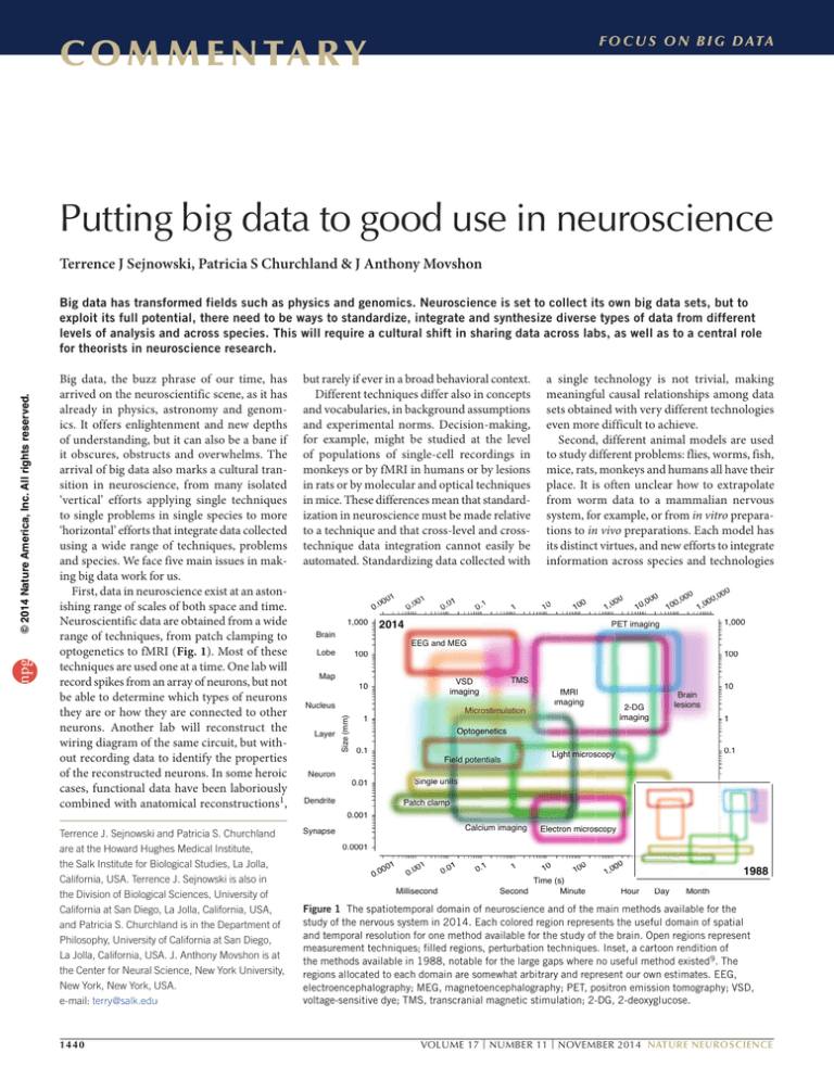
First, data in neuroscience exist at an astonishing range of scales of both space and time.
Neuroscientific data are obtained from a wide
range of techniques, from patch clamping to
optogenetics to fMRI (Fig. 1). Most of these
techniques are used one at a time. One lab will
record spikes from an array of neurons, but not
be able to determine which types of neurons
they are or how they are connected to other
neurons. Another lab will reconstruct the
wiring diagram of the same circuit, but without recording data to identify the properties
of the reconstructed neurons. In some heroic
cases, functional data have been laboriously
combined with anatomical reconstructions1,
but rarely if ever in a broad behavioral context.
Different techniques differ also in concepts
and vocabularies, in background assumptions
and experimental norms. Decision-making,
for example, might be studied at the level
of populations of single-cell recordings in
monkeys or by fMRI in humans or by lesions
in rats or by molecular and optical techniques
in mice. These differences mean that standardization in neuroscience must be made relative
to a technique and that cross-level and crosstechnique data integration cannot easily be
automated. Standardizing data collected with
Terrence J. Sejnowski and Patricia S. Churchland
are at the Howard Hughes Medical Institute,
the Salk Institute for Biological Studies, La Jolla,
California, USA. Terrence J. Sejnowski is also in
the Division of Biological Sciences, University of
California at San Diego, La Jolla, California, USA,
and Patricia S. Churchland is in the Department of
Philosophy, University of California at San Diego,
La Jolla, California, USA. J. Anthony Movshon is at
the Center for Neural Science, New York University,
New York, New York, USA.
e-mail: terry@salk.edu
Synapse
1440
1
00
0.0
Brain
1,000
000
Lobe
Map
01
0.0
1
0.1
0.0
Neuron
10
0
10
00
1,0
0
,00
10
00
0,0
10
0
,00
00
1,0
1,000
1,
000
PET imaging
EEG and MEG
100
10
00
TMS
VSD
ima
imag
ag
ging
g
imaging
10
0
Nucleus
Layer
1
a single technology is not trivial, making
meaningful causal relationships among data
sets obtained with very different technologies
even more difficult to achieve.
Second, different animal models are used
to study different problems: flies, worms, fish,
mice, rats, monkeys and humans all have their
place. It is often unclear how to extrapolate
from worm data to a mammalian nervous
system, for example, or from in vitro preparations to in vivo preparations. Each model has
its distinct virtues, and new efforts to integrate
information across species and technologies
2014
100
0
Size (mm)
Siz
npg
© 2014 Nature America, Inc. All rights reserved.
Big data has transformed fields such as physics and genomics. Neuroscience is set to collect its own big data sets, but to
exploit its full potential, there need to be ways to standardize, integrate and synthesize diverse types of data from different
levels of analysis and across species. This will require a cultural shift in sharing data across labs, as well as to a central role
for theorists in neuroscience research.
fM
fM
MR
R
RI
fMRI
imag
im
ging
imaging
Mi
Micro
Micr
o timula
osti
mulati
l tion
Microstimulation
1
10
0
Brain
lesions
2
2-DG
im
imaging
1
Optogenetics
O
Optoge
Opto
pt ge
gene
enetics
eneti
0.1
1
0.1
Lig
ght microscopy
miccrrossco
opyy
Light
Field
d potentials
Single
Sing
ngle
le units
u
un
nits
nits
0.01
0
.01
1
Dendrite
0 01
0.01
1
Patch clam
mp
clamp
0.001
0.0
001
1
0.001
0
0.00
.0
0
00
01
Calcium imaging
Electron
n microscopy
microssco
s opy
Electron
0.0001
0.0001
0
0.0
0001
1
00
0.0
1
.00
0
Millisecond
0
.01
0.1
1
10
0
10
Time (s)
Second
Minut
Minute
tte
e
00
1,0
10
0
00
Hourr
00
00
10
Day
00
00
00
1
1988
Month
Figure 1 The spatiotemporal domain of neuroscience and of the main methods available for the
study of the nervous system in 2014. Each colored region represents the useful domain of spatial
and temporal resolution for one method available for the study of the brain. Open regions represent
measurement techniques; filled regions, perturbation techniques. Inset, a cartoon rendition of
the methods available in 1988, notable for the large gaps where no useful method existed9. The
regions allocated to each domain are somewhat arbitrary and represent our own estimates. EEG,
electroencephalography; MEG, magnetoencephalography; PET, positron emission tomography; VSD,
voltage-sensitive dye; TMS, transcranial magnetic stimulation; 2-DG, 2-deoxyglucose.
VOLUME 17 | NUMBER 11 | NOVEMBER 2014 NATURE NEUROSCIENCE
npg
© 2014 Nature America, Inc. All rights reserved.
C O M M E N TA R Y
may pay off handsomely. But this will require
a deepened appreciation of comparative and
evolutionary neurobiology.
It has been said that “nothing in neuroscience makes sense except in the light of
behavior”2. Traditionally, neuroscientists
have restricted the range and richness of
behavioral measurements to keep the collection and interpretation of correlated data from
neurons manageable. This strategy constrains
our understanding of how the brain supports
the full range of behaviors. Big data is making it possible to record from the same set of
neurons while the subject engages in a much
richer set of behaviors. Behavioral research
will greatly benefit from the application of
machine learning techniques that allow fully
automated analysis of behavior in freely moving animals3–5. The challenge is to discover the
causal relationships between big neural data
and big behavioral data.
Third, as things stand in neuroscience, integration of functional data is mainly tackled by
individual labs and by those with whom they
collaborate. Such a strategy of ‘every tub on
its own bottom’ depends on individuals to
absorb information, communicate with others in the same subfield, and otherwise keep
up. Meetings, lab visits, publications, review
articles and so forth have been the mainstay
of this form of integration. Although powerful and productive and a source of innovation,
this style has limits. With increases in numbers
of laboratories and publications, it is hard for
individuals to keep up with the latest technology and harder still to keep data from slipping
into oblivion, including data whose significance can be appreciated only later when the
science catches up with the technology. This
will require a cultural shift in the way that data
are shared across labs.
Note too that this kind of integration is
essentially vertical, in the sense that it is largely
directed toward one particular problem, going
up and down the organizational levels on that
problem. Horizontal integration of data across
a range of problems—for example, learning,
decision-making, perception, emotion and
motor control—is even harder to achieve in
one laboratory. There is just too much data for
one laboratory to get its collective head around.
A goal of the BRAIN Initiative6 is to record
and manipulate a large number of neurons
during extended, behavioral experiments,
to identify the neurons recorded from, to
reconstruct the circuit that gave rise to the
activity, and to relate the combined data
to behavior—all in the same individual.
Although this may seem like a pie-in-the-sky
experiment, it is within reach in some species,
such as the nematode worm Caenorhabditis
elegans, whose neuronal connectivity is already
known, and the transparent larval zebrafish,
where it is possible to record simultaneously
from most of its 100,000 or so neurons. To
accomplish these ambitious goals will take
teams of closely coordinated researchers with
complementary expertise.
Fourth, as data sets grow and become more
complex, it will become more and more difficult to analyze and extract conclusions. In
the worst case scenario, the data may not be
reducible to simpler descriptions. Here we
need to rely on new approaches to analyzing data in high-dimensional spaces using
pattern-searching algorithms that have been
developed in statistics and machine learning.
To illustrate, consider the project of
Vogelstein et al.7, whose aim was to understand in Drosophila larvae the causal role of
each of 10,000 neurons in producing a simple
behavior in the animal’s repertoire, such as
turning or going backwards. Drawing on over
1,000 genetic lines and using optogenetic techniques to stimulate individual neurons in each
line, they generated a basic data set consisting
of correlations between stimulated identified
neurons and a behavioral output. (Notice that
the data set would have been far more massive had they stimulated neurons two or three
or more at a time.) To find patterns in their
huge accumulation of correlational data, they
fed the data to an unsupervised learning program, which yielded a potential understanding of links between neurons and behavior.
Correlational data could enhance understanding of the connectional structure to address
questions of circuitry. Nevertheless, the methodological significance of the project is that is
shows how new tools can be put to work to
find patterns in data obtained from networks of
neurons, patterns that emerge only from using
new analytic tools on very large data sets.
The statistical design of these experiments
will be critical to insure that data sets are carefully calibrated, are of sufficient power to admit
NATURE NEUROSCIENCE VOLUME 17 | NUMBER 11 | NOVEMBER 2014
analysis, and can be used by other researchers
who want to ask different questions. This is not
an easy process and requires a level of planning and quality control that goes beyond most
exploratory experiments that are undertaken in
most laboratories8. Here again, a modest cultural change can make a large impact.
Fifth, at some point along the Baconian rise
of ever larger and more complex data sets, a
deeper understanding should emerge from the
accumulated knowledge, as it has in other areas
of science. What we have today is a lot of small
models that encompass limited data sets. These
models are more descriptive than explanatory.
Theory has been slow in coming. One obstacle
is that sometimes theorists do not clearly convey what they propose, perhaps because they
seek safety in needlessly complex mathematics or because they are too remote from the
experimental base to undergird their theoretical ideas. Any of these issues can detract from
productive ideas. This can change.
What we contemplate are modest cultural
changes, wherein some neuroscientists are
mainly theorists, with appropriate grant support to make the research feasible. The term
“theorist” enjoys an uneven reputation in neuroscience, but serious scholars with this portfolio do now exist, although they tend to be in
short supply. We need to cultivate a new generation of computationally trained researchers
who are aware of the richness of data and can
draw on knowledge from many laboratories,
courageous enough to make judicious simplifications and to have their ideas tested, and
imaginative enough to generate interesting,
testable large-scale ideas.
COMPETING FINANCIAL INTERESTS
The authors declare no competing financial interests.
1. Bock, D.D. et al. Nature 471, 177–182 (2011).
2. Shepherd, G.M. Neurobiology. 8 (Oxford Univ. Press,
1988).
3. Dankert, H., Wang, L., Hoopfer, E.D., Anderson, D.J. &
Perona, P. Nat. Methods 6, 297–303 (2009).
4. Falkner, A.L., Dollar, P., Perona, P., Anderson, D.J. &
Lin, D. J. Neurosci. 34, 5971–5984 (2014).
5. Wu, T. et al. IEEE Trans. Syst. Man Cybern. B Cybern.
42, 1027–1038 (2012).
6. National Institutes of Health. BRAIN 2025: a scientific vision. http://www.nih.gov/science/brain/2025/
(2014).
7. Vogelstein, J.T., Park, Y. & Ohyama, T. Science 344,
386–392 (2014).
8. Mountain, M. Phys. Today 67, 8–10 (2014).
9. Churchland, P.S. & Sejnowski, T.J. Science 242,
741–745 (1988).
1441


