II. Electrons and Their Interaction with Matter
advertisement

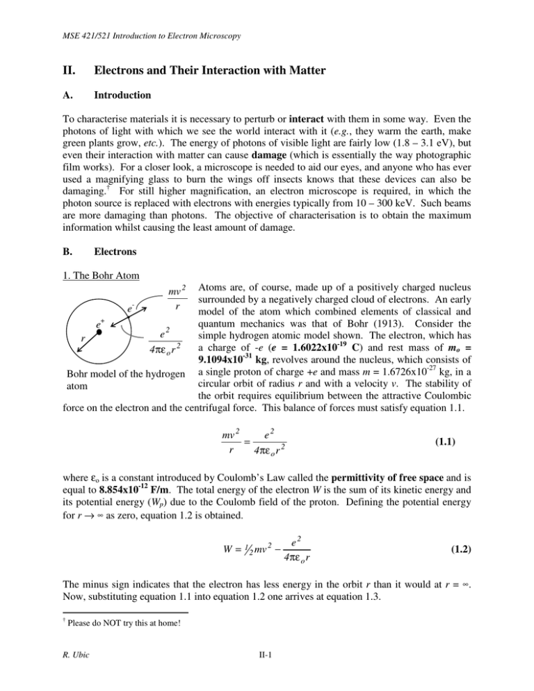
MSE 421/521 Introduction to Electron Microscopy II. Electrons and Their Interaction with Matter A. Introduction To characterise materials it is necessary to perturb or interact with them in some way. Even the photons of light with which we see the world interact with it (e.g., they warm the earth, make green plants grow, etc.). The energy of photons of visible light are fairly low (1.8 – 3.1 eV), but even their interaction with matter can cause damage (which is essentially the way photographic film works). For a closer look, a microscope is needed to aid our eyes, and anyone who has ever used a magnifying glass to burn the wings off insects knows that these devices can also be damaging.† For still higher magnification, an electron microscope is required, in which the photon source is replaced with electrons with energies typically from 10 – 300 keV. Such beams are more damaging than photons. The objective of characterisation is to obtain the maximum information whilst causing the least amount of damage. B. Electrons 1. The Bohr Atom Atoms are, of course, made up of a positively charged nucleus surrounded by a negatively charged cloud of electrons. An early emodel of the atom which combined elements of classical and quantum mechanics was that of Bohr (1913). Consider the e+ 2 e simple hydrogen atomic model shown. The electron, which has r 2 a charge of -e (e = 1.6022x10⁻¹⁹ C) and rest mass of mo = 4πε o r 9.1094x10⁻³¹ kg, revolves around the nucleus, which consists of -27 Bohr model of the hydrogen a single proton of charge +e and mass m = 1.6726x10 kg, in a circular orbit of radius r and with a velocity v. The stability of atom the orbit requires equilibrium between the attractive Coulombic force on the electron and the centrifugal force. This balance of forces must satisfy equation 1.1. mv 2 r mv 2 e2 = r 4πε o r 2 (1.1) where εo is a constant introduced by Coulomb’s Law called the permittivity of free space and is equal to 8.854x10⁻¹² F/m. The total energy of the electron W is the sum of its kinetic energy and its potential energy (Wp) due to the Coulomb field of the proton. Defining the potential energy for r → ∞ as zero, equation 1.2 is obtained. W= 1 2 2 mv − e2 4πε o r (1.2) The minus sign indicates that the electron has less energy in the orbit r than it would at r = ∞. Now, substituting equation 1.1 into equation 1.2 one arrives at equation 1.3. † Please do NOT try this at home! R. Ubic II-1 MSE 421/521 Introduction to Electron Microscopy W= e2 e2 −e 2 − = 8 πε o r 4πε o r 8 πε o r (1.3) Equation 1.3 states that the electron is bound to the nucleus, and the energy required to excite it away is the positive quantity -W. W decreases as r decreases, but, of course, that is not to imply negative energy! Bohr realised that this classical model of the atom would be unstable because the electron’s energy would dissipate, sending the electron crashing into the nucleus. According to classical electromagnetic theory, an electric charge e undergoing harmonic oscillation with angular frequency ωo (rad/s)† and amplitude xo along a line, its coordinate x being given by the equation x = xocosωot, where t is time, loses energy W by emitting radiation (light) at the rate − dW ω 4o e 2 xo2 = dt 3c 3 An electron in a circular orbit in the xy plane is equivalent to a linear oscillator along the x-axis and one along y, and should consequently radiate twice as fast. This energy loss is a simple way of understanding Bohr’s dilemma. In order to retain the orbit’s stability, Bohr suggested that the electron can only radiate energy of a certain size. He postulated that only those circular orbits are stable for which the angular momentum is an integer multiple of h/2π, where h = 6.6261x10⁻³⁴ Js is Planck’s constant. Mathematically, this requirement takes on the form of equation 1.4. mvr = nh 2π n = 1,2,3... (1.4) Substituting equation 1.4 into equation 1.1, one obtains an expression for the radii of all the allowed circular orbits in hydrogen. rn = εoh 2 πme 2 n 2 ≈ 5.29n 2 Å (1.5) Now, combining equations 1.3 and 1.5 one obtains an expression for the binding energy for electrons in various circular orbits n. − e 2 πme 2 − me 4 1 −13.6 = 2 2 2 ≈ W = eV 2 2 n2 8 πε o ε o h n 8 ε o h n (1.6) According to equation 1.6, when n = 1, the electron is bound to the nucleus with 13.6 eV (1 eV = 1.6022x10⁻¹⁹ J). It can be further deduced that as n increases (r increases), less energy is required to strip the electron from the nucleus. One final postulate to complete this view of the † The frequency ν in Hz is simply given by ν = ωo/2π. R. Ubic II-2 MSE 421/521 Introduction to Electron Microscopy atom states that the transition of an electron from one energy level Wn1 to another Wn2 is accompanied by the emission or absorption of electromagnetic radiation of frequency ν such that: hν = Wn2 − Wn1 (1.7) Thus, the energy involved in electronic transitions is quantized. approximation, n determines the energy of the electron. To a very good first 2. Properties of Waves a. Definitions The general definition of a wave is a fluctuation in one or more properties over space or time. A simple form of wave is a periodic oscillation. Such a wave can be described by its amplitude, A, which is the height of each peak above the baseline (or the depth of each trough). The wavelength is defined as the distance between peaks (or the distance between troughs) and is usually given the symbol λ (lambda). The frequency is defined as the number of peaks (or troughs) which pass a given point in a given time. The common symbol for frequency is ν, and a common unit of frequency is the Hertz (Hz), which is cycles per second. The period, T, of a wave is the inverse of its frequency (1/ν), that is, the time interval between peaks (or troughs) as seen from a stationary point. A A time, t distance, x λ R. Ubic A T II-3 A MSE 421/521 Introduction to Electron Microscopy Visible Spectrum The Electromagnetic Spectrum Radio Waves 0.1 Microwaves 10 Infrared 1000 10 5 Ultraviolet Xrays 107 109 Frequency (GHz) Gamma Rays 10 11 Cosmic Rays 1013 The electromagnetic spectrum Although frequency ranges are often cited for the various forms of EM radiation, they are only a guide. The difference between the various forms of natural radiation is really governed by the source of the radiation rather than its frequency. Low-frequency radio waves are caused naturally by the reversal of electron or nuclear spins in atoms and generally have wavelengths of more than a metre - and more than half a kilometre for longwave. This part of the spectrum is illustrated below. Radio Longwave 0.1 AM 1 VHF TV (2-4) VHF TV (5-6) FM UHF TV (7-13) Shortwave 10 100 Frequency (MHz) 1000 The radio spectrum Microwave frequencies are usually defined as those between 300 MHz and 300 GHz, or wavelengths of 1 mm to 1 m. Radiation of this type is excited by transitions between molecular rotational energy levels. The microwave spectrum is broken down into various bands, as shown below R. Ubic II-4 0.1 1 U-Band Ka-Band Ku-Band K-Band X-Band C-Band S-Band L-Band UHF TV (14-83) Microwaves Microwave Ovens MSE 421/521 Introduction to Electron Microscopy 10 100 Frequency (GHz) The microwave spectrum. The infrared band occurs at still higher frequencies, with wavelengths typically on the order of a few hundred microns. Radiation in this part of the spectrum is typically produced by molecular vibrations (phonon energy or heat). The visible spectrum is tiny by comparison, with wavelengths ranging only from about 400 - 700 nm. Ultraviolet radiation can have wavelengths down to just 2 nm. Both visible and ultraviolet light are produced naturally by transitions involving valence (outer) electrons. Far IR 0.1 1 Visible Near UV Far UV Middle IR 10 Near IR Infrared, Visible, and Ultraviolet 100 Extreme UV 1000 10 4 10 5 10 6 Frequency (THz) The infrared, visible, and ultraviolet spectra With wavelengths of only 1-20 Å, x-rays are the result of transitions of more tightly held inner electrons. It is interesting to note that in 1883 Lord Kelvin, then president of the Royal Society, predicted that “X-rays will prove to be a hoax.” Finally, gamma and cosmic rays, with wavelengths of less than 1 Å, are produced from very high-energy re-arrangements of nuclear particles. The product of wave frequency and wavelength is a constant which is equal to the speed of the wave. In a vacuum, the speed of light (EM radiation) is c = 2.9979X10⁸ ms-1 so: νλ = c Inside media other than vacuum (e.g., transparent glass), the speed of light is slowed down by a factor equal to the square root of the dielectric constant (εr), which implies that the wavelength decreases by the same amount (the frequency is unaffected). Strangely, photons can be described in terms of a wave or a particle (in reality they must therefore be neither in the conventional sense). R. Ubic II-5 MSE 421/521 Introduction to Electron Microscopy The energy, E, of a photon is described by E = hν where h = Planck’s constant = 6.6261 x 10⁻³⁴ Js. This energy only comes in discrete packets or quanta (hence particle representation), yet can also easily be described as a wave with frequency ν (hence the wave representation). The rate of flow of this energy through a unit area perpendicular to the direction of propagation is called intensity, I I ∝ A2 which is often expressed as relative intensity (e.g., Iscattered beam / Iincident beam). 3. Electrons as waves Credit for discovering the electron is usually given to J.J. Thomson, who was the first to quantify the ratio of mass to charge of what he termed negatively-charged “corpuscles.”† The ratio he published in 1897 was 0.4 - 0.5 X 10⁻⁷ g/C (the modern value is 0.569 X 10⁻⁷ g/C); however, Thomson did not have the whole picture. H.A. Lorentz and A. Einstein determined in 1905 that the mass of any object varies with its velocity, especially at very high (relativistic) speeds, according to: m = mo 1− v 2 c2 where c is the speed of light (2.9979 X 10⁸ m/s), mo is the rest mass of an electron (9.1091 X 10⁻³¹ kg), and v is velocity. A. Einstein published his famous equation, E = mc2, in the same year; however, this is actually a shortened form of a longer equation valid only in the case of stationary objects. The full relativistic equations is: E = mc 2 = ρ 2 c 2 + mo2 c 4 where ρ is momentum. In the case of photons (mo = 0) then the equation reduces to: E = ρc The energy of a photon can also be defined by the photoelectric effect, which points to the particle properties of light: E = hν or E = hc/λ † Curiously, Thomson studiously avoided the word “electron,” which had been invented by a wily and less respectable scientist named G.J. Stoney in 1891. R. Ubic II-6 MSE 421/521 Introduction to Electron Microscopy where h is Planck’s constant (6.6261X10⁻³⁴ Js), ν is frequency, and λ is wavelength. Combining these two equations, we obtain: λ = h/ρ = h/mv In his 1924 Ph.D. dissertation in Paris, Louis deBroglie postulated that particles may also have wave-like properties and that the above equation may also be true for particles like electrons as well as for photons - the wave nature of matter. Using this equation, Einstein’s relativistic one, and a simple energy balance, it is possible to derive the wavelength of the electron as a function of accelerating voltage, V, only. To start, the kinetic energy of the electron is: KE = eV = mc 2 − mo c 2 ∴ m = mo + eV c2 where moc2 is the rest energy (mass energy) and e is the elemental charge of an electron (1.6022 X 10⁻¹⁹ C). Setting the above two equations equal to each other and solving for v: mo + eV mo = 2 2 c 1− v 1 and taking the inverse : c mo + 2 1− v eV c2 = 2 c2 mo Now multiplying both sides by mo and squaring: mo2 eV mo + 2 c ∴v2 = c2 − 2 = 1− v 2 c2 mo2 c 2 eV mo + 2 c 2 ⇒ v = c 1− mo2 eV mo + 2 c 2 = c 1− Now we can calculate the momentum, ρ: eV mo2 ρ = mv = mo + 2 c 1 − 2 c eV mo + 2 c and squaring both sides gives us: R. Ubic II-7 1 eV 1 + 2 mo c 2 MSE 421/521 Introduction to Electron Microscopy 2 2 2 2 eV 2 mo eV 2 mo c 2 ρ = m o + 2 c 1 − = mo + 2 c − 2 2 c c eV eV mo + 2 mo + c 2 c 2 2 2 eV 2 eV mo c = m o + 2 c − mo + 2 2 c c eV mo + c 2 2 2 2 eV 2mo eV e 2V 2 = c mo + 2 − mo2 c 2 = c 2 mo2 + + 4 − mo2 2 c c c 2 2mo eV e 2V 2 = c + 4 2 c c 2 1 = 2 2mo c 2 eV + e 2V 2 c ( ) Therefore, ρ= 1 2mo c 2 eV + e 2V 2 c Now, substituting this into deBroglie's relation: λ= h h = = hc 2mo c 2 eV + e 2V 2 mv ρ ( ) −1 2 The wave nature of electrons makes possible interference and diffraction. The eventual discovery of electron diffraction was made jointly by C.J. Davisson and L.H. Germer at Bell Labs, USA and G.P. Thomson (son of J.J.) and A. Reid in the UK in 1927. Both teams used Ni crystals in their experiments, and Davisson and Thomson later shared the Nobel Prize for their discovery. C. Electron generation The illumination system in any electron microscope consists of a source of electrons (the electron gun) and a lens system to focus and control emitted electrons. They work in one of two basic ways. The first method is to overcome the work function by resistance heating; the second method is to use a strong electric field. 1. Thermionic emission A thermionic electron gun consists essentially of a heated wire or crystal from which electrons are given enough thermal energy to overcome the work function combined with an electric potential to give the newly freed electrons velocity. The heated source is usually held at some R. Ubic II-8 MSE 421/521 Introduction to Electron Microscopy potential (~500V to ~100kV) negative relative to ground, so that a sample (as well as the rest of the microscope) can be kept at ground. a. Tungsten Wires are made of W because it has a very high melting temperature (3422°C), so more thermal energy can be made available, although its work function (4.5 eV) is fairly high even at the ~2500°C operating temperature. Tungsten hairpin filaments are used in standard machines like the LEO 1430 VP SEM. A wire of tungsten metal is heated (T > 2500°C) under a large bias field, stripping electrons off according to the Edison effect. b. LaB6/CeB6 It is easier to strip electrons off lanthanum hexaboride, LaB6, or cerium hexaboride, CeB6, and such crystals can produce beams 30 times as bright as a tungsten one which are stable over long periods. The disadvantages of using such crystals are that they require a higher vacuum, they suffers from thermal shock (and so must be heated and cooled slowly), and they are expensive ($1000 compared to $30 for tungsten). The JEOL 2100 uses a LaB6 crystal. LaB6 melts at 2530°C and CeB6 at 2540°C. Both are used at ~1500°C because at this temperature they both have a low work function (~2.5 eV). Higher temperatures cause increased evaporative loss of the crystal and premature failure. 2. Field-effect emission Field emission guns (FEGs) offer a further increase in brightness, but at the penalty of requiring an ultrahigh vacuum system. The cold field emission gun, such as the one on the Hitachi 4500, uses a tungsten single crystal and an intense electric field to extract the electrons. This field lowers the height of the potential barrier via the Schottky effect. If the electric field is increased sufficiently, the width of the potential barrier becomes small enough to allow electrons to escape through the surface potential barrier by quantum-mechanical tunnelling, a process known as field-emission. A high vacuum is required to reduce contamination of the crystal and the gun must be flashed (heated to high temperature) every few hours to drive off contaminants. In the thermally assisted field emission gun (Schottky emitter), a pointed crystal of W is welded to the end of a V-shaped W filament, coated with ZrO2, and heated to 1500°C. An intense electric field is still used to extract the electrons, but the stability is greatly improved and the gun does not require flashing. Such thermal field emitters enhance the pure field emission effect by giving some thermal energy to the electrons in the metal, so that the required tunneling distance is shorter for successful escape from the surface. As a result, the emission current density is typically increased by a factor of 10. R. Ubic II-9 MSE 421/521 Introduction to Electron Microscopy A comparison of electron emission characteristics of W, LaB6, CeB6 and FEG guns LaB6 CeB6 FEG FEG W wire <100> <100> (cold) (Schottky) Operating temperature (°C) 2500 1500 1500 27 1500 Work function (eV) 4.5 ~2.5 ~2.5 4.5 4.5 2 5 Emission current (A/cm ) 1 100 100 10 105 -2 -1 6 7 7 9 Brightness (A cm sr ) 10 10 10 10 109 30 10 10 0.01 0.01 Crossover diameter (µm) Short-term beam current <1 <1 <1 3-5 >5 stability (%RMS) Energy spread (eV) 2 1.5 1.5 0.2 - 0.4 0.2 - 0.4 Typical service life (hr) 30-100 1000 1,500 1000 100 Operating vacuum (torr) 10-5 10-7 10-7 10-8 10-9 Evaporation rate (g cm-2 sec-1) NA 2.2 x 10⁻⁹ 1.6 x 10⁻⁹ NA NA For a given source, the brightness of the beam increases linearly with accelerating voltage. Thermionic guns have large areas of emission and are therefore insensitive to changes in filament current and contamination. They are very stable. The beam size (spot size) is reduced by 104 times for analysis to just ≈1nm, further reducing the effects of instability. The cold field emission gun has a very small area of emission and so is highly sensitive to contamination, resulting in short-term fluctuations in beam current and gradual long-term build-up of contaminants. The beam size is reduced only 5-10 times from the source, to ≈1nm for analysis; therefore, the beam is still very sensitive to contamination. Also, as the electrons come from only a tiny area on the crystal in a field emission gun, the beams produced are nearly coherent (i.e., same wavelength and phase), which is why they can be so intense. R. Ubic II-10 MSE 421/521 Introduction to Electron Microscopy 3. Triode electron gun In this triode electron gun, the filament is contained in a Wehnelt cylinder with a small (< 1mm) hole directly below the filament. The electrons thermionically emitted are accelerated towards the anode as a result of the large potential difference between the filament assembly (cathode) and the anode. The anode is usually at earth potential so that the rest of the microscope column is at earth. Consequently, the Wehnelt cap is kept at a large negative potential compared to the anode. In addition, there is a bias resistor between the filament heating circuit and the Wehnelt cap so that the cap is biased slightly negative with respect to the filament to ensure that it does not act as a sink for electrons. This arrangement ensures that most of the electrons are emitted from the filament tip and that the Wehnelt repels the electrons and causes a focusing action. The gap between the filament tip and the Wehnelt cap is critical to this focusing action and filament lifetime. The triode gun produces a beam of electrons which comes to a focus (crossover) at a point just below the Wehnelt cap with a diameter of about 50 µm for W and 1 µm for LaB6 or CeB6. D. Magnetic Lenses Magnetic fields can displace electrons and so can be used to focus electron beams. A magnetic field can be produced by passing a current through a coil consisting of many turns of wire. The field strength can be increased by using a ferromagnetic core like Fe. Varying the current to the coil will alter R. Ubic II-11 MSE 421/521 Introduction to Electron Microscopy the strength of the magnetic field generated. The direction and magnitude of the force felt by the electron is: v v v F = e(v × B ) v v where e is the electron charge, v is the velocity of the electron, and B is the magnetic field strength. The geometry of magnetic lenses in electron microscopes is slightly more complicated, but the principle is the same. A coil of wire is wound on an iron core (the pole piece), which has a very small gap across which the field is produced. By varying the current in the coil, typically 0 – 1A, the magnetic field strength and hence the focal length of the lens can be varied. The lens is designed to produce a magnetic field almost parallel to the electron trajectory. This magnetic field will have components both along the axis of the microscope, Bax, as well as in the radial direction, Brad. Initially, the electron is unaffected by Bax, which is parallel to its direction of travel, but experiences a small force, Bradev, from the small radial component, which pushes it into the plane of the paper (in fact, since the actual lens is circular, the path is a helix). With this motion, the velocity of the electron now has a component perpendicular to the axis, and so is affected by Bax, which pushes it to the right (again, because the lens is circular, motion is actually towards the axis). The net result of Bax and Brad on the electron is to make it spiral down the gap in a helical path. Because Bax grows as the electron approaches the middle of the gap, its path narrows and becomes focussed around the optic axis. This spiralling of electrons through magnetic lenses is an important difference between these lenses and optical ones. As the strength of the lens changes, so does the rotation; but this effect can be eliminated (as it is in the JEOL 2100) by using oppositely poled subsequent lenses. Another difference is, of course, that these lenses can be switched off simply by stopping the current through them. Electromagnetic lenses only approximate to a Maxwell lens when electrons pass close to the lens axis. It is impossible to produce a perfect electromagnetic lens according to the Maxwell criteria. There will always be aberrations and distortions present, which ultimately limit the resolution in transmission electron microscopes. R. Ubic II-12 MSE 421/521 Introduction to Electron Microscopy E. Interaction of Electrons with Atoms 1. Electron Scattering The probability that a particular electron will be scattered in a particular way is defined by its mean free path, λ, or scattering cross section, σ. This cross section represents the area which the scattering particle appears to present to the electron. If there are N such particles per volume, then the probability of a single electron being scattered in a distance dx through the specimen is Nσdx. The mean free path is: λ = 1/Nσ. Here, λ is essentially the average distance an electron will travel through the specimen before being scattered in this way. Mean free paths are often similar to the typical thickness of a TEM sample, which implies that most electrons are scattered just once or not at all in the specimen. On the other hand, thicker samples like those used in an SEM provide sufficient opportunity for an electron to be scattered many times. The probability of an electron undergoing n scattering events while travelling a distance x is given by the Poisson equation: p(n) = (1/n!)(x/λ)nexp(-x/λ) This equation assumes multiple scattering by the same process (same λ), and so does not very accurately predict multiple scattering in real materials. 2. Interaction volume The primary or incident electron beam enters the specimen and can potentially undergo numerous scattering events. The scattering means that the sample is excited below its surface over a region much larger than the incident beam diameter. The region into which the electrons penetrate the specimen is known as the interaction volume. In a scanning electron microscope (SEM) sample this interaction volume is much larger than in a transmission electron microscope (TEM) sample because TEM samples are generally only ≈100nm thick. Incident (primary) beam SEM Secondary electrons Backscattered electrons ≈1µm X-rays R. Ubic II-13 TEM ≈100nm MSE 421/521 Introduction to Electron Microscopy The maximum diameter, B, of the truncated interaction volume in a TEM specimen can be estimated as: B = 0.198(Z/E)(ρ/A)1/2t3/2 where Z = atomic number, E = electron energy (keV), ρ = density (g/cm3), and t = thickness (nm). The penetration depth is a function of two factors. First, it increases as the accelerating voltage is increased. Secondly, it decreases as the atomic number of the sample atoms increases. primary electrons Backscattered electron Characteristic x-ray cathodoluminescence or Auger electron ejected secondary electron ejected secondary electron primary electron primary electron 3. Inelastic Scattering Inelastic scattering is a general term which refers to any process which causes the primary electron to lose a detectable amount of energy, ∆E. These processes all involve an interaction between the primary beam electrons and orbital electrons of the atoms in the sample. Most of the exchanged energy is lost as heat; however, it may also be released in other ways. a. Phonon scattering Phonons are quanta of atomic vibrations in a solid. Primary electrons can lose energy by exciting phonons, which result in heating of the solid. The amount of energy arising from each interaction is small, about 1eV; however, the heating effects can be strong particularly at high voltages. In addition, the scattering event results in a large deflection of the electrons, typically 10°, which gives rise to increased diffuse intensity in electron diffraction patterns. The heating amounts to only about 10°C in a good metallic conductor like Al; however, in poor conductors like polymers or ceramics the heating can be large. It is possible to melt Al2O3 (Tm > 2000°C) in a TEM. R. Ubic II-14 MSE 421/521 Introduction to Electron Microscopy b. Plasmon scattering A plasmon is a wave in the sea of electrons in the conduction band of a metal. This type of scattering is a result of the interaction of the primary electrons with the free electron gas in a material. Primary electrons lose 5 – 30 eV per event and have a small mean free path (they do not travel far into the sample before causing plasmon scattering). These events are common and are important in Auger and EELS studies. They also contribute to the diffuse intensity around the transmitted spot in an electron diffraction pattern. c. Single valence electron excitation It is also possible, but less likely, that a primary electron will transfer some energy to a single conduction-band electron rather than the entire sea. The mean free path for this process is quite large (microns), the energy loss is small (≈1eV), and the typical scattering angle is also small; therefore, this process is not exploited in electron microscopy. d. Inner shell excitation leading to characteristic x-ray production A primary electron may transfer enough energy to a K or L shell electron to knock it out of the atom. Because the binding energies of such electrons is typically high, the amount of energy lost by the primary beam is quite large. It requires 283eV to remove a carbon K electron and 69,508eV to remove a tungsten K electron. The mean free path for this process is quite large, so it is relatively infrequent. The probability of this type of scattering occurring decreases both as the incident voltage increases (high-energy electrons are more likely to pass through the sample without interacting) and as atomic number increases (the critical energy required to produce an x-ray increases with atomic number). The hole left in the inner shell can be filled by one of the out electrons dropping down, releasing a quantum of energy characteristic of that particular transition in that particular atom. Virtually all x-rays which are emitted escape from the surface, and they are produced throughout the entire interaction volume. These x-rays may therefore have come from well below the sample surface. For this reason, the smallest region which can easily be analysed in an SEM using x-rays is about 1µm. e. Inner shell excitation leading to characteristic (Auger) electron production An alternative to x-ray emission is the ejection of an outer electron carrying away the transferred energy of the primary electron as kinetic energy. This process is called Auger emission. Three electrons are involved: the primary electron (which loses some of its energy to the inner shell electron), the outer electron (which fills the hole in the inner shell), and the other outer electron (which carries off the surplus energy). Auger electrons have low energies and are easily absorbed by the specimen or any gaseous atoms in the vacuum; therefore, their detection requires very high vacuum systems. Nonetheless, their easy absorption means that those which escape the sample to be detected have come from the outer ≈1nm of the sample surface and so give detailed information about this region. Auger electron spectroscopy is an important surface analysis technique. They are outer electrons and so associated with bonding and can give information on the local atom environment. The probability of Auger electron emission instead of x-ray emission increases for light elements. f. Outer shell excitation leading to cathodoluminescence If the primary electron ejects an outer electron (one from between valence and conduction bands) from an atom, that hole will also be filled by a higher-energy electron (one from the conduction R. Ubic II-15 MSE 421/521 Introduction to Electron Microscopy band); but the difference in energy levels is likely to be small and so the characteristic radiation emitted will be of low frequency, typically in the visible spectrum. This effect is called cathodoluminescence. Certain semiconductors and insulators will emit ultraviolet or visible light when bombarded with high-energy electrons. The intensity of these emissions is modified by the presence of impurities or defects like dislocations. This imaging technique is used extensively to examine defects in semiconductors like GaAs and AlN. g. Excitation of outer electrons leading to emission of low-energy secondary electrons Secondary electrons describe those electrons which escape from the specimen surface with energies below about 50eV. They could conceivably be primary electrons which have undergone numerous collisions eventually escaping from the surface with a little energy left over; however, they are much more likely to be electrons ejected by one of the processes above. This interaction forms the main method of image formation in the SEM. Because secondary electrons have low energies, they can only escape from regions near the surface (≈10nm), i.e., only from a small proportion of the interaction volume; however, the yield of secondary electrons can be ≥1 (each primary electron produces at least one secondary electron). primary beam high yield shadowing low yield specimen Consequently, sharp edges and corners appear bright, due to their greater surface areas, while flat regions appear darker. Similarly, parts of the sample tilted towards the detector appear bright because electrons can escape easily whereas those parts not facing the detector appear dim, not because fewer electrons are emitted, but because the distance the electrons must travel to the detector is much further. Since these secondary electrons arise from the surface, they produce what are termed topographical images showing the hills and valleys of a sample’s surface. h. Brehmsstrahlung x-rays When the primary electrons slam into the specimen, they are slowed and deflected by the Coloumb field of the atoms and their kinetic energy is transformed into photons of x-ray radiation. These x-ray emissions are made up of a mixture of rays with many wavelengths. decelerated electron emits radiation The primary electrons can collide with any atom at any angle any number of times, such that a whole range of energy losses and hence wavelengths of x-rays are produced. These x-rays are of no use in microanalysis and are simply unwanted background radiation. R. Ubic II-16 MSE 421/521 Introduction to Electron Microscopy i. Absorption The penetration depth or mean free path of the incident beam determines the depth and volume of material that will be sampled. In many cases the radiation detected is a different type to that used in the probe (e.g., EDS and XPS). Visible light is used mainly to obtain a visual image of the surface of a material, although it can also be used to probe the optical properties of a (usually transparent) specimen. Ultraviolet radiation (3.1 ≤ E ≤ 620 eV) can be used to investigate the electron distribution in surface atoms. After visible light, x-rays (620 eV < E ≤ 12.4 keV) are the next most utilised part of the spectrum to characterise materials. They are useful for probing the atomic arrangement (crystallography) of matter. The penetration distance varies with wavelength and material and is typically several microns for x-rays but much shorter for electrons. The linear absorption coefficient, µ, which increases with atomic number, determines the depth of penetration according to: I = I o exp(−µx) where I is the intensity of radiation transmitted through the material, Io is the intensity of radiation originally incident, and x is the thickness of the material. For a substance with mass density ρ, the mass absorption coefficient is µ/ρ and is independent of the physical state of the material (solid, liquid, or gas), and the equation can be re-written as: Mass Absorption Coefficient (cm2/g) I = I o exp[−( µ ρ )ρx] Wavelength (Å) Absorption coefficients (µ/ρ) of Pb showing K and L absorption edges When x-ray photons of wavelength 0.4 Å are incident on a sheet of Pb, the absorption coefficient is about 30 cm2/g. As the wavelength decreases (frequency increases), the absorption coefficient decreases because photons of higher energy pass more easily through a material. When the wavelength is reduced to just below the critical value, which is 0.14088 Å for Pb, the absorption R. Ubic II-17 MSE 421/521 Introduction to Electron Microscopy coefficient suddenly rises by a factor of about 5. Photons now have sufficient energy to knock K electrons out of the atoms. True K absorption is now occurring and a large fraction of the incident energy is converted into K fluorescent radiation and kinetic energy of ejected photoelectrons. The five-fold increase in µ/ρ means a tremendous decrease in transmitted intensity because of the exponential dependence of I. If the percentage of photons transmitted in a given Pb sheet is 0.1 for x-rays with wavelengths just longer than 0.14088 Å, it is only 2.2X10⁻⁵ for wavelengths just shorter. The value of 0.14088 Å is called the K absorption edge for Pb. Three closely spaced L edges exist at longer wavelengths, five M edges, etc. Each of these discontinuities marks the wavelength of photons which have just sufficient energy to eject an L, M, N, etc. electron from the atom. Absorption of x-rays occurs in two distinctly different ways, by scattering and by true absorption, and these two processes together make up the total absorption measured by µ. Compton scattering of x-rays occurs in all directions and thus reduces the number of photons in the transmitted beam. True absorption is an inelastic process caused by the production of photoelectrons and fluorescent radiation. 4. Elastic Scattering Rutherford scattering or back scattering These electrons have undergone billiard-ball type collisions with nuclei of specimen atoms and have as much energy as the primary electrons (10’s of eV) and are scattered from atoms up to 2µm deep in the sample. About 30% of the primary beam is reflected in this way. The contrast in such images is very sensitive to the average atomic number, Z, of the phases being imaged. Atoms with a high Z have large nuclei, so there is a greater chance of scattering an incoming electron. The more electrons being detected from a given area, the brighter the image will be; therefore, phases with higher average atomic numbers appear brighter than those with lower Z. Diffraction As we have already seen, electrons can be diffracted by crystal samples. Such interactions involve no change in electron energy and so are elastic. In a TEM, the result is typically a spot pattern (or a pattern of discs as in convergent beam electron diffraction). Electron diffraction can also take place in the SEM, and this technique is called electron backscattered diffraction (EBSD). 5. Electron Microscopes All electron microscopes have an electron gun (source of electrons), a system of condenser lenses, and some form of signal detector (e.g., phosphor screen, scintillator, etc.). An SEM additionally requires scanning coils. It becomes an analytical SEM by the addition of an x-ray detector, and a microprobe analyser (EPMA) when x-ray spectrometers are added. A TEM also requires objective and projector lenses and usually a viewing screen. The addition of an x-ray detector or electron energy spectrometer make the instrument an analytical TEM. The addition of scanning coils creates a scanning TEM (STEM). The combination of x-ray detector or electron energy spectrometer with scanning coils creates an analytical STEM. R. Ubic II-18


