Notes - Science
advertisement
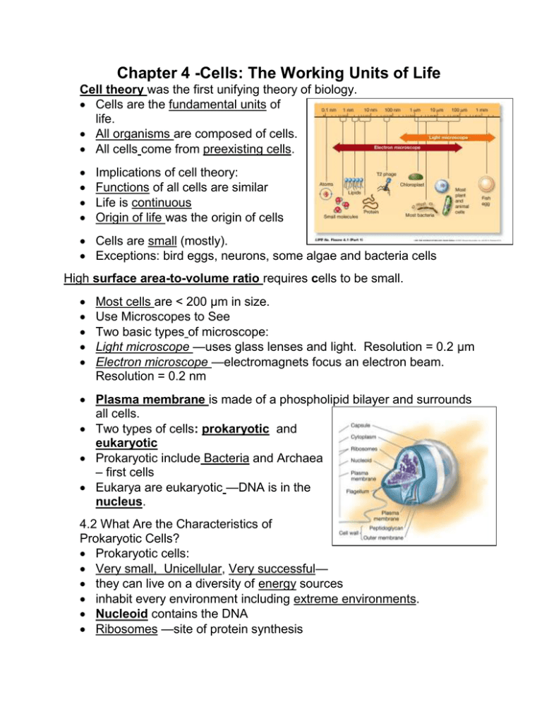
Chapter 4 -Cells: The Working Units of Life Cell theory was the first unifying theory of biology. Cells are the fundamental units of life. All organisms are composed of cells. All cells come from preexisting cells. Implications of cell theory: Functions of all cells are similar Life is continuous Origin of life was the origin of cells Cells are small (mostly). Exceptions: bird eggs, neurons, some algae and bacteria cells High surface area-to-volume ratio requires cells to be small. Most cells are < 200 μm in size. Use Microscopes to See Two basic types of microscope: Light microscope —uses glass lenses and light. Resolution = 0.2 μm Electron microscope —electromagnets focus an electron beam. Resolution = 0.2 nm Plasma membrane is made of a phospholipid bilayer and surrounds all cells. Two types of cells: prokaryotic and eukaryotic Prokaryotic include Bacteria and Archaea – first cells Eukarya are eukaryotic —DNA is in the nucleus. 4.2 What Are the Characteristics of Prokaryotic Cells? Prokaryotic cells: Very small, Unicellular, Very successful— they can live on a diversity of energy sources inhabit every environment including extreme environments. Nucleoid contains the DNA Ribosomes —site of protein synthesis Most have a rigid cell wall made of peptidoglycan. Some have a slimy capsule of polysaccharides outside the cell wall. In photosynthetic bacteria, the plasma membrane folds into the cytoplasm to form an internal membrane system where photosynthesis occurs. Some prokaryotes swim by means of flagella, made of the protein flagellin. Some bacteria have pili —hair-like structures projecting from the surface. They help bacteria adhere to other cells. Some rod-shaped bacteria have a cytoskeleton made of the protein actin. 4.3 What Are the Characteristics of Eukaryotic Cells? Eukaryotic cells much larger than prokaryotes. Have membrane-enclosed compartments with organelles. Compartmentalization allowed eukaryotic cells to specialize—forming tissues and organs into multicellular organisms. Organelles have been studied using different techniques: microscopy and cell fractionation. The nucleus contains DNA, surrounded by double membranes —the nuclear envelope. Nuclear pores in the envelope control passage of molecules. Large molecules such as proteins need a signal—a certain sequence of amino acids— to pass through. Ribosomes—sites of protein synthesis in both prokaryotic and eukaryotic cells. The endomembrane system includes the endoplasmic reticulum and the Golgi apparatus. The outer membrane of the nuclear envelope is continuous with the endomembrane system. Rough endoplasmic reticulum (RER): ribosomes are attached and make proteins for transport Smooth endoplasmic reticulum (SER): more tubular, no ribosomes Chemically modifies small molecules such as drugs and pesticides Hydrolysis of glycogen in animal cells Synthesis of lipids and steroids Source of peroxisomes The Golgi apparatus: Modifies, concentrates, packages, sorts proteins from RER In plant cells, polysaccharides for cell walls are synthesized Cis region receives vesicles and trans sends to plasma membrane Lysosomes originate from the Golgi apparatus. They contain digestive enzymes and fuse with phagosome from phagocytosis Autophagy - Lysosomes also digest cell materials Peroxisomes: collect toxic by-products of metabolism such as H2O2, using specialized enzymes. Glyoxysomes: only in plants—lipids are converted to carbohydrates for growth In the mitochondria, generate ATP from food molecules using a large surface area from many folds. Numerous mitochondria in cells that require a lot of energy. The mitochondrial matrix contains DNA and ribosomes. Plastids occur only in plants and some protists. Chloroplasts: Site of photosynthesis—have a double membrane – inner stroma contains DNA and ribosomes. Chromoplasts contain red, orange, and yellow pigments—gives color to flowers. Leucoplasts store starches and fats. Plant and protist cells have vacuoles: Store waste products and toxic compounds—may deter herbivores Provide structure for plant cells with water vacuole—turgor Store anthocyanins (pink and blue pigments) in flowers and fruits Digestion in seeds —vacuoles have enzymes to hydrolyze stored food for early growth Many protists have food vacuoles—formed by phagocytosis. Freshwater protists may have contractile vacuoles to expel excess water. The cytoskeleton: Supports and maintains shape Allows some types of movement Positions organelles Some fibers act as support for motor proteins Interacts with extracellular structures to hold cell in place Three components: Microfilaments, Intermediate filaments, and Microtubules Cell biologists use two approaches to show cause and effect —that a structure is responsible for a function: Inhibition and Mutation Microfilaments: Help a cell or parts of a cell to move Determine cell shape Made from the protein actin Actin has + and – ends and polymerizes to form long helical chains (reversible). Intermediate filaments: Many different kinds Made of fibrous proteins of the keratin family Stabilize cell structure and resist tension Disruption of intermediate filament networks causes blistering Microtubules: Form rigid internal skeleton in some cells Act as tracks along which motor proteins move Made from the protein tubulin—a dimer Have + and – ends Can change length rapidly by adding or losing dimers Cilia and eukaryotic flagella: made of microtubules in “9 + 2” array Cilia—short, usually many present, move with stiff power stroke and flexible recovery stroke Flagella—longer, usually one or two present, movement is snake-like The nine microtubule doublets extend into the basal body in the cytoplasm. In the basal body, each doublet is joined by another microtubule, making nine triplets. Centrioles are identical to basal bodies. Involved in formation of the mitotic spindle. Motor proteins: undergo reversible shape changes powered by ATP Dynein binds to microtubule doublets and allows them to slide past each other. Kinesin binds to a vesicle and “walks” it along by changing shape. Extracellular structures are outside the plasma membrane Example: peptidoglycan cell wall of bacteria Plant cell walls: cellulose fibers embedded in other complex polysaccharides and proteins Adjacent plant cells are connected by plasma membrane-lined channels called plasmodesmata. Many animal cells are surrounded by an extracellular matrix, composed of fibrous proteins such as collagen, gel- like proteoglycans (glycoproteins), and other proteins. The extracellular matrix: Holds cells together in tissues Contributes to properties of bone, cartilage, skin, etc. Orient cell movements in development and tissue repair Plays a role in chemical signaling 4.5 How Did Eukaryotic Cells Originate? Eukaryotic cells appeared about 1.5 billion years ago. Endosymbiosis theory explains how eukaryotes could evolve from prokaryotes. Cells engulfed other cells that became mitochondria and chloroplasts. Shared chemistry suggests that eukaryotes evolved from prokaryotes: Both use nucleic acids as hereditary material Both use the same 20 amino acids Both use D sugars and L amino acids
