SBI 4UI Dissections: Brain and Eye The Brain Examine the sheep
advertisement
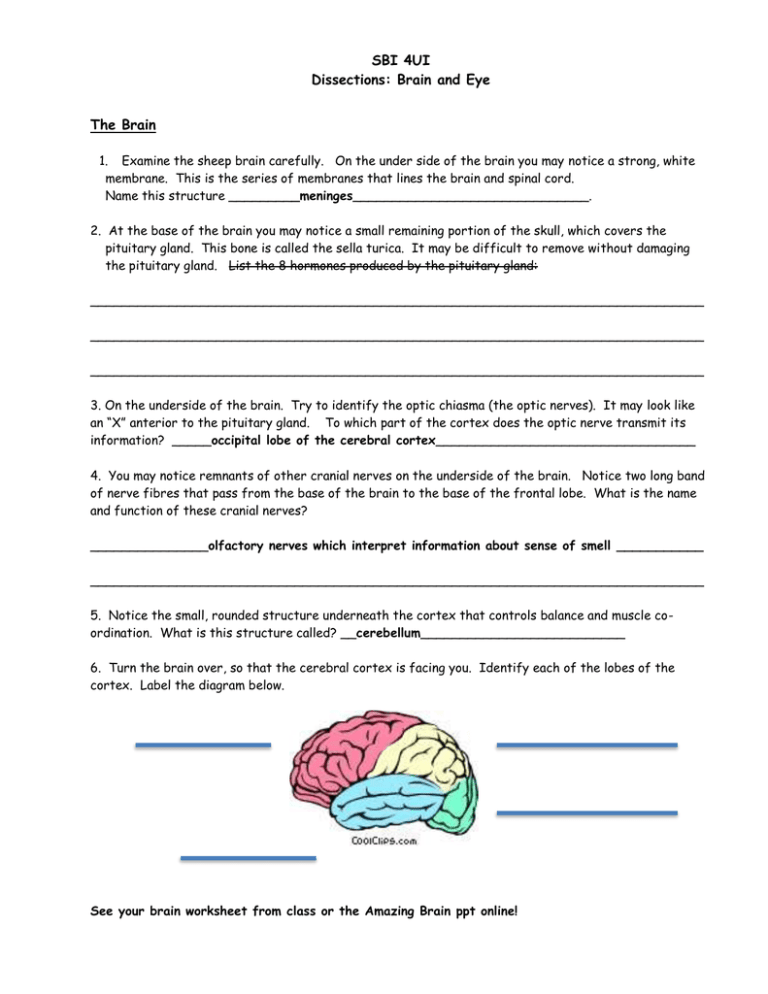
SBI 4UI Dissections: Brain and Eye The Brain 1. Examine the sheep brain carefully. On the under side of the brain you may notice a strong, white membrane. This is the series of membranes that lines the brain and spinal cord. Name this structure _________meninges______________________________. 2. At the base of the brain you may notice a small remaining portion of the skull, which covers the pituitary gland. This bone is called the sella turica. It may be difficult to remove without damaging the pituitary gland. List the 8 hormones produced by the pituitary gland: ______________________________________________________________________________ ______________________________________________________________________________ ______________________________________________________________________________ 3. On the underside of the brain. Try to identify the optic chiasma (the optic nerves). It may look like an “X” anterior to the pituitary gland. To which part of the cortex does the optic nerve transmit its information? _____occipital lobe of the cerebral cortex_________________________________ 4. You may notice remnants of other cranial nerves on the underside of the brain. Notice two long band of nerve fibres that pass from the base of the brain to the base of the frontal lobe. What is the name and function of these cranial nerves? _______________olfactory nerves which interpret information about sense of smell ___________ ______________________________________________________________________________ 5. Notice the small, rounded structure underneath the cortex that controls balance and muscle coordination. What is this structure called? __cerebellum__________________________ 6. Turn the brain over, so that the cerebral cortex is facing you. Identify each of the lobes of the cortex. Label the diagram below. See your brain worksheet from class or the Amazing Brain ppt online! 7. Note the folded nature of the cortex. What is the name of the raised areas? __gyri______ What is the name of the lower, depressed areas of the cortex? ____sulci________________ 8. Use your fingers to pull apart the hemispheres. Use dissecting scissors to cut the structure that joins the two hemispheres together. What is this structure called? _corpus collosum___________ 9. Use the knife to dissect the cerebellum and brain stem along the saggital plane (the midline). 10. Examine the brain in cross-section. With your group, try to identify as many regions as possible. Label the following diagram to review the parts. See your brain worksheet from class or the Amazing Brain ppt online! 11. Use the knife to cut the cortex perpendicular to the midline, (also called a “coronal section”). Why do some areas appear lighter in colour than others? Lighter areas contain axons, many of which have myelin covering them. The fatty myelin has a white appearance. The darker “grey matter” contains mostly dendrites, and cell bodies. The Eye ** MUST WEAR EYE PROTECTION 1. Examine the sheep eye. Find the site where the optic nerve enters the eye. What does it look like? Flat, round disc-like structure. 2. Remove the eyelid and any fat tissue. Examine the outer surface of the eye. What is the white structure called? ________the sclera_____________________ 3. You may notice four small muscles (on the top, bottom, and sides). These muscles control the movement of the eyeball. 4. Using dissecting scissors, cut around the outer edge of the cornea. (BEWARE when making the first cut.) What is the name of the fluid in the anterior (front) part of the eye? _____________the aqueous humor___________________ 5. Using forceps, take out the lens and examine it. In a living eye, the lens is very flexible and can adjust to see at different distances. 6. What is the name of the fluid in the posterior (back) part of the eye? The vitreous humor. 7. Using scissors, cut through the eyeball and examine the retina. What cells of the retina are sensitive to colour? ____________cone cells_____________ What cells of the retina are sensitive to bright or dim light? ______rod cells________________ Label the following diagram to review the parts of the eye. See your eye diagram from the class notes.
