Neural Tube Defects
advertisement
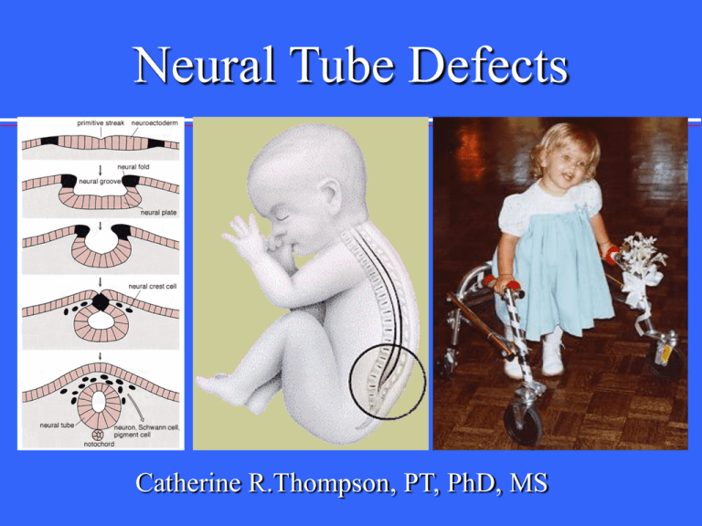
Neural Tube Defects Catherine R.Thompson, PT, PhD, MS Directions This PowerPoint presentation should be used in conjunction with the following resources: • The “Neural Tube Defects” handout • Case Study - Spina Bifida handout • Chapter 5 in Pediatric Physical Therapy (Tecklin, 1999) Introduction This presentation offers some illustrations of key concepts listed on the handouts and described in the text. Follow this presentation as you use the additional resources to gain an appreciation of your role as a PT working with children and adults with these types of impairments. When do neural tube defects occur? Neural Tube Development Normal embryological development Neural plate development -18th day Cranial closure 24th day (upper spine) Caudal closure 26th day (lower spine) What is Spina Bifida? A midline defect of the bone, skin, spinal column, &/or spinal cord. Clinical Considerations Note this chart illustrates WEEKS of gestation (pregnancy). Does the mother generally know she is pregnant when the neural tube is developing? (See Tecklin, page 166.) Clinical considerations: At what point could health professionals prevent the development of neural tube defects? (See Tecklin, page166.) Consider the role of the PT in health promotion and prevention through education. Preventive Care • The United States Public Health Service recommends that: "All women of childbearing age in the United States who are capable of becoming pregnant should consume 0.4 mg of folic acid per day for the purpose of reducing their risk of having a pregnancy affected with spina bifida or other neural tube defects." Folic acid is a "B" vitamin that can be found in such foods as: cereals, broccoli, spinach, corn and others, and also as a vitamin supplement. Clinical Considerations What factors contribute to neural tube defects? (See Tecklin, pages 163-164) Types of Myelodysplasia* • Spina bifida occulta • Lipomeningocele • Meningocele • Myelomeningocele = Spina Bifida *defective development of the spinal cord Neurologic pathology Spina bifida occulta (occulta = closed) A condition involving nonfusion of the halves of the vertebral arches without disturbance of the underlying neural tissue Neurologic pathology Lipomeningocele (lipo = fat) lipoma or fatty tumor located over the lumbosacral spine. Associated with bowel & bladder dysfunction Lipomeningocele Neurologic pathology Meningocele (cele = sac) Fluid-filled sac with meninges involved but neural tissue unaffected Types of Myelodysplasia Myelomeningocele or spina bifida: meninges and spinal tissue protruding through a dorsal defect in the vertebrae The spinal defect with myelomeningocele Incidence and Prevalence • Incidence – 1/1000 • Prevalence – Increased incidence in families of Celtic and Irish heritage (genetic or environmental?) – Increased incidence in minorities (genetic or environmental?) – Increased incidence in families Etiology Neural Tube defects may result from: • Combination of environmental and genetic causes • Teratogens – Remember what these are? • Nutritional deficiencies - notably, folic acid deficiency Diagnosis and Detection Amniocentesis AFP - indication of abnormal leakage Blood test Maternal blood samples of AFP Ultrasonography For locating back lesion vs. cranial signs (See Tecklin, pages 167-168.) Prognosis Spina bifida is a: static non-progressive defect with worsening from secondary problems. The prognosis for a normal life span is generally good for a child with good health habits and a supportive family/caregiver. Impairments associated with Spina Bifida Physiological changes below the level of the lesion generally include: abnormal nerve conduction, resulting in: somatosensory losses motor paralysis, including loss of bowel and bladder control Impairments associated with Spina Bifida Physiological changes below the level of the lesion generally include: abnormal nerve conduction, resulting in: changes in muscle tone* *Note: Muscle tone can range from flaccid to normal to spastic; may have UMN signs with/without true spastic paraparesis; progression of neurologic dysfunction or change in neurologic status most concerning Impairments associated with Spina Bifida Anatomical changes below the level of lesion: musculoskeletal deformities (scoliosis) joint and extremity deformities (joint contractures, club foot, hip subluxations, diminished growth of non-weight bearing limbs) osteoporosis abnormal or damaged nerve tissue Impairments associated with Spina Bifida Anatomical changes associated with a cervical lesion: An enlarged head caused by hydrocephalus (“water on the brain” Hydrocephalus Arnold Chiari Malformation Arnold Chiari type II Malformation: cerebellar hypoplasia (hypoplasia = reduced growth) with caudal displacement of the hindbrain through the foramen magnum usually associated with hydrocephalus Health Problems associated with the Arnold Chiari syndrome Cranial Nerve Palsies Visual Deficits Pressure from the enlarged ventricles affecting adjacent brain structures (See Tecklin, page 166, for symptoms associated with Arnold Chiari syndrome.) Health Problems associated with the Arnold Chiari syndrome Cognitive and perceptual problems: Potential for lower intellect Memory deficits Distractibility “Cocktail party personality” (chattering speech - with limited content) Visual perceptual deficits Health Problems associated with Arnold Chiari syndrome Motor dysfunction: Upper limb incoordination: halting and deliberate movement instead of smooth continuous movement Spasticity: related to upper motor neuron lesions Complications leading to progressive neurological dysfunction Tubular cavitation called a syrinx • Syringobulbia (syringes occurring in the brainstem) • Syringomyelia (syringes anywhere in the spinal cord) • Bowel and/or Bladder Dysfunction: potential for neurogenic bowel and/or bladder (requires clean, intermittent catheterization on a regularly timed schedule) • (Refer to Tecklin, pages 209-212.) Other Complications Hydrocephalus Hydromyelia Tethering of the spinal cord: fixation or tethering of the distal end of the spinal cord causing intermittent bowstringing of the spinal cord between the normal cephalic attachment and the point of tether Seizures Related Problems Skin Breakdown Decubitus ulcers and other types of skin breakdown Obesity Latex Allergy Medical Management • Surgical closure of back lesion 24-48 hrs after birth with shunt insertion within 6 months Medical Management • Neurosurgical goals • Orthopedic goals • Urologic goals With the potential of numerous complications in sight, medical management has a variety of important goals. (See Tecklin page 180 for tables listing goals.) Physical Therapy Management Pre-closure: – MMT, ROM assessment, therapeutic positioning for sleeping. Post-closure: – MMT, sensory assessment, home program instruction (PROM exercises, handling and carrying positions, and therapeutic positioning for sleeping). Newborn Therapeutic positioning pre- and post-surgery for repair of myelomeningocele. Keep an eye out for shunt malfunction. (See Table 5-1 on page 178 in Tecklin.) (Refer to Tecklin for details of appropriate handling & positioning, pages 177-178. Keep in mind the risk of infection to the back and problems with pressure on the lesion site.) The Young Toddler Typically seen in a transdisciplinary clinic for management of multiple and varied medical, surgical needs, and therapeutic needs. Transdisciplinary teamwork enhances communication, prevents delays in care, coordinates management. Transdisciplinary team consists of: neurosurgeon, orthopedist, urologist, PT, OT, nurse, social worker, and may include others. Concerns for the Young Toddler Developmental delay: delayed and abnormal head and trunk control, righting, and equilibrium responses (See Tecklin, pages 180-182.) Handling/Positioning: The child needs to develop upright head control in many positions Structural Problems: Club Foot • Congenital deformity with the following components: adductus, equinus, varus, and medial rotation Structural Problems: Club Foot Equinus: due to combination of a plantar-flexed talus, posterior ankle capsular contracture and shortening of the gastrocnemius Varus: frontal plane parallelism of talus and calcaneus, contracture of the medial subtalar joint capsules and contracture of the posterior tibialis Adductus and Medial rotation: metatarsus adductus, medial deviation of the neck of the talus, and medial displacement of the talonavicular joint. Structural Problems Low Lumbar Paralysis: “sloppy knees” from absent lateral hamstrings (and active medial hamstrings and quads) Consider the nerves that innervate these muscles. Orthoses and Equipment typical for children with SB • Total contact orthosis • A-frame (Toronto standing frame) • parapodium (Orlau swivel walker) • Star Cart • RGO (new isocentric RGO) • HKAFO • rollator walker • floor reaction AFO (a.k.a. anti-crouch orthosis) • articulating ankle joints in S1-level lesions • twister cables Example of a Parapodium • Commonly used for children with high lesions (T12-L3) • Offers support to the hips, knees, and ankles. • (See Tecklin for additional descriptions and illustrations of orthoses used for various lesion levels.) Activities for the Young Toddler Stimulate automatic balance responses against gravity in all positions to activate responses in the lower extremities. Encourage brief periods of well-aligned weight-bearing throughout the day to stimulate acetabular development (reducing likelihood for hip dysplasia) and prevent osteoporosis. Avoid infant walkers, jumper seats, swings, bouncer chairs, excessive use of infant car seats. The Adolescent Psychosocial issues: • dependency on parents or caretakers • poor personal hygiene from lack of independence and motivation, • need for vocational training • loss of “cure fantasy” during adolescence Wheelchair Issues MY OPINION: I disagree with the statement that the family should wait until the child is age 5 or 6 to obtain the first wheelchair (p. 181). Consider the child’s health & quality of life with and without a wheelchair. Consult with the family & interdisciplinary team experts (physicians, Seating Clinic staff, PT with seating experience,vendors.) before making wheelchair decisions. Errors are costly. The Adult Need to focus on health promotion and fitness. Watch for overuse syndrome, especially in upper extremities. Also, low back pain. Monitor for safe and properly fitting equipment (wheelchair, bathroom devices, supportive & protective shoes Model advocacy to improve access to community-based resources. The Adult Need to change the status quo: Despite 21st century medicine and treatment advances, many children with SB never achieve independence - many never marry, never live away from parents. There is not necessarily a correlation between the level of independence and level of lesion. The Challenge • Look at the case study in your “Neural Tube Defects” handout. Can you answer the questions posed for the case presented? • Contact your instructor for answers to questions posed in the handout. Sample Documentation • Look at the sample documentation of a case featuring a child with spina bifida. Can you relate the content of the medical chart to the development of a PT diagnosis and appropriate plan of care? (Give it a try.) Summary There are several types of neural tube defects with myelomeningocele (or spina bifida) being the most commonly seen by physical therapists. Summary A physical therapist examines the individual with spina bifida for sensory and motor deficits as well as perceptual motor deficits that might result from brain injury secondary to hydrocephalus or other neurological complications. Summary Common health problems that require monitoring include: • musculoskeletal deformities (scoliosis), joint and extremity deformities (joint contractures, club foot, hip subluxations, diminished growth of non-weight bearing limbs), osteoporosis Summary Neurological/integumentary complications: abnormal or damaged nerve tissue (tethering of spinal cord with growth),skin breakdown, decubitus ulcers and other types of skin problems Summary Cardiopulmonary problems: risk for poor cardiovascular fitness other health concerns: obesity, latex allergy psychosocial problems: diminished self-esteem, poor body image, learned helplessness, potentially limited social interaction Summary • The role of the PT in the care of an individual with spina bifida is to promote functional independence, prevent complications, and promote optimal health across the life span. Illustrations Slide 1: http://images.google.com/imgres?imgurl=www.abbottlabs.com.au/images/medcond/spinalcord.gif&imgrefurl =http://www.abbottlabs.com.au/html/add/medcond/afp.html&hl=en&h=343&w=242&start=16&prev=/i mages%3Fq%3Dneural%2Btube%2Bdefects%26svnum%3D10%26hl%3Den%26lr%3D%26ie%3DUTF -8%26oe%3DUTF-8%26sa%3DG http://www.riken.go.jp/engn/r-world/research/lab/nokagaku/develop/develop/image/01b.gif http://www.sbagno.org/sb-5.jpg Slide 4: http://medicine.ucsd.edu/peds/Pediatric%20Links/Links/Neonatology/Neural%20Tube%20Defects%20N EJM%20Nov%201999_files/image005.gif Slide 5: http://www.med.umich.edu/lrc/coursepages/M1/embryology/embryo/images/neural_crest_and_notocord.gif Slide 7: http://www.ohiorepromed.com/images/preg_23.jpg Illustrations Slide 10 http://www.stocktonbeachbackpackers.com.au/images/sauna.jpg Slide 12: http://www.mercksource.com/ppdocs/us/common/dorlands/dorland/images/fig_s_0040.jpg .com/displaygraphic.php/411/shutack_fig4-BB.gif Slide 13: http://www.kinderchirurgie.ch/atlas/atlasnervensystem/lipomeningocele.JPG Slide 25: http://ped1.med.uth.tmc.edu/spinabifida/acmal_files/image002.jpg Slide 29: http://images.google.com/imgres?imgurl=www.uottawa.ca/academic/med/hendelman/rscreview/syrinx_mr i.jpg&imgrefurl=http://www.uottawa.ca/academic/med/hendelman/diagnosis/diagnosissyringomyelia.htm&hl=en&h=235&w=139&start=3&prev=/images%3Fq%3DSyringobulbia%26svnu m%3D10%26hl%3Den%26lr%3D%26ie%3DUTF-8%26oe%3DUTF-8%26sa%3DG Slide 40: http://www.physioroom.com/images/anatomy/hamstring_1.jpg
