File - leavingcertbiology.net
advertisement
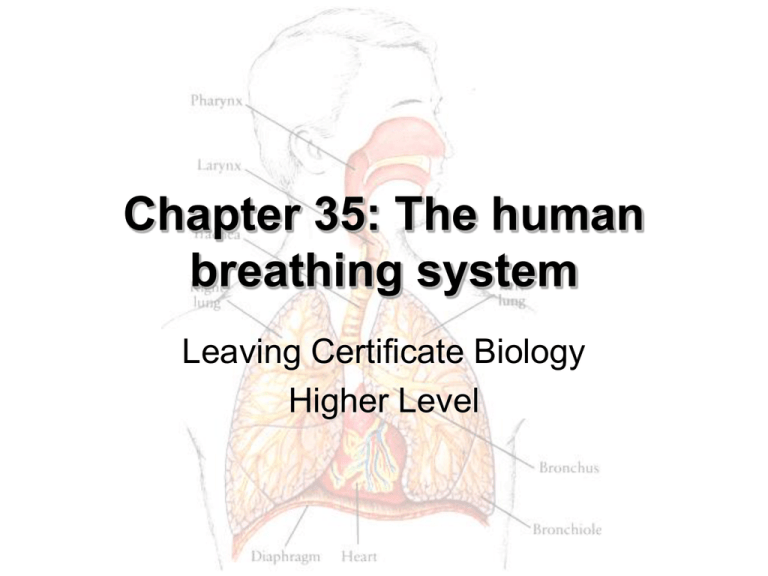
Chapter 35: The human breathing system Leaving Certificate Biology Higher Level Macrostructure of the Human Breathing System • Nasal and buccal cavities: – Warm and moisten air entering lungs – Mucus and small hairs filter the air and then transport the dirt-loaded mucus to the pharynx where it is swallowed • Pharynx (throat): – Junction between oesophagus and windpipe (trachea) – Pharynx has a sphincter (epiglottis) that closes over the opening to the trachea (glottis) that prevents food travelling into the trachea Macrostructure of the Human Breathing System • Glottis: – Opening to the trachea • Epiglottis: – Sphincter that closes over the glottis to prevent food getting into the trachea during swallowing – Swallowing causes the vocal cords to pull on the glottis and the larynx to be pulled upwards thereby closing the epiglottis over the glottis – Put your finger on your larynx and swallow Macrostructure of the Human Breathing System • Larynx (voice box): – Made of cartilage and sits on top of the trachea – Three functions: • Produces sound • Controls air flowing into and out of the trachea • Directs food into oesophagus • Trachea (windpipe): – Directs inhaled air into the lungs – Contains c-shaped rings of cartilage that keeps the trachea open – Cilia of trachea carry dirt-laden mucus up to the pharynx Macrostructure of the Human Breathing System • Bronchi: – Two divisions of the trachea – Directs air into respective lung – Supported by cartilage • Bronchioles: – Tiny divisions of the bronchi – Air passages that are less then 1 mm in diameter – Not supported by cartilage Macrostructure of the Human Breathing System • Alveoli: – Tiny air sacs at the end of the bronchioles where gas exchange occurs – Walls of alveoli are only 1 cell thick to maximise diffusion – Each alveolus has rich blood supply as many capillaries surround each – There are ~700 million alveoli with a total surface are of 90 m2 (surface area is increased in people who take part in high intensity sports) Essential Features of Alveoli and Capillaries • • • • • • Alveoli are extremely numerous Alveoli have rich blood supply Alveoli have walls only one-cell thick Alveoli surface is moist Alveoli walls are elastic Capillaries that surround each alveolus are only one-cell thick Gaseous Exchange O2 Exchange of gases in lungs Alveolus Exchange of gases in alveolus • Gaseous exchange occurs by diffusion. Heart Exchange of gases between Energy Cellular blood and cells CO + 2 respiration Glucose + H2O Transport of Gases • Inhaled O2: • Exhaled O2: 21% (atmospheric oxygen) 16% • Inhaled CO2: • Exhaled CO2: 0.04% 4% • Inhaled H2O(g): • Exhaled H2O(g): variable 100% humidity Transport of Gases • Oxygen is transported mostly (97%) by haemoglobin as oxyhaemoglobin • Remaining oxygen (3%) is carried dissolved in solution by the plasma • Carbon dioxide is transported mostly (80%) by the plasma as either hydrogen carbonate ions, HCO3– (70%) or as dissolved carbon dioxide (10%) • Remaining carbon dioxide (20%) is carried by the haemoglobin in red blood cells Macrostructure of the Human Breathing System • Lungs: – Composed of spongy, elastic tissue that expands easily during inhalation and recoils rapidly as exhalation occurs • Pleural membranes: – Thin pair of membranes covering and separating the lungs from other organs, such as the heart – The lungs are stuck to the rib cage and diaphragm by the pleural fluid (think of a layer of water between a table and a piece of glass and how difficult it is to lift it off the table) Macrostructure of the Human Breathing System • Rib cage: – Composed of 12 thoracic vertebrae, 12 ribs, and the sternum – Muscles are located between each rib – called intercostal muscles that contract causing the rib cage to move upwards and outwards, drawing air into the lungs Mechanism of Breathing • Inhalation: – Active process where the brain sends signal to the inspiratory muscles (intercostals and diaphragm) to contract – Rib cage expands upwards and outwards and the diaphragm pulls downwards – The movements of the rib cage and the diaphragm reduce the pressure within the thoracic cavity and air rushes in – Inhalation can be consciously and subconsciously (during sleeping) controlled Mechanism of Breathing • Exhalation: – Passive process where there is normally no signal sent to the inspiratory muscles – Can be an active process during strenuous activity when the brain send signal to the abdominals to contract forcibly expelling air from the thoracic cavity – During exhalation intercostals and diaphragm relax – Rib cage moves down and inwards and the diaphragm pushed upwards – The movements of the rib cage and the diaphragm during exhalation increase the pressure within the thoracic cavity and air rushes out Carbon Dioxide and Breathing Control (Higher Level Ext.) • Carbon dioxide dissolved in the blood is the most powerful stimulant for an increase in the rate of breathing • Receptors in the brain sense the levels of carbon dioxide in the blood and respond by increasing or decreasing the rate and depth of breathing Effect of Exercise on Breathing Rate • Exercise stimulates increased respiration which produces more carbon dioxide which diffuses into the bloodstream • The brain is extremely sensitive to changes in the carbon dioxide concentration within the bloodstream and acts on this by increasing breathing rate and heart rate to excrete the excess carbon dioxide Breathing Disorders • Asthma: – One possible cause: immune reaction to an external allergen (e.g. pollen) – Symptoms: difficulty breathing (‘asthma attack’) due to constriction of the airways – One possible preventative measure: avoid the allergen (e.g. pollen) by avoiding area where the allergen is present in high quantities – One possible treatment: most common treatment for asthma is the inhaler that has drugs in it that stimulate the airways (bronchi and bronchioles) to widen and dilate Breathing Disorders • Bronchitis: – One possible cause: smoking, air pollution, dust, viral infection, bacterial infection – Symptoms: laboured breathing, episodes of constant coughing, excessive production of mucus and inflamed airways – One possible preventative measure: do not smoke, avoid second-hand smoke, pollutants and dust – One possible treatment: stop smoking, avoid polluted air, use of bronchiodilating drugs or antibiotics if pathogenic bacteria are the cause
