10. Molecular and genetic mechanisms of ontogenesis
advertisement
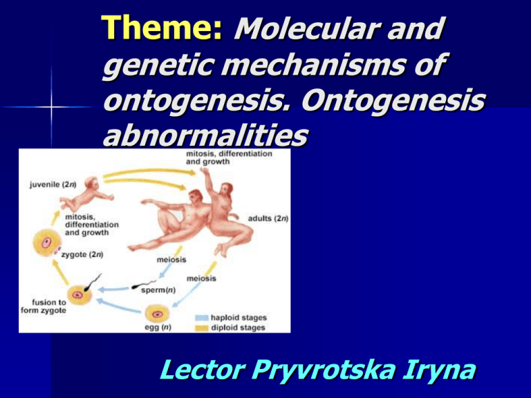
Theme: Molecular and genetic mechanisms of ontogenesis. Ontogenesis abnormalities Lector Pryvrotska Iryna Plan of the lecture: Asexual reproduction in unicellular and multicellular organisms Sexual reproduction in unicellular and multicellular organisms The periods of ontogenesis Spermatogenesis and oogenesis Morphogenetic specialization of sex cells: a sperm and an ovum Fertilization. Parthenogenesis Biological peculiarities of human reproduction Reproduction is the method by which individuals give rise to other individuals of same type. There are two types of reproduction : asexual and sexual Types of reproduction asexual Organisms single-cellular organisms, multi-cellular organisms: one ore more somatic cells of parental organisms; Parents One person Offspring's genetically identical to its parent like a xero copy. Cell formation Mitosis sexual Parental organisms forms gametes Two persons genetically different from parents Meiosis Forms of asexual reproduction in single-cellular organisms: Asexual is a reproduction without the fusion of sexual cells, identical offspring grows directly from a one or few body cells, which divides mitotically binary fission – parent cell splits in two cells by mitosis Asexual reproduction endogony - in parent cell forms only two daughter cells by internal budding schizogony - formation a great amount of daughter cells in parent cell Asexual reproduction budding - after karyokinesis the special region in parent cell rapid grows and organized into new organism. sporogony - is reproduction by the spores Forms of asexual reproduction in multi-cellular organisms vegetative (regeneration) – a group of cells from the parent organism separates and new organism forms Asexual reproduction polyembriony the production of two or more embryo's from the one zygote sporogony - is reproduction by the spores Sexual - is a reproduction by fusion of sexual cells and formation of zygote In sexual reproduction, genetic material from two individuals combines to begin the life of a third individual who has a new combination of inherited traits Sexual reproduction Forms of sexual reproduction in single-cellular organisms are Conjugation – a cytoplasm bridge forms Copulation - two individuals acquire the between two organisms, the nuclei transfer across this bridge and after exchange ones forms a new gene combination but no new offspring. gametes properties, fuse and form a zygote – the life of a new individual begins. In multicellular organism sexual reproduction may be two forms: - with fertilization - without fertilization Parthenogenesis is the development of new organism from an egg without fertilization. Ontogenesis the development of the individual organism. It includes the set of morphological, phisiological and biochenical transformations from the moment of germing up to death. Types of ontogenesis in animals: The larval type of an ontogenesis is characterized by development of an organism by metamorphosis. Metamorphosis – is change of shape or structure of an organism from one developmental stage to another. F/e mosquito: ovum- larva- pupa - imago; louses – ovum- larva - imago; pincers – ovum- larva- nymph - imago. 2. The non-larval type of an ontogenesis is characterized by formation of an organism in an egg (birds) 3. Intrauterine ontogenesis – is development of an organism inside a maternal organism. (mammalian). 1. The ontogenesis of multicellular organisms is divided in two periods 1 embryonic 2 postembryonic. For higher animals and man there are: 1) prenatal (before birth) 2) posnatal (after birth) periods of development Germ cells The ovum nourishes the embryo with yolk, which contains rich stores of lipids. It provides the machinery for protein synthesis An ovum is enormous in size. It protects the developing embryo inside jellylike protein coatings and strong membranes (zona pellucida), sacs of fluid, and sometimes hard or leathery shells. Corona radiata outside the cell consists of the great amount of follicular cells, which produce follicular fluid for attracting the sperms. Distinguish the following types of ovum cells: The isolecythal ovum contain a little yolk. It is distributed in regular intervals on all cytoplasm of an ovum. (ovum of mollusca, lancelet, mammalian). The telolecythal ovum have much yolk of grains. They collect at a vegetative pole. On animal pole there is cytoplasm without yolk and with nucleus.(ovum of fishes, amphibians, reptilie). The centrolecythal ovum has the central nucleus and around it settles down yolk as grains. (insects). Unlike the egg, the sperm is one of the smallest cells in the body. Each sperm consists of: a head region a body or midpiece a tail or flagellum. The head has a haploid nucleus. An acrosome – a small bump on the front end of the head contains enzymes that help the cell penetrate the ovum’s outer membrane. Sperm cell The body or midpiece has mitochondria to provide the cell energy and centrioles. A tail consists of microtubules for propulsion. The sperm’s streamlined size and shape effect its narrow objective: to reach the egg, penetrate its coating, and deliver a haploid nucleus into the egg’s cytoplasm Oogenesis This process begins in a haploid oogonium. An oogonium accumulates cytoplasm, replicates its chromosomes- primary oocyte. In meiosis I, the primary oocyte divides to form a small polar body and a large, haploid secondary oocyte. Oogenesis Ovulation – dischange (going out) of a secondary oocyte from a follicule of the ovary. In meiosis II, the secondary oocyte divides to yield another small polar body and a mature ovum. Therefore, each cell undergoing meiosis in female can potentially divide to yield a maximum of four cells, only one of which will become the ovum Spermatogenesis Sperm development begins with spermatogonia. A diploid spermatogonium divides mitotically and becomes a primary spermatocyte as it moves toward the lumen of the tubule. In meiosis I- to form two secondary spermatocytes. In meiosis II, each secondary spermatocyte divides to yield two equal – sized spermatids. Therefore, each cell undergoing meiosis in male can potentially divide to yield a maximum of four spermatids. Gametogenesis Periods Spermatogenesis Cells Genetic formula Oogenesis Cells Genetic formula Reproduction Cells divides mitoticaly ♂ - from puberty to death ♀- strongly at 3-7 m. in embryogeny completed at 3-d year spermatog onia 2n2c 2n4c oogonia 2n2c 2n4c Cells Genetic formula Cells Genetic formula Growth Cell growth and increase in size. Duplication of DNA. primary 2n4c primary spermatoc oocyte yte 2n4c Cells Genetic formula Cells Genetic formula Maturation Meiosis I- two cells halves genetically material secondary spermatocyt es Meiosis II two spermatids 1n2c Secondary 1n2c 1n1c mature 1n1c Oocyte ovum Cells Genetic formula Cells Formation (spematogenesis) Spiralization sperms 1n1c of chromosome s, formation of acrosome,cel l centre, tail Genetic formula Gametogenesis Ontogenesis abnormalities 1) multynuclear oocytes; 2) 10% of sperms can be abnormal; 3) development of germ cells with 22 or 24 chromosomes The fusion of haploid gametes to form a new diploid cell is called fertilization or syngamy Fertilization may be external and internal Fertilization During fertilization two processes take place: Egg’s activation – a wave of chemical reactions sweeps across the surface of the newly aroused egg, causing that surface to harden and present a barrier to the entry of any additional sperm. The egg’s oxygen consumption skyrockets, as does its rate of protein synthesis. Syngamy – male and female haploid nuclei converge and fuse to form the zygote’s single diploid nucleus Fertilization 3 functions – transmission of genes – restoration of the diploid number of chromosomes reduced during meiosis – initiation of development in offspring Fertilization internal fertilization – capacitation – sperm must penetrate cumulus and zona pellucida extracellular matrix consisting of 3 types of glycoproteins one of these, ZP3, acts as a sperm receptor Fertilization activates the egg, initiating metabolic processes acrosomal reaction – sperm are activated – acrosomal process – sperm and egg membranes fuse – ion channels open, allowing Na+ to flow in – fast block to polyspermy cortical reaction – egg’s ER releases Ca2+ into the cytosol at site of sperm entry Fertilization – slow block to polyspermy Ca2+ causes cortical granules underneath the plasma membrane to fuse mucopolysaccharides draw water into the space, swelling it vitelline layer becomes the fertilization membrane Cleavage rapid divisions following fertilization – often skip G1 and G2 phases – blastomeres result most animal eggs have polarity – substances are heterogeneously distributed in cytoplasm vegetal pole animal pole Kinds of cleavage: Holoblastic (total cleavage) – the zygote is divided Meroblastic (incomplete cleavage) – the part of cytoplasm of completely. There are 1) uniform and 2) irregular holoblastic cleavage. They are haracteristic for isolecythal and telolecythal cells an zygote is divided where yolk is absence. There are: 1) discoidal and 2) superficial meroblastic cleavage. In discoidal meroblastic cleavage the segmentation occurs on an animal pole in telolecythal cells. Birds’ eggs contain so much yolk that the small disc of cytoplasm on the surface is dwarfed by compasion. No cleavage of the massive yolk is possible, and all cell division is restricted to the small cytoplasmic disc, or blastodisc. In superficial meroblastic cleavage the segmentation occurs on an peripheric zone of cytoplasm in centrolecital cells. In the man the cleavage of zygote is holoblastic, irregular and asynchronous Mammalian Development implantation – ICM forms flat disk with 2 layers (epiblast and hypoblast) – embryo develops from epiblast cells, hypoblast forms yolk sac gastrulation Gastrulation series of cell migrations to positions where they will form the three primary cell layers: 1) ectoderm (outside germinal layer); 2) endoderm (inside germinal layer) and 3) mesoderm (medium germinal layer) The germinal layers give rice to various tissues and organs of animals. It is called as histogenesis and organogenesis The fate of primary germinal layers is given bellow: Ectoderm Mesoderm Endoderm Skin (epidermis), hair, nails, the eye lens, the pituitary gland, the epithelium of the nasal cavity, mouth, anal canal, nervous system, sense organs Connective tissue, bones, muscles, dermis, heart, blood vessels, gonads, excretory organs (kidneys) and the notochord (the dorsally located supportive rod found in all chordates, at least in embryonic stages) Digestive tract, lungs, liver, pancreas, thyroid gland, urinary bladder Mammalian Development 4 extraembryonic membranes form: – chorion- from trophoblast, surrounds embryo and all other membranes – amnion- from epiblast, encloses embryo in amniotic fluid – yolk sac- from hypoblast, site of early blood cell formation – allantois- outpocketing of embryo’s gut, incorporated into umbilical cord, forms blood vessels of umbilical cord Provisional organs are present during embryonic period and absent after birth. Provisional organs in human embryo are: an youlk sac an amnion an allantois a chorion. The youlk sac – the extraembryonic membrane that connects with the midgut. In human it is the first site of blood cell formation, but has no nutritive function. Chorion [Gr. «membane»] – in human it is cellular, outermost extraembryonic membrane, composed of trophoblast lined with mesoderm, it develops villi about 2 weeks after fertilization, is vascularized by allantoic vessels a week later. Amnion [Gr. «bowl» ; «membrane enveloping the fetus»] – the thin but tough extraembryonic membrane of reptilies, birds, and mammals that lines the chorion and contains the embryo and later the fetus, with the amniotic fluid around it. Placenta By the 23-rd day of human embryonic development, two other embryonic membranes – the chorion and the allantois to give rise to a placenta Exchange of material takes place in the placenta by diffusion between the blood of the mother and that of the embryo Within the placenta there is no mixing of maternal and fetal blood Human Gestation – organogenesis – fetus- all major structures are present – human chorionic gonadotropin (hCG)produced by embryo, maintains corpus luteum – mucous plug formation in cervix – negative feedbackcessation of ovulation and menstrual cycles Human Gestation 2nd trimester – increased movement of fetus – hCG levels decline, leading to deterioration of corpus luteum – placenta secretes progesterone – uterus increases to visible size Human Gestation 3rd trimester – rapid growth of fetus – estrogens and oxytocin initiate labor – positive feedback- oxytocin stimulates prostaglandin secretion by placenta, leading to increased contractions In embrionic development of man there are following critical periods: Implantation (7 day after a fertilization) – introduction of a zygote in a wall of an uterus Placentation (the end of 2 week of pregnancy) – form at embryo of a placenta Perinatal period (from 28 week of pregnancy to 7 day after birth) – is transferring a fetus out of aqueous into air (on the average, about 280 days after the beginning of the mother’s last regular menstrual period). Intrauterine pregnancy Any substance that can causes abnormal development of the egg in the mother's womb is called a teratogen. Growth is rapid, and each body organ has a critical period in which it is especially sensitive to outside influences. About 7% of all congenital defects are caused by exposure to teratogens. Drugs Alcohol. Ontogenesis abnormalities Chances of Down syndrome rapidly increase with parental age starting at about age 35 in women and 55 in men. Previously, it was thought there might be a tendency toward nondisjunction as a women ages because her eggs age as she does a women is born with all the eggs she will ever have and they remain in suspended animation until one matures each month. Ontogenesis abnormalities References: 1. Biology. – Sylvia S. Mader, Wm. C. Brown Publishers: Dubuque, Lowa – Melbourne, Australia – Oxford, England, IV edition. – p.169-181, 694-705. 2. Biology. Art notebook – Sylvia S.Mader, Wm. C. Brown Publishers: Dubuque, Lowa – Melbourne, Australia – Oxford, England, IV edition. – p.42-47. 3. Human biology. – McMillan, Beverly. – p. 310-327.
