Brain - Paul Trapnell
advertisement
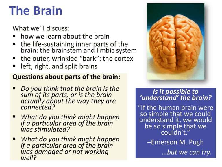
The Brain What we’ll discuss: how we learn about the brain the life-sustaining inner parts of the brain: the brainstem and limbic system the outer, wrinkled “bark”: the cortex left, right, and split brains Questions about parts of the brain: Do you think that the brain is the sum of its parts, or is the brain actually about the way they are connected? What do you think might happen if a particular area of the brain was stimulated? What do you think might happen if a particular area of the brain was damaged or not working well? Is it possible to ‘understand’ the brain? “If the human brain were so simple that we could understand it, we would be so simple that we couldn’t.” –Emerson M. Pugh …but we can try. Investigating the Brain and Mind: How did we move beyond phrenology? How did we get inside the skull and under the “bumps”? by finding what happens when part of the brain is damaged or otherwise unable to work properly by looking at the structure and activity of the brain: CAT, MRI, fMRI, and PET scans Strategies for finding out what is different about the mind when part of the brain isn’t working normally: case studies of accidents (e.g. Phineas Gage) case studies of split-brain patients (corpus callosum cut to stop seizures) lesioning brain parts in animals to find out what happens chemically numbing, magnetically deactivating, or electrically stimulating parts of the brain Neuroscience Techniques ©2001 Prentice Hall Intentional brain damage: Lesions (surgical destruction of brain tissue) performed on animals has yielded some insights, especially about less complex brain structures no longer necessary, as we now can chemically or magnetically deactivate brain areas to get similar information 4 Studying cases of brain damage When a stroke or injury damages part of the brain, we have a chance to see the impact on the mind. Phineas Gage (1850s) Dynamite accident Big changes in his personality Became impulsive, aggressive Insight into frontal lobe Executive functions Planning, impulse control Emotion regulation Phineas Gage (1850s) Phineas Gage (1850s) 1861 Paul Broca Hears of patient with progressive loss of speech Comprehension OK Dies. Broca does autopsy LESION found on right frontal lobel “Broca’s area” Split-Brain Patients “Split” = surgery in which the connection between the brain hemispheres is cut in order to end severe full-brain seizures Study of split-brain patients has yielded insights discussed at the end of the chapter We can stimulate parts of the brain to see what happens Parts of the brain, and even neurons, can be stimulated electrically, chemically, or magnetically. This can result in behaviors such as giggling, head turning, or simulated vivid recall. Researchers can see which neurons or neural networks fire in conjunction with certain mental experiences, and even specific concepts. Monitoring activity in the brain Tools to read electrical, metabolic, and magnetic activity in the brain: EEG: electroencephalogram PET: positron emission tomography MRI: magnetic resonance imaging fMRI: functional MRI EEG: electroencephalogram An EEG (electroencephalogram) is a recording of the electrical waves sweeping across the brain’s surface. An EEG is useful in studying seizures and sleep. 13 EEG EEG Electrical Stimulation Wilder Penfield Chemical Stimulation Transcranial Magnetic Stimulation (TMS) “The God Helmet” Brain Imaging PET: positron emission tomography The PET scan allows us to see what part of the brain is active by tracing where a radioactive form of glucose goes while the brain performs a given task. Brain Imaging PET ● Inject radioactive sugars MRI ● Magnet spin H atoms -> radio pulse f-MRI (functional MRI) ● Series of quick scans (20 ms) ● BOLD: “Blood oxygen level dependent” signal Larmor Frequency (Definitely NOT on the exam. ) Enlarged Ventricles Unaffected Persons Persons with Schizophrenia Psychopaths Normals 25 Cerebral Arteries Human Connectome Project Pink = Corpus Callosum circuits connecting hemisph. Green = Frontal Lobe circuits going to Hippocampus Structural Connectome by gender on a very large population (949) youths. RESULTS M optimized for intrahemispheric communication. F optimized for interhemispheric communication. Shows up very early ages Wide differences by adolescence and adulthood. CONCLUSION 1. MALE brains are structured to facilitate connectivity between perception and coordinated action 2. FEMALE brains are structured to facilitate communication between analytical and intuitive processing modes. More Parts of the Nervous System Peripheral nervous system Somatic/Skeletal afferent nerves efferent nerves (incoming) (outgoing) Autonomic sympathetic (engine) parasympathetic (brakes) The Peripheral Nervous System The Autonomic Nervous System: The sympathetic NS arouses (fight-or-flight) The parasympathetic NS calms (rest and digest) The Central Nervous System The brain is a web of neural networks. The spinal cord is full of interneurons that sometimes have a “mind of their own.” Interneurons in the Spine Your spine’s interneurons trigger your hand to pull away from a fire before you can say OUCH! This is an example of a reflex action. Neural Networks These complex webs of interconnected neurons form with experience. Remember: “Neurons that fire together, wire together.” Central Nervous System Spinal Cord relay station reflexes Brain a crude grouping via evolutionary stages 1. basic bodily functions HINDBRAIN 2. motivation and emotion LIMBIC SYSTEM 3. reasoning and foresight FOREBRAIN Areas of the brain and their functions The brainstem and cerebellum: The limbic (border) system: The cortex (the outer covering): • coordinates the body • manages emotions, and connects thought to body • integrates information The Brainstem: Pons and Medulla The Brainstem: Pons and Medulla The Base of the Brainstem: The Medulla The medulla controls the most basic functions such as heartbeat and breathing. Someone with total brain damage above the medulla could still breathe independently, but someone with damage in this area could not. The Brainstem: The Pons The pons helps coordinate automatic and unconscious movements. Cerebellum (“little brain”) The cerebellum helps coordinate voluntary movement such as playing a sport. The cerebellum has many other functions, including enabling nonverbal learning and memory. The Thalamus (“Inner Chamber”) The thalamus is the “sensory switchboard” or “router.” All sensory messages, except smell, are routed through the thalamus on the way to the cortex (higher, outer brain). The thalamus also sends messages from the cortex to the medulla and cerebellum. Reticular (“Netlike”) Formation The reticular formation is a nerve network in the brainstem. It enables alertness, (arousal) from coma to wide awake (as demonstrated in the cat experiments). It also filters incoming sensory information. The Midbrain tectum ("roof") superior colliculus inferior colliculus substantia nigra dopamine factory The Forebrain Thalamus Midbrain Practice Questions! After suffering an accidental brain injury, Kira has difficulty walking in a smooth and coordinated manner. It is most probable that she has suffered damage to her: A. B. C. D. amygdala. angular gyrus. cerebellum. corpus callosum. What disease is related to degeneration of the neuron’s myelin sheath? A. B. C. D. Parkinson’s disease multiple sclerosis Alzheimer’s disease schizophrenia Antidepressants such as Prozac target which neurotransmitter? A. B. C. D. serotonin glutamate GABA acetylcholine Which technique is most useful for seeing which regions of the brain are most active while a person reads a poem? A. B. C. D. EEG fMRI EKG PET The Limbic (“Border”) System The limbic system coordinates: emotions such as fear and aggression. basic drives such as hunger and sex. the formation of episodic memories. The hippocampus (“seahorse”) processes conscious, episodic memories. works with the amygdala to form emotionally charged memories. The Amygdala (“almond”) consists of two lima beansized neural clusters. helps process emotions, especially fear and aggression. The Amygdala Electrical stimulation of a cat’s amygdala provokes aggressive reactions. If you move the electrode very slightly and cage the cat with a mouse, the cat will cower in terror. The Hypothalamus: lies below (“hypo”) the thalamus. regulates body temperature and ensures adequate food and water intake (homeostasis), and is involved in sex drive. directs the endocrine system via messages to the pituitary gland. Thalamus The Hypothalamus as a Reward Center Riddle: Why did the rat cross the grid? Why did the rat want to get to the other side? Pushing the pedal that stimulated the electrode placed in the hypothalamus was much more rewarding than food pellets. Review of Brain Structures The Cerebral Cortex The lobes consist of: outer grey “bark” structure that is wrinkled in order to create more surface area for 20+ billion neurons. inner white stuff—axons linking parts of the brain. 180+ billion glial cells, which feed and protect neurons and assist neural transmission. 300 billion synaptic connections The brain has left and right hemispheres The Lobes of the Cerebral Cortex: Preview Frontal Lobes involved in speaking and muscle movements and in making plans and judgments Parietal Lobes include the sensory cortex Occipital Lobes include the visual areas; they receive visual information from the opposite visual field Temporal Lobes include the auditory processing areas 61 Functions of the Brain: The Motor and Sensory Strips Output: Motor cortex (Left hemisphere section controls the body’s right side) Input: Sensory cortex (Left hemisphere section receives input from the body’s right side) Axons receiving motor signals FROM the cortex Axons sending sensory information TO the cortex Sensory Functions of the Cortex The sensory strip deals with information from touch stimuli. The occipital lobe deals with visual information. Auditory information is sent to the temporal lobe. The Visual Cortex This fMRI scan shows increased activity in the visual cortex when a person looks at a photograph. Association function of the cortex More complex animals have more cortical space devoted to integrating/associating information Association Areas: Frontal Lobes The frontal lobes are active in “executive functions” such as judgment, planning, and inhibition of impulses. The frontal lobes are also active in the use of working memory and the processing of new memories. Phineas Gage (1823-1860) Case study: In a work accident, a metal rod shot up through Phineas Gage’s skull, destroying his eye and part of his frontal lobes. After healing, he was able to function in many ways, but his personality changed; he was rude, odd, irritable, and unpredictable. Possible explanation: Damage to the frontal lobes could result in loss of the ability to suppress impulses and to modulate emotions. Parietal Lobe Association Areas This part of the brain has many functions in the association areas behind the sensory strip: managing input from multiple senses performing spatial and mathematical reasoning monitoring the sensation of movement Temporal Lobe Association Areas Some abilities managed by association areas in this “by the temples” lobe: recognizing specific faces managing sensory input related to sound, which helps the understanding of spoken words Whole-brain Association Activity Whole-brain association activity involves complex activities which require communication among association areas across the brain such as: memory language attention meditation and spirituality consciousness Specialization and Integration Five steps in reading a word aloud: Plasticity: The Brain is Flexible If the brain is damaged, especially in the general association areas of the cortex: the brain does not repair damaged neurons, BUT it can restore some functions it can form new connections, reassign existing networks, and insert new neurons, some grown from stem cells This 6-year-old had a hemispherectomy to end lifethreatening seizures; her remaining hemisphere compensated for the damage. Our Two Hemispheres Lateralization (“going to one side”) The two hemispheres serve some different functions. How do we know about these differences? Brain damage studies revealed many functions of the left hemisphere. Brain scans and split brain studies show more about the functions of the two hemispheres, and how they coordinate with each other. The intact but lateralized brain Right-Left Hemisphere Differences Left Hemisphere Thoughts and logic Details such as “trees” Language: words and definitions Linear and literal Calculation Pieces and details Right Hemisphere Feelings and intuition Big picture such as “forest” Language: tone, inflection, context Inferences and associations Perception Wholes, including the self SplitTo end severe whole-brain seizures, some people have had surgery to cut the corpus callosum, a band of axons connecting the hemispheres. Brain Studies Researchers have studied the impact of this surgery on patients’ functioning. Separating the Hemispheres: Factors to Keep in Mind Each hemisphere controls the opposite side of the body AND is aware of the visual field on that opposite side. Without the corpus callosum, the halves of the body and the halves of the visual field do not work together. Only the left half of the brain has enough verbal ability to express its thoughts out loud. Split visual field Each hemisphere does not perceive what each EYE sees. Instead, it perceives the half of the view in front of you that goes with the half of the body that is controlled by that hemisphere. Split Brain Experiments 4-78 Divided Awareness in the Split Brain Try to explain the following result: 80 The divided brain in action Talent: people are able to follow two instructions and draw two different shapes simultaneously Drawback: people can be frustrated that the right and left sides do different things A blue circle is briefly flashed in the left visual field, and a red square is flashed in the right visual field. Based on Roger Sperry’s work with split-brain patients, what would split-brain patients say that they saw? A) B) C) D) blue circle. nothing. blue circle around a red square. red square. Split Brain Video Results of Split Brain Research • Left side of the brain: – seems to control language, writing, logical thought, analysis, and mathematical abilities, – processes information sequentially, – can speak. • Right side of the brain – controls emotional expression, spatial perception, recognition of faces, patterns, melodies, and emotions, – processes information globally, – cannot speak. Neural Plasticity Synaptic plasticity – Learning (e.g., long-term potentiation) – Pruning (e.g., during development) Neurogenesis Structural mod. of neural circuits – Functional modification of brain circuitry – for example, after a stroke or trauma Neural Plasticity https://www.dynamicb rain.ca/the-nature-ofthings.html http://www.cbc.ca/nat ureofthings/episodes/c hanging-your-mind

