 Leona Navratil
-
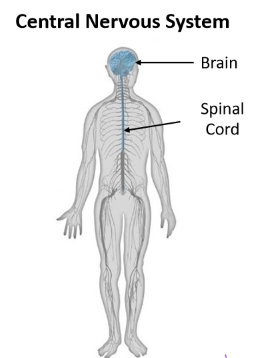
The central nervous system is composed of the ______and __________.
brain; spinal cord
-
The peripheral nervous system is composed of_____________, _________ and ____________.
sensory receptors, nerves; motor nerves
-
Brain

the most complex organ in the human body
-
What is the computing hardware of the brain?
Neurons!
-
How many neurons does the brain consist of alone?
86 billion
-
How many synapses are there between brain neurons?
150 trillion synapses
-
What is the average brain mass?
1.3-1.5kg in adults
-
What percent of our basal metabolism does our brain use to power itself?
20-25%
**It uses primarily glucose!
-
Spinal cord
transmits electrical impulses to and from the brain
-
What are the two types of tracts in the spinal cord?
Afferent and Efferent tracks (also we have sensorimotor integration)
-

Afferent tracts
transmit electrical impulses from the body to the brain
(inward)
-
Efferent tracts
transmit electrical impulses from the brain to the body
(outward)
-
Sensorimotor integration
Transform sensory into basic motor activity (e.g. tendon tap reflex)
-incorporating sensation about one's body and the external environment to shape movement
- link between nerves and muscle
-
Example of a polysynaptic reflex:
redraw reflex
-
Nerves

send electrical impulses to and from the spinal cord
-
Sensory nerves

send sensory information from active sensory receptors to the spinal cord
-
Motor nerves

send motor commands from the spinal cord to activate skeletal muscle
-
Receptors

modified ending of sensory nerves. First step in processing that leads to our sense of touch and proprioception (sense of body position, motion and force in the absence of vision)
-
What are the 4 receptor cells?
Merkle, Meissner, Ruffini, Pacinian
-
What is in charge of proprioception?
- Muscle spindles
- Golgi tendon organ = force through muscle
-
What are the two divisions of the peripheral nervous system?
- The autonomic division: controls self-regulated action of internal organs and glands
- Somatic division: controls voluntary movement of skeletal muscles
-
What does the PNS provide information about? What role does the CNS have?
the state of the body and environment.
**The CNS processes this information and generates appropriate responses
-
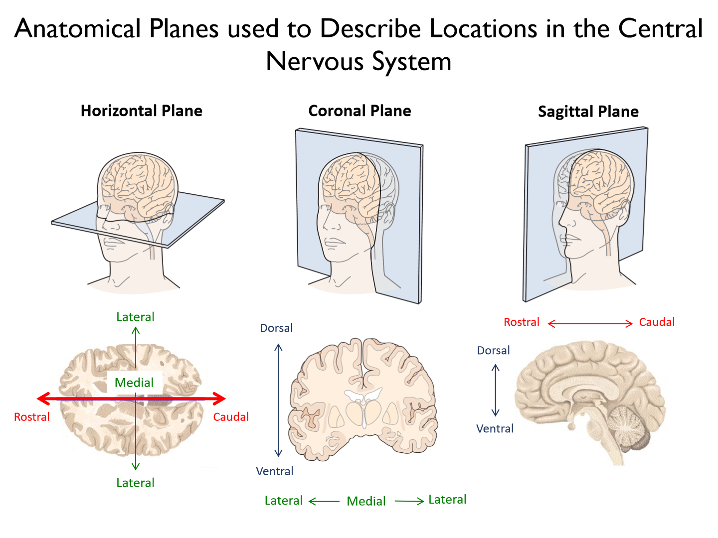
Rostral

towards the nose
-
Caudal

towards the coccyx (tail bone)
-
Ventral

(anterior) towards the belly
-
Dorsal
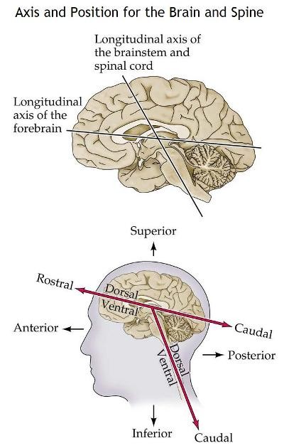
(posterior) towards the back or top of head
-
Coronal slice

(frontal plane slice)
-
Medial

Towards the midline
-
Lateral
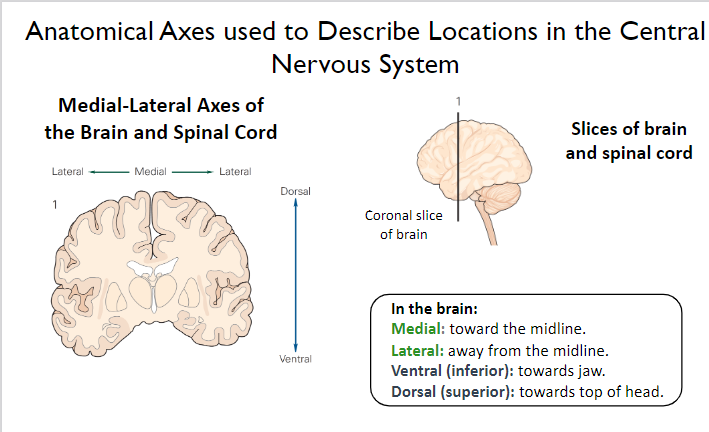
away from the midline
-
Look at the picture
-
What are the 4 subdivided areas of the brain?

1. Cerebrum (thoughts)
2. Diencephalon (relay center)
3. Brainstem (life functions, e.g. breathing, swallowing)
4. Cerebellum (posture & balance, corrects error)
-
What does the cerebrum consist of?

1. 2 hemispheres (left - R hand and right - L hand)
2. Cerebral cortex (gray matter - upper level processing)
3. White matter (including corpus callosum)
-
Corpus callosum

communication between the hemispheres
-
What are the three deep-lying neural structures of the cerebrum?
1. Basal ganglia
2. Amygdala
3. Hippocampus
-
Basal ganglia

movement (vigor, amplitude) and motor learning
-
Amygdala

Expression of emotion
-
Hippocampus

memory formation
Cool fact: there are place cells in the hippocampus to remember familiar environments and when you are reminded of a place (smell, vision, etc) these cells fire
-
What are the largest part of the brain?
Cerebral hemispheres
-
What are cerebral hemispheres responsible for?

perceptual, motor (mostly planning) and cognitive function.
-
Do the subdivisions of the cerebrum only exist in one hemisphere?
NO. All of these structures are in both hemispheres of the brain, with slightbilateral differences (e.g., handedness, especially in cortex). Each structureis subdivided into several anatomically and functionally distinct areas.
- Hand dominance = motor cortex has more connection and larger SA for specific digits
-
What are the 4 lobes of the cerebral cortex

1. Frontal lobe
2. Parietal lobe
3. Occipital lobe
4. Temporal lobe
-
What is each lobe named after?
overlying cranial bones that form the skull
-
Frontal lobe functions

motor planning, organizing behavior, decision making, working memory.
*lots of higher functions
-
Parietal lobe functions
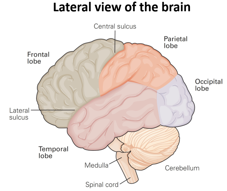
sensory guidance of motor actions, multisensory integration, spatial awareness.
*lots of vestibular cues (balance and spatial orientation for the purpose of coordinating movement with balance)
-
Occipital lobe functions

early visual processing (direction, orientation, colour, motion)
-
Temporal lobe functions

vision, hearing, memory, language
-
Are the lobes as far as the brain subdivides?
NO. Each lobe of cerebral cortex is further subdivided into several anatomically and functionally distinct regions.
-
Corpus callosum description
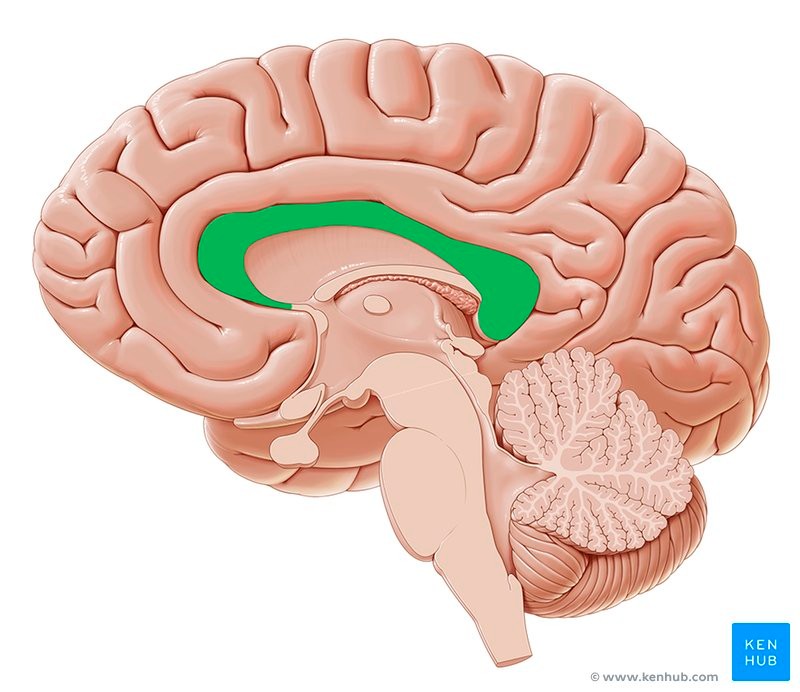
The corpus callosum (also known as callosal commissure) is a wide, flatbundle of axons that connects and allows communication betweenstructures in the two cerebral hemispheres.
* allows for a balance of forces and integrated movement (e.g. belly rub and head pat)
-
What does the diencephalon consist of?

1. Thalamus
2. Hypothalamus
-
Thalamus

• Essential link in sensory pathways(aside from olfactory receptors) from periphery to brain. (temp., cutaneous, pain, etc.)
• Interconnects cerebellum and basal ganglia with regions of cerebral cortex involved in cognition and movement. (info both in and out)
**Relay center
-
Hypothalamus
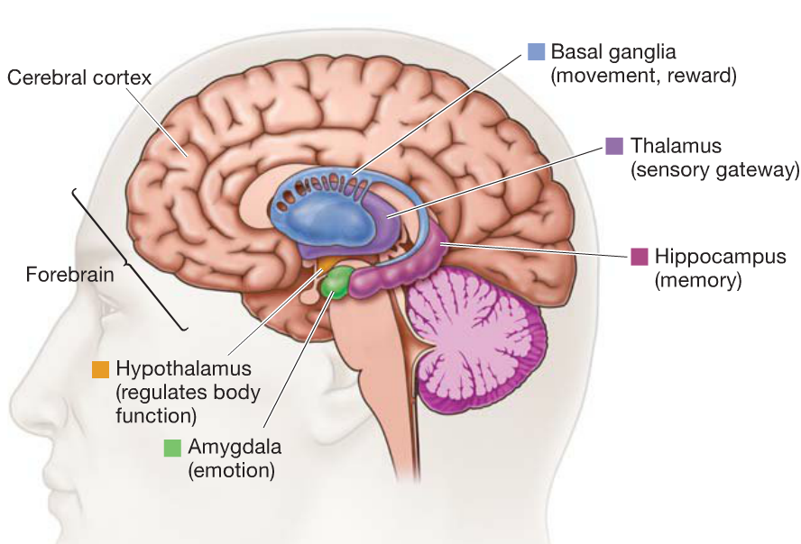
• Somatic growth, eating, drinking.
• Essential component of neuro-endocrine system (pituitary gland). (GH, FSH, etc.)
• Motivation (release dopamine, norepi., epi., etc.) and arousal
-
What does the brainstem consist of?
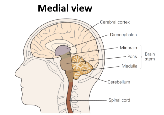
1. Midbrain
2. Pons
3. Medulla
-
Midbrain

rostral to pons
• Linkages between cerebellum, basalganglia, and cerebral cortex.
• Involved in hearing and control of eye movements (oculomotor control).
(Vestibular function = VOR reflex)
-
Pons
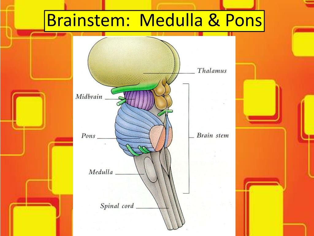
rostral to medulla, protrudes from ventral surface of spinal cord.
• Pontine nucleus: Relay info about sensation and movement from cerebral cortex to cerebellum.
• Sleep, respiration, taste.
Crossing over of controls
-
Medulla

caudal part of brainstem and direct extension of spinal cord.
• Regulates blood pressure and respiration.
• Early pathways of taste, hearing, balance, and control of head, neck and limb muscles.
-
Cerebellum functions

1. Maintaining posture and balance.
2. Coordinating head, eye and arm movements.
3. Error-based motor learning.
4. Receives somatosensory information from the spinal cord.
5. Receives copies of motor commands from cerebral cortex.
-
What is a cool fact about neurons in the cerebellum?

Despite accounting for less than 10% of brain volume, the cerebellumcontains more than 50% of the brain’s neurons. It has more neurons thanany other brain structure, including cerebral cortex!
-
What tells us we have deviated from our initial path?
muscle spindles, golgi tendon organ, eyes and cutaneous receptors
-
What are the 3 main functions of the spinal cord?

1. Receives sensory information from skin, muscles, and joints. (free nerve endings, thermoreceptors, etc.)
2. Contains motor neurons responsible for voluntary movements. (ventral horn)
3. Performs basic integration of somatosensory feedback and motor responses (reflexes).
- reflex is also integrated and higher centers can override
-
What are the three divisions of the spinal cord?

1. dorsal horn
2. ventral horn
3. intermediate zones
-
Dorsal horn

Sensory neurons (cell bodies in the dorsal route ganglia) from muscles and skin terminate in the dorsal horn of the spinal cord. (input)
-
Ventral horn
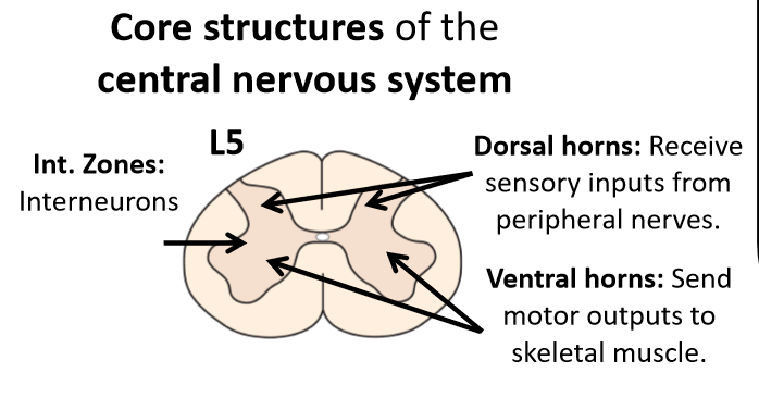
Cell bodies and axons of motor neurons that influence muscle firing patterns. (output)
-
Intermediate zones

Interneurons that influence firing patterns of sensory (dorsal horn) neurons and motor neurons (ventral horn)
- aid in the decussation of pain and temp. receptors when entering the spine
- aid in polysynaptic reflexes such as the tendon tap (interneuron splits so that alpha motor neuron will cause contraction and inhibitory interneuron will allow other muscle to relax.
-
Topdown control
prevent reflex through anticipation
-
What is the largest and simplest component of the CNS
the spinal cord
-
What is the spinal cord composed of?
31 pairs of sensory and motor nerves
-
What does sensory division in the spine carry?
somatosensory, touch, pain, temp., and information from visceral sensory organs
-
What are spinal motor neurons?
the 'final common pathway' through which all higher brain areas controlling movement must act
-
does the thickness of the spinal cord vary?
Yes! it varies along its length
-
What is cervical enlargement caused by?

Large number of sensory and motor fibers entering and leaving the spinal cord to innervate the upper limbs
-
What is lumbar enlargement caused by?
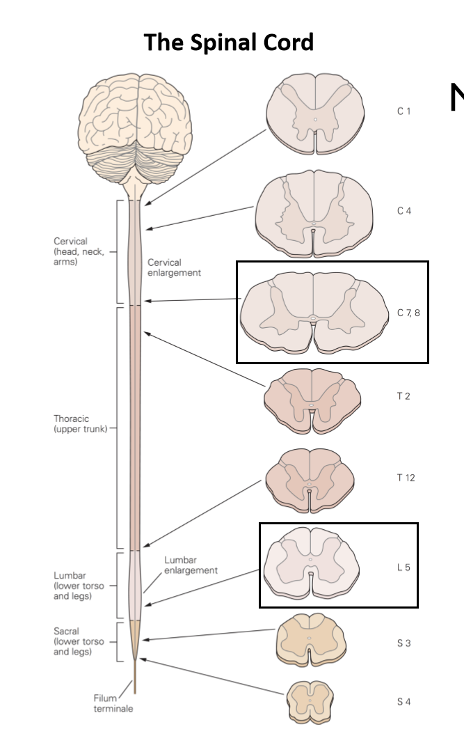
Large number of sensory and motor fibers entering and leaving the spinal cord to innervate the lower limbs
-
As you move up the spine, what do you see?
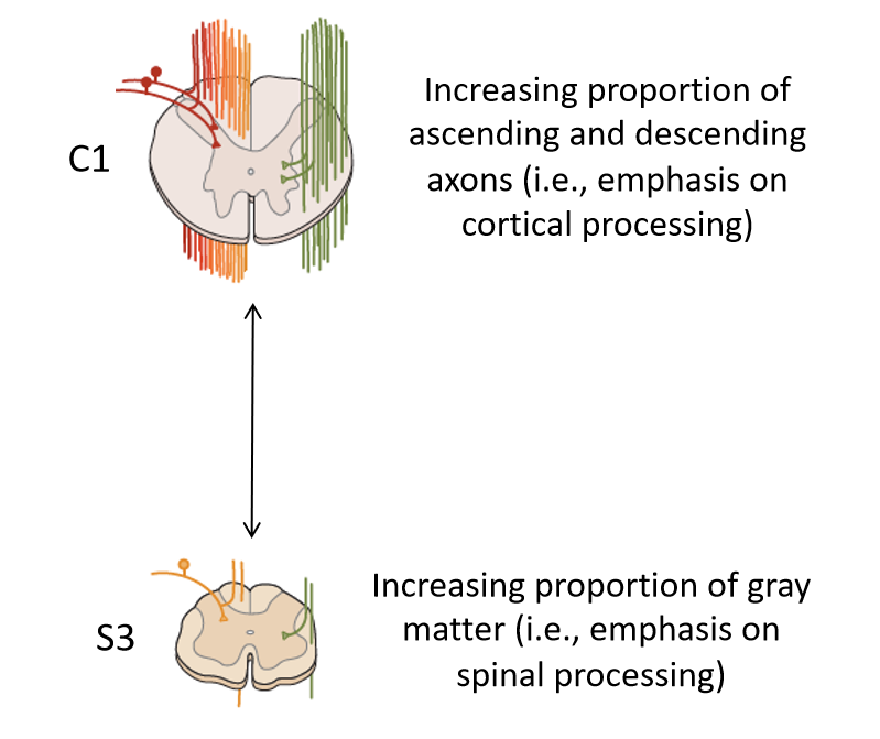
An increasing proportion of ascending and descending axons (i.e. emphasis on cortical processing)
- based on level of spine
- more and more information as it also must carry lower portion
- more white matter to carry all the nerves
-
As you move down the spine what

increasing proportion of gray matter (i.e. emphasis on spinal processing)
-
Why does the cerebellum receive copies of motor commands from the cerebral cortex?
So that we consciously know what the motor plan was so that we may fix mistakes by comparing it to our somatosensory information being received from the spinal cord.
-
Reciprocal inhibition
e.g. reflex: one muscle contracts, the opposing one relaxes
- The interneuron turns the excitatory impulse into an inhibitory through GABA neurotransmitter
-
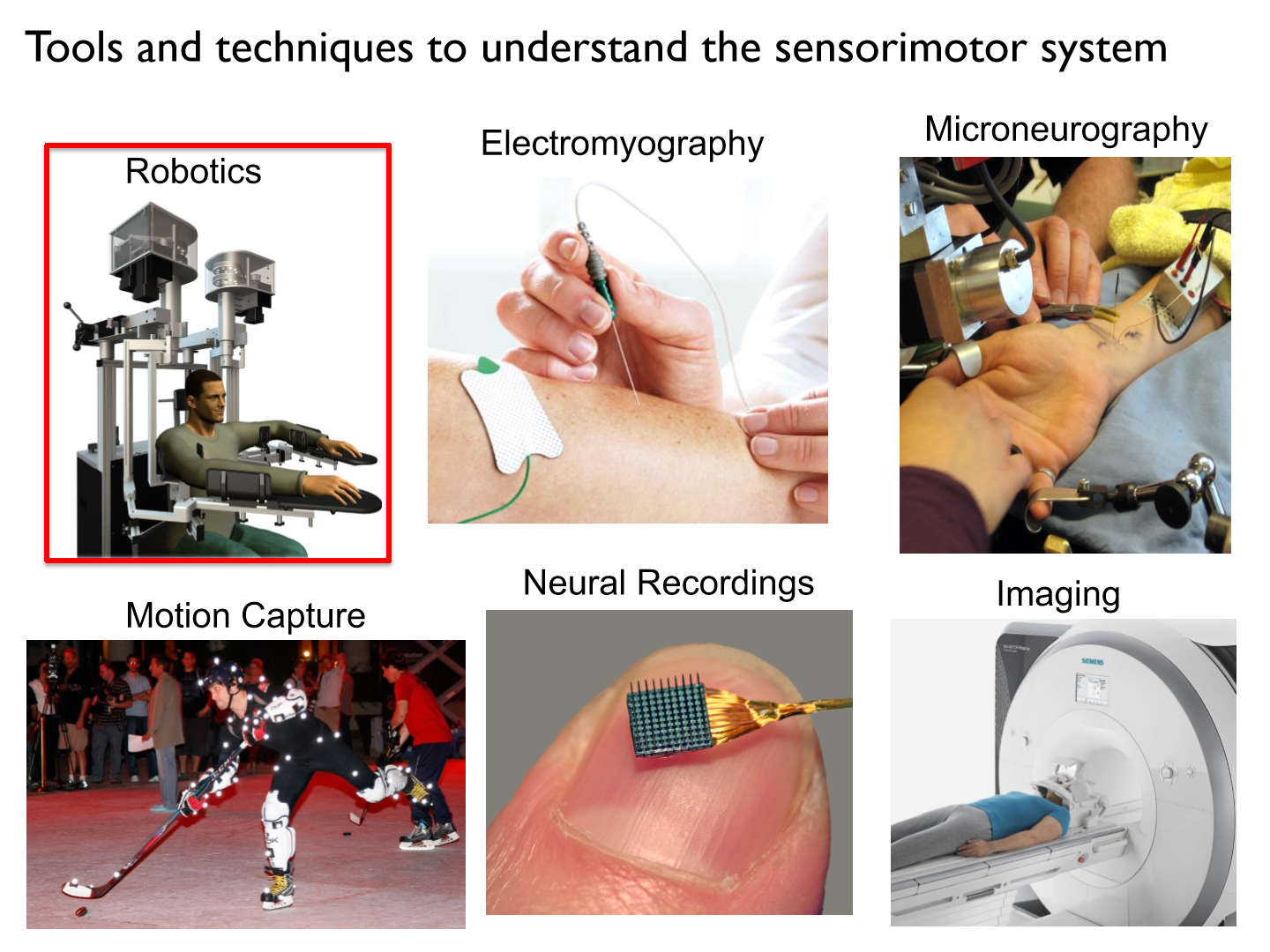
What are some tools/techniques to understand the sensorimotor system?
-
Robotics
a non-invasive tool that can be used to monitor and perturb movements to examine sensory and motor functions.
-
Torque motors

can be used to apply independent loads at the shoulder and/or elbow
-
How do we adjust robots to fit the participant's arm?
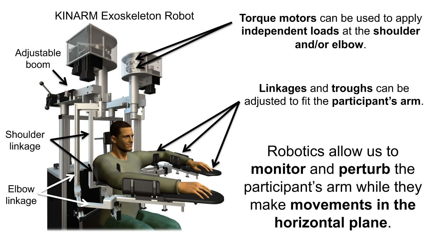
linkages and troughs
-
What do robotics allow us to do?
to monitor and perturb the participant's arm while they make movements in the horizontal plane
*ONLY reaching in 2D (forward/backwards, left/right)
• Monitor and record arm movements
• Quantify how the nervous system responds to unexpected perturbations
• Quantify sensory and motor deficits in clinical populations
-
How do robotics monitor and record arm movements? (draw)
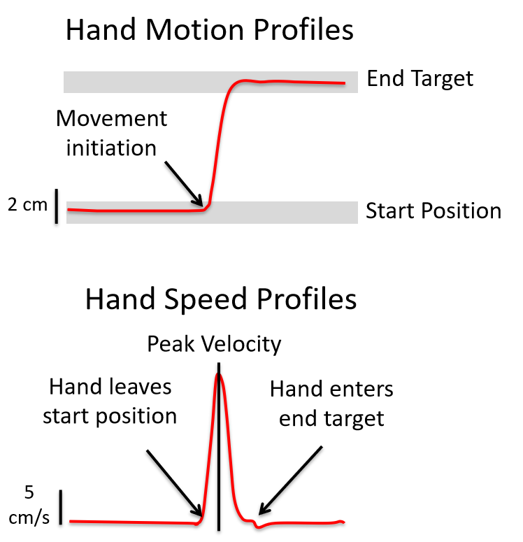
Hand motion profiles and hand speed profiles
*record arm motion while participants perform sensory and motor tasks with their arm
-
What happens if someone bumps your arm while you are reaching to a door knob vs. a push bar?
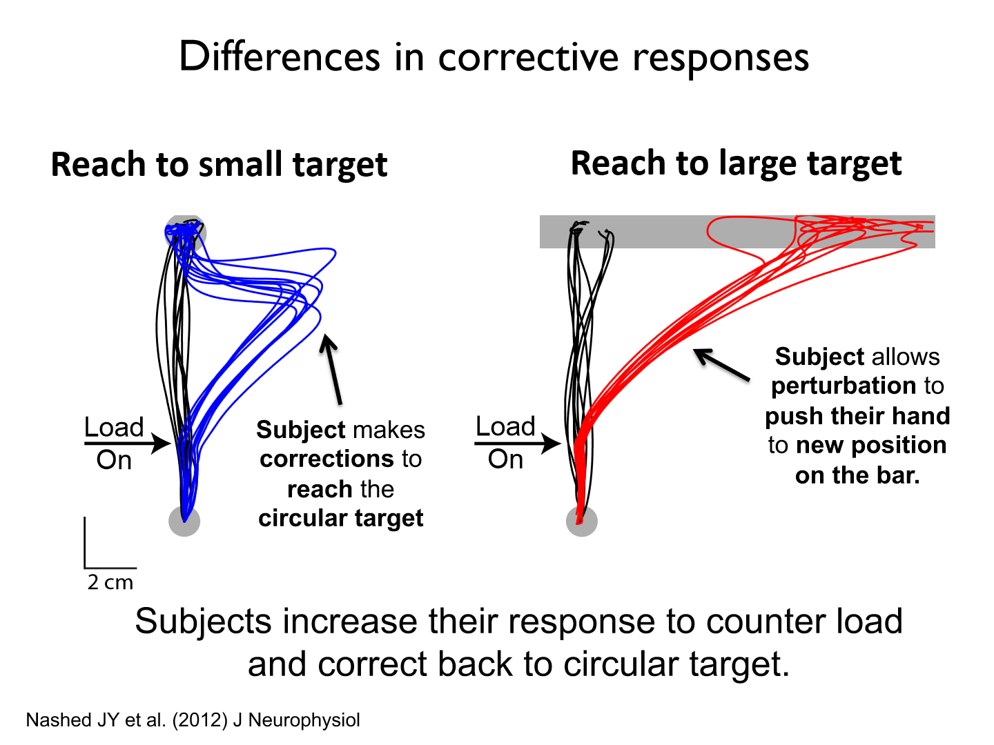
- Reaching to the door knob is much more precise as it is a small target
- The push bar is a much larger target therefore, when it is perturbed participants won't go to the middle of the bar, they will simple go somewhere along the length
*Subjects increase their response to counter load and correct back to circular target
-
Task description for how target shape influences corrective response
• Subjects instructed to reach the spatial goal.
• no perturbations (loads) applied on most trials.
• perturbations on 20% of trials
• visual-feedback removed at perturbation onset (sensory feedback comes from muscles).
-
How do we quantify sensory and motor deficits in stroke patients?
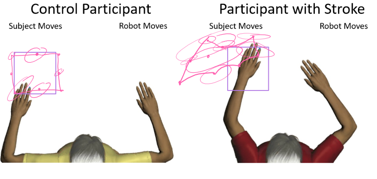
We assess the motor cortex and cerebellum by using position sense (no vision)
*match position that the robot moves their arm to
-
KINARM Endpoint robot difference
Only controls endpoint/hands
-
3D Robotic Exoskeletons

devices being used to assist in movement rehabilitation
-
Pros of robotics
1. Easy to use and non-invasive.
2. Useful for quantifying and perturbing body motion.
3. Easy to integrate with other tools such as EMG.
-
Cons of robotics
1. Expensive
2. Can require long setup times. (have to calibrate based on different people)
3. Can't replicate many features of how humans move freely in space (unnatural?)
-
Microneurography
an invasive technique used to record the activity of peripheral neurons
-
How does microneurography locate and characterize neurons?
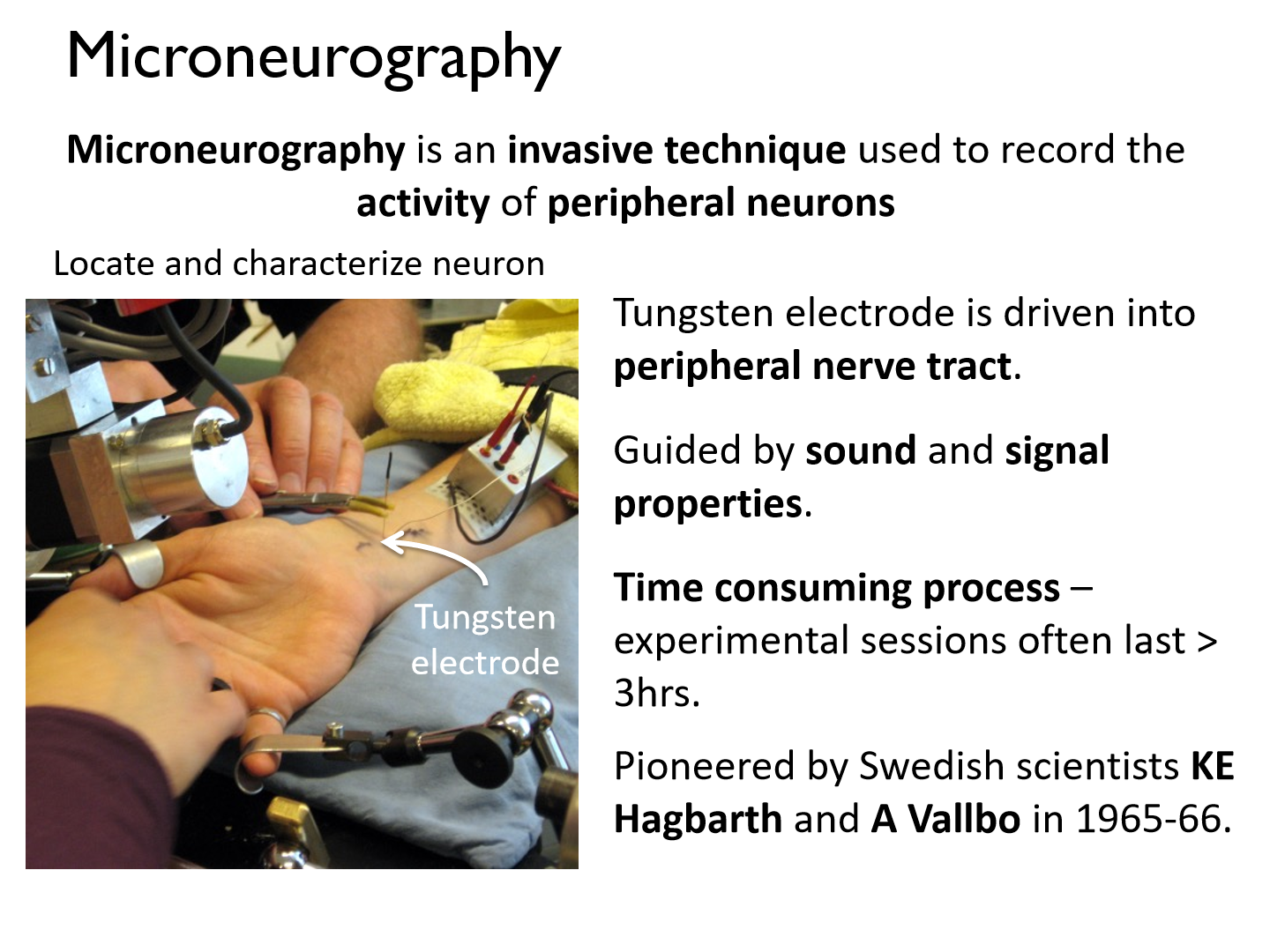
- Tungsten electrode is driven into peripheral nerve tract. (single cell recordings)
- Guided by sound and signal properties.
- Time consuming process – experimental sessions often last > 3hrs.
- Pioneered by Swedish scientists KE Hagbarth and A Vallbo in 1965-66.
-
Nerve fibre

the axon of a sensory neuron and its myelin sheath (if present)
-
Fascicle

a bundle of individual nerve fibers
-
Endoneurium, perineurium, and epineurium

connective tissue surrounding nerve fibers, fascicles, and nerves, respectively.
-
Nerve
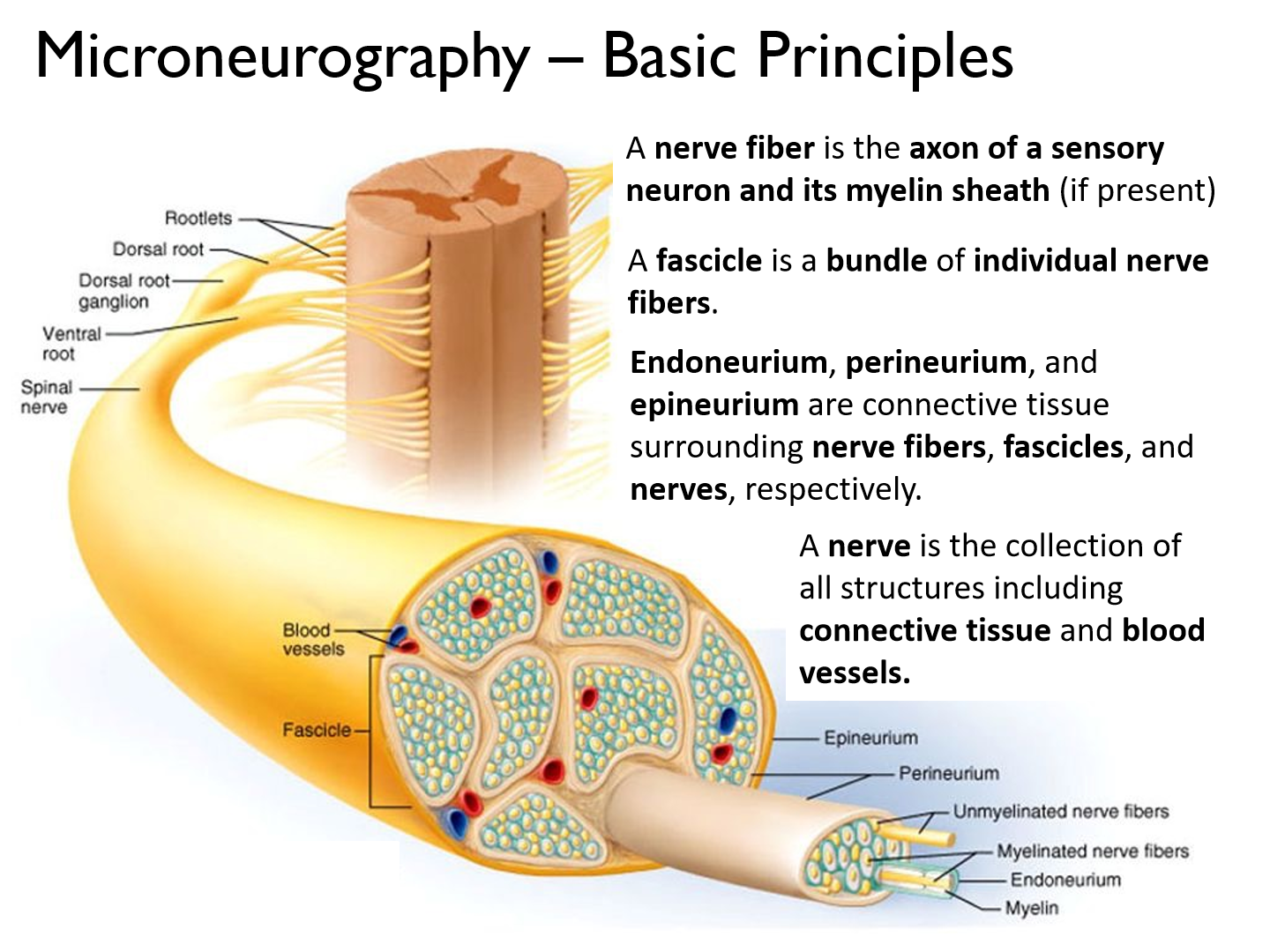
is the collection of all structures including connective tissue and blood vessels.
-
Microneurography electrode insert
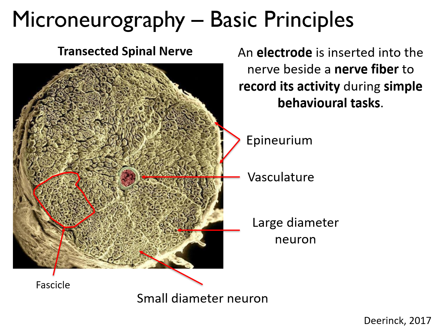
An electrode is inserted into the nerve beside a nerve fiber to record its activity during simple behavioural tasks
-
Difference between large and small diameter neurons.

Large diameter:
- large group of muscles controlled
- higher recruitment group (more F produced)
- less precise muscle control
- fast twitch
Small diameter:
- smaller group of muscles controlled
- smaller recruitment group (less F produced)
- more fine movement
- slow twitch
-
Microneurography recording electrode
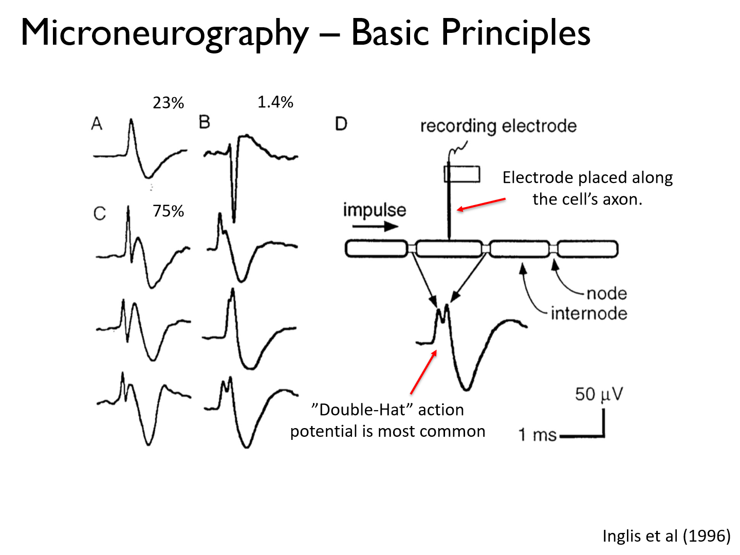
- probably won't hit a node because they are so small
- produce a "double-hat" AP because you are reading the peaks @ both nodes of Ranvier (signals are sent in both directions)
-
What are the 4 typical steps to a microneurography experiement?
1. Search (find the nerve)
2. Characterize (motor/sensory, what type of receptor, where on skin/muscle)
3. Position
4. Stimulate
-
Step 1 of microneurography (search for peripheral nerve fiber)
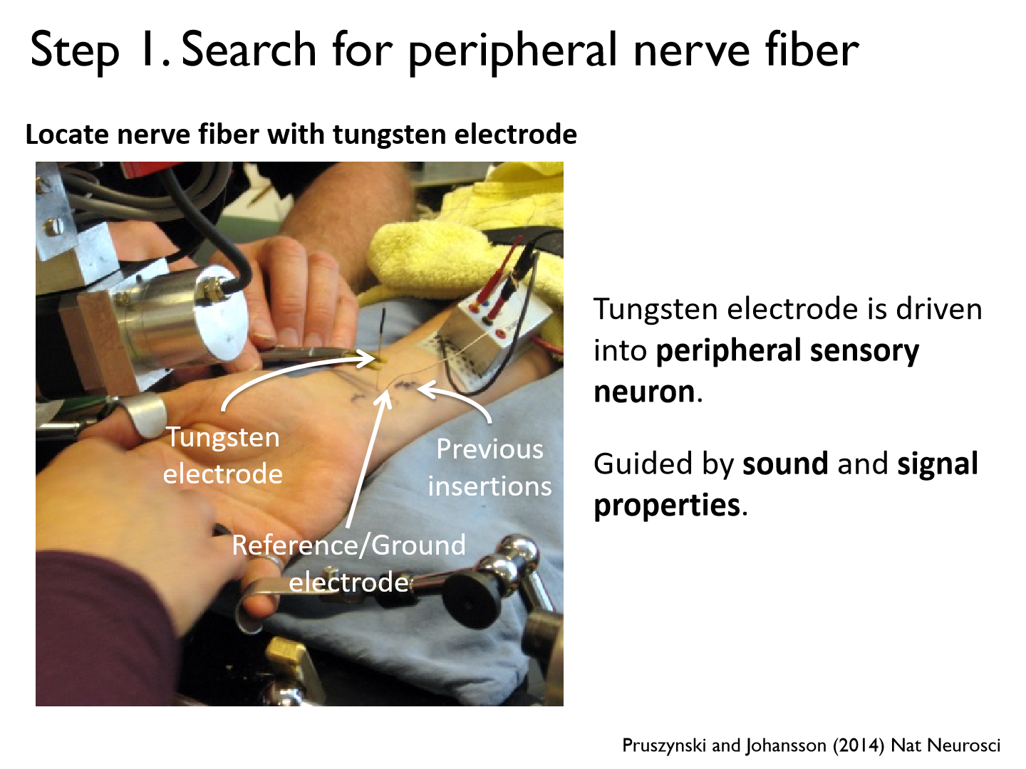
Tungsten electrode is driven into peripheral sensory neuron.
Guided by sound and signal properties.
-
What are reference/ground electrodes for?

There is a lot of electrical stimuli in the human body, so it takes the average to filter out noise

