11.2 Muscles and Movement
advertisement
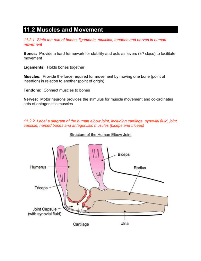
11.2 Muscles and Movement 11.2.1 State the role of bones, ligaments, muscles, tendons and nerves in human movement Bones: Provide a hard framework for stability and acts as levers (3rd class) to facilitate movement Ligaments: Holds bones together Muscles: Provide the force required for movement by moving one bone (point of insertion) in relation to another (point of origin) Tendons: Connect muscles to bones Nerves: Motor neurons provides the stimulus for muscle movement and co-ordinates sets of antagonistic muscles 11.2.2 Label a diagram of the human elbow joint, including cartilage, synovial fluid, joint capsule, named bones and antagonistic muscles (biceps and triceps) Structure of the Human Elbow Joint 11.2.3 Outline the function of the structures in the human elbow joint named in 11.2.2 Biceps: Bends the arm (flexor) Triceps: Straightens the arm (extensor) Humerus: Anchors muscle (muscle origin) Radius / Ulna: Acts as forearm levers (muscle insertion) - radius acts as a lever for the biceps, ulna acts as a lever for the triceps Cartilage: Allows easy movement (smooth surface), absorbs shock and distributes load Synovial Fluid: Provides food, oxygen and lubrication to the cartilage Joint Capsule: Seals the joint space and provides passive stability by limiting range of movement 11.2.4 Compare the movement of the hip joint and knee joint Similarities: Both Both are synovial joints are involved in the movement of the leg Differences: Comparison of Hip Joint and Knee Joint 11.2.5 Describe the structure of striated muscle fibres, including the myofibrils with light and dark bands, mitochondria, the sarcoplasmic reticulum, nuclei and the sarcolemma Each muscle fibre has the following specialised features designed to facilitate muscular contraction Many nuclei (fibres are long and were formed from many muscle cells fusing together, hence the fibres are multinucleated) Large number of mitochondria (muscle contraction requires a lot of ATP) Tubular myofibrils, divided into sections called sarcomeres, and made of two different myofilaments (proteins responsible for contraction) Where thin (actin) and thick (myosin) filaments overlap, a dark band occurs, and this is flanked by light regions containing thin filament only The membrane surrounding a muscle fibre is called the sarcolemma The internal membranous network is called the sacroplasmic reticulum, it is analogous to endoplasmic reticulum but is specialised for muscle contraction (it contains high levels of Ca2+ ions) Structure of a Striated Muscle Fibre 11.2.6 Draw and label a diagram to show the structure of the sarcomere, including Z lines, actin filaments, myosin filaments with heads, and the resultant light and dark bands The The The The H zone is the area only occupied by the thick filaments (myosin) I bands (light) are the regions occupied by only thin filaments (actin) A bands (dark) are the regions occupied by both filaments (overlap) Z lines represent the extremities of a single sarcomere 11.2.7 Explain how skeletal muscles contract, including the release of calcium ions from the sarcoplasmic reticulum, the formation of cross-bridges, the sliding of actin and myosin filaments, and the use of ATP to break cross-bridges and reset myosin heads action potential from a motor neuron triggers the release of Ca 2+ ions from the sarcoplasmic reticulum Calcium ions expose the myosin heads by binding to a blocking molecule (troponin complexed with tropomyosin) and causing it to move The myosin heads form a cross-bridge with actin binding sites ATP binds to the myosin heads and breaks the cross-bridge The hydrolysis of ATP causes the myosin heads to change shape and swivel this moves them towards the next actin binding site An The movement of the myosin heads cause the actin filaments to slide over the myosin filaments, shortening the length of the sarcomere Via the repeated hydrolysis of ATP, the skeletal muscle will contract Contraction of a Skeletal Muscle 11.2.8 Analyse electron micrographs to find the state of contraction of muscle fibres Muscle fibres can be fully relaxed, slightly contracted, moderately contracted and fully contracted The sarcomere gets shorter when the muscle contracts, however the A band does not, showing that the filaments are not themselves contracting Instead, the filaments are sliding over each other and increasing their overlap, which can be seen as the gradual reduction in the H zone Electron Micrographs of Relaxed and Contracted Muscle Fibres

