Organ Notes
advertisement
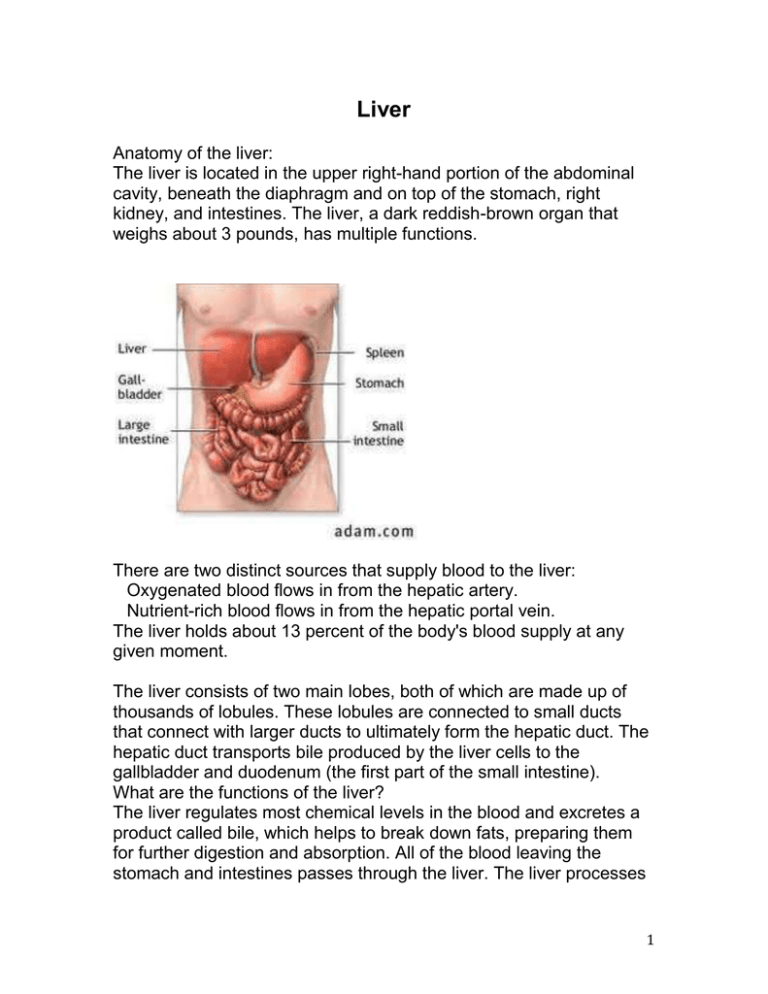
Liver Anatomy of the liver: The liver is located in the upper right-hand portion of the abdominal cavity, beneath the diaphragm and on top of the stomach, right kidney, and intestines. The liver, a dark reddish-brown organ that weighs about 3 pounds, has multiple functions. There are two distinct sources that supply blood to the liver: Oxygenated blood flows in from the hepatic artery. Nutrient-rich blood flows in from the hepatic portal vein. The liver holds about 13 percent of the body's blood supply at any given moment. The liver consists of two main lobes, both of which are made up of thousands of lobules. These lobules are connected to small ducts that connect with larger ducts to ultimately form the hepatic duct. The hepatic duct transports bile produced by the liver cells to the gallbladder and duodenum (the first part of the small intestine). What are the functions of the liver? The liver regulates most chemical levels in the blood and excretes a product called bile, which helps to break down fats, preparing them for further digestion and absorption. All of the blood leaving the stomach and intestines passes through the liver. The liver processes 1 this blood and breaks down the nutrients and drugs in the blood into forms that are easier to use for the rest of the body. More than 500 vital functions have been identified with the liver. Some of the more well-known functions include the following: production of bile, which helps carry away waste and break down fats in the small intestine during digestion production of certain proteins for blood plasma production of cholesterol and special proteins to help carry fats through the body conversion of excess glucose into glycogen for storage (This glycogen can later be converted back to glucose for energy.) regulation of blood levels of amino acids, which form the building blocks of proteins processing of hemoglobin for use of its iron content (The liver stores iron.) conversion of poisonous ammonia to urea (Urea is one of the end products of protein metabolism that is excreted in the urine.) clearing the blood of drugs and other poisonous substances regulating blood clotting resisting infections by producing immune factors and removing bacteria from the bloodstream When the liver has broken down harmful substances, they are excreted into the bile or blood. Bile by-products enter the intestine and ultimately leave the body in the feces. Blood by-products are filtered out by the kidneys and leave the body in the form of urine. Kidney The kidneys are a pair of vital organs that perform many functions to keep the blood clean and chemically balanced. Understanding how the kidneys work can help a person keep them healthy. 2 What do the kidneys do? The kidneys are bean-shaped organs, each about the size of a fist. They are located near the middle of the back, just below the rib cage, one on each side of the spine. The kidneys are sophisticated reprocessing machines. Every day, a person's kidneys process about 200 quarts of blood to sift out about 2 quarts of waste products and extra water. The wastes and extra water become urine, which flows to the bladder through tubes called ureters. The bladder stores urine until releasing it through urination. Wastes in the blood come from the normal breakdown of active tissues, such as muscles, and from food. The body uses food for energy and self-repairs. After the body has taken what it needs from food, wastes are sent to the blood. If the kidneys did not remove them, these wastes would build up in the blood and damage the body. The actual removal of wastes occurs in tiny units inside the kidneys called nephrons. Each kidney has about a million nephrons. In the 3 nephron, a glomerulus-which is a tiny blood vessel, or capillaryintertwines with a tiny urine-collecting tube called a tubule. The glomerulus acts as a filtering unit, or sieve, and keeps normal proteins and cells in the bloodstream, allowing extra fluid and wastes to pass through. A complicated chemical exchange takes place, as waste materials and water leave the blood and enter the urinary system. At first, the tubules receive a combination of waste materials and chemicals the body can still use. The kidneys measure out chemicals like sodium, phosphorus, and potassium and release them back to the blood to return to the body. In this way, the kidneys regulate the body's level of these substances. The right balance is necessary for life. In addition to removing wastes, the kidneys release three important hormones: erythropoietin, or EPO, which stimulates the bone marrow to make red blood cells renin, which regulates blood pressure calcitriol, the active form of vitamin D, which helps maintain calcium for bones and for normal chemical balance in the body What is renal function? The word "renal" refers to the kidneys. The terms "renal function" and "kidney function" mean the same thing. Health professionals use the term "renal function" to talk about how efficiently the kidneys filter blood. People with two healthy kidneys have 100 percent of their kidney function. Small or mild declines in kidney function-as much as 30 to 40 percent-would rarely be noticeable. Some people are born with only one kidney but can still lead normal, healthy lives. Every year, thousands of people donate one of their kidneys for transplantation to a family member or friend. For many people with reduced kidney function, a kidney disease is also present and will get worse. Serious health problems occur when people have less than 25 percent of their kidney function. When kidney function 4 drops below 10 to 15 percent, a person needs some form of renal replacement therapy—either blood-cleansing treatments called dialysis or a kidney transplant-to sustain life. Heart The heart is an amazing organ. It continuously pumps oxygen and nutrient-rich blood throughout the body to sustain life. This fist-sized powerhouse beats (expands and contracts) 100,000 times per day, pumping five or six quarts of blood each minute, or about 2,000 gallons per day. How Does Blood Travel Through the Heart? As the heart beats, it pumps blood through a system of blood vessels, called the circulatory system. The vessels are elastic, muscular tubes that carry blood to every part of the body. Blood is essential. In addition to carrying fresh oxygen from the lungs and nutrients to the body's tissues, it also takes the body's waste products, including carbon dioxide, away from the tissues. This is necessary to sustain life and promote the health of all parts of the body. There are three main types of blood vessels: Arteries. They begin with the aorta, the large artery leaving the heart. Arteries carry oxygen-rich blood away from the heart to all of the body's tissues. They branch several times, becoming smaller and smaller as they carry blood further from the heart and into organs. Capillaries. These are small, thin blood vessels that connect the arteries and the veins. Their thin walls allow oxygen, nutrients, carbon dioxide, and other waste products to pass to and from our organ's cells. Veins. These are blood vessels that take blood back to the heart; this blood has lower oxygen content and is rich in waste products that are to be excreted or removed from the body. Veins become larger and larger as they get closer to the heart. The superior vena cava is the large vein that brings blood from the head and arms to the heart, and the inferior vena cava 5 brings blood from the abdomen and legs into the heart. This vast system of blood vessels -- arteries, veins, and capillaries -is over 60,000 miles long. That's long enough to go around the world more than twice! Blood flows continuously through your body's blood vessels. Your heart is the pump that makes it all possible. Where Is Your Heart and What Does It Look Like? The heart is located under the rib cage, slightly to the left of your breastbone (sternum) and between your lungs. Looking at the outside of the heart, you can see that the heart is made of muscle. The strong muscular walls contract (squeeze), pumping blood to the rest of the body. On the surface of the heart, there are coronary arteries, which supply oxygen-rich blood to the heart muscle itself. The major blood vessels that enter the heart are the superior vena cava, the inferior vena cava, and the pulmonary veins.The pulmonary artery and the aorta exit the heart and carry oxygen-rich blood to the rest of the body. Where Is Your Heart and What Does It Look Like? continued... On the inside, the heart is a four-chambered, hollow organ. It is divided into the left and right side by a muscular wall called the septum. The right and left sides of the heart are further divided into two top chambers called the atria, which receive blood from the veins, and two bottom chambers called ventricles, which pump blood into the arteries. The atria and ventricles work together, contracting and relaxing to pump blood out of the heart. As blood leaves each chamber of the heart, it passes through a valve. There are four heart valves within the heart: Mitral valve, Tricuspid valve, Aortic valve, Pulmonic valve The tricuspid and mitral valves lie between the atria and ventricles. The aortic and pulmonic valves lie between the ventricles and the 6 major blood vessels leaving the heart. The heart valves work the same way as one-way valves in the plumbing of your home. They prevent blood from flowing in the wrong direction. Each valve has a set of flaps, called leaflets or cusps. The mitral valve has two leaflets; the others have three. The leaflets are attached to and supported by a ring of tough, fibrous tissue called the annulus. The annulus helps to maintain the proper shape of the valve. The leaflets of the mitral and tricuspid valves are also supported by tough, fibrous strings called chordae tendineae. These are similar to the strings supporting a parachute. They extend from the valve leaflets to small muscles, called papillary muscles, which are part of the inside walls of the ventricles. How Does Blood Flow Through the Heart? The right and left sides of the heart work together. The pattern described below is repeated over and over, causing blood to flow continuously to the heart, lungs, and body. Right Side of the Heart Blood enters the heart through two large veins, the inferior and superior vena cava, emptying oxygen-poor blood from the body into the right atrium of the heart. As the atrium contracts, blood flows from your right atrium into your right ventricle through the open tricuspid valve. When the ventricle is full, the tricuspid valve shuts. This prevents blood from flowing backward into the atria while the ventricle contracts. As the ventricle contracts, blood leaves the heart through the pulmonic valve, into the pulmonary artery and to the lungs where it is oxygenated. Left Side of the Heart The pulmonary vein empties oxygen-rich blood from the lungs into the left atrium of the heart. 7 As the atrium contracts, blood flows from your left atrium into your left ventricle through the open mitral valve. When the ventricle is full, the mitral valve shuts. This prevents blood from flowing backward into the atrium while the ventricle contracts. As the ventricle contracts, blood leaves the heart through the aortic valve, into the aorta and to the body. How Does Blood Flow Through Your Lungs? Once blood travels through the pulmonic valve, it enters your lungs. This is called the pulmonary circulation. From your pulmonic valve, blood travels to the pulmonary artery to tiny capillary vessels in the lungs. Here, oxygen travels from the tiny air sacs in the lungs, through the walls of the capillaries, into the blood. At the same time, carbon dioxide, a waste product of metabolism, passes from the blood into the air sacs. Carbon dioxide leaves the body when you exhale. Once the blood is purified and oxygenated, it travels back to the left atrium through the pulmonary veins. What Are the Coronary Arteries of the Heart? Like all organs, your heart is made of tissue that requires a supply of oxygen and nutrients. Although its chambers are full of blood, the heart receives no nourishment from this blood. The heart receives its own supply of blood from a network of arteries, called the coronary arteries. Two major coronary arteries branch off from the aorta near the point where the aorta and the left ventricle meet: Right coronary artery supplies the right atrium and right ventricle with blood. It branches into the posterior descending artery, which supplies the bottom portion of the left ventricle and back of the septum with blood. Left main coronary artery branches into the circumflex artery and the left anterior descending artery. The circumflex artery supplies blood to the left atrium, side and back of the left 8 ventricle, and the left anterior descending artery supplies the front and bottom of the left ventricle and the front of the septum with blood. These arteries and their branches supply all parts of the heart muscle with blood. Coronary artery disease occurs when plaque builds up in the coronary arteries and prevents the heart from getting the enriched blood it needs. If this happens, a network of tiny blood vessels in the heart that aren't usually open called collateral vessels may enlarge and become active. This allows blood to flow around the blocked artery to the heart muscle, protecting the heart tissue from injury. How Does the Heart Beat? The atria and ventricles work together, alternately contracting and relaxing to pump blood through your heart. The electrical system of the heart is the power source that makes this possible. Your heartbeat is triggered by electrical impulses that travel down a special pathway through the heart. The impulse starts in a small bundle of specialized cells called the SA node (sinoatrial node), located in the right atrium. This node is known as the heart's natural pacemaker. The electrical activity spreads through the walls of the atria and causes them to contract. A cluster of cells in the center of the heart between the atria and ventricles, the AV node (atrioventricular node) is like a gate that slows the electrical signal before it enters the ventricles. This delay gives the atria time to contract before the ventricles do. The His-Purkinje network is a pathway of fibers that sends the impulse to the muscular walls of the ventricles, causing them to contract. At rest, a normal heart beats around 50 to 99 times a minute. Exercise, emotions, fever, and some medications can cause your heart to beat faster, sometimes to well over 100 beats per minute. UNDERSTANDING HOW THE BRAIN WORKS It's important to understand the complexity of the human brain. The human brain weighs only three pounds but is estimated to have about 9 100 billion cells. It is hard to get a handle on a number that large (or connections that small). Let's try to get an understanding of this complexity by comparing it with something humans have created--the entire phone system for the planet. If we took all the phones in the world and all the wires (there are over four billion people on the planet), the number of connections and the trillions of messages per day would NOT equal the complexity or activity of a single human brain. Now let's take a "small problem"--break every phone in Michigan and cut every wire in the state. How long would it take for the entire state (about 15 million people) to get phone service back? A week, a month, or several years? If you guessed several years, you are now beginning to see the complexity of recovering from a head injury. In the example I used, Michigan residents would be without phone service while the rest of the world had phone service that worked fine. This is also true with people who have a head injury. Some parts of the brain will work fine while others are in need of repair or are slowly being reconnected. AN ELECTRICAL AND CHEMICAL MACHINE Let's start looking at the building blocks of the brain. As previously stated, the brain consists of about 100 billion cells. Most of these cells are called neurons. A neuron is basically an on/off switch just like the one you use to control the lights in your home. It is either in a resting state (off) or it is shooting an electrical impulse down a wire (on). It has a cell body, a long little wire (the "wire" is called an axon), and at the very end it has a little part that shoots out a chemical. This chemical goes across a gap (synapse) where it triggers another neuron to send a message. There are a lot of these neurons sending messages down a wire (axon). By the way, each of these billions of axons is generating a small amount of electrical charge; this total power has been estimated to equal a 60 watt bulb. Doctors have learned that measuring this electrical activity can tell how the brain is working. A device that measures electrical activity in the brain is called an EEG (electroencephalograph). Each of the billions of neurons "spit out" chemicals that trigger other neurons. Different neurons use different types of chemicals. These 10 chemicals are called "transmitters" and are given names like epinephrine, norepinephrine, or dopamine. Pretty simple, right? Well, no. Even in the simplified model that I'm presenting, it gets more complex. IS THE BRAIN ONE BIG COMPUTER? Is the brain like a big phone system (because it has a lot of connections) or is it one big computer with ON or OFF states (like the zeros and ones in a computer)? Neither of the above is correct. Let's look at the brain using a different model. Let's look at the brain as an orchestra. In an orchestra, you have different musical sections. There is a percussion section, a string section, a woodwind section, and so on. Each has its own job to do and must work closely with the other sections. When playing music, each section waits for the conductor. The conductor raises a baton and all the members of the orchestra begin playing at the same time playing on the same note. If the drum section hasn't been practicing, they don't play as well as the rest of the orchestra. The overall sound of the music seems "off" or plays poorly at certain times. This is a better model of how the brain works. We used to think of the brain as a big computer, but it's really like millions of little computers all working together. GETTING INFORMATION IN AND OUT OF THE BRAIN How does information come into the brain? A lot of information comes in through the spinal cord at the base of the brain. Think of a spinal cord as a thick phone cable with thousands of phone lines. If you cut that spinal cord, you won't be able to move or feel anything in your body. Information goes OUT from the brain to make body parts (arms and legs) do their job. There is also a great deal of INCOMING information (hot, cold, pain, joint sensation, etc.). Vision and hearing do not go through the spinal cord but go directly into the brain. That’s why people can be completely paralyzed (unable to move their arms and legs) but still see and hear with no problems. 11 Information enters from the spinal cord and comes up the middle of the brain. It branches out like a tree and goes to the surface of the brain. The surface of the brain is gray due to the color of the cell bodies (that's why it's called the gray matter). The wires or axons have a coating on them that's colored white (called white matter). TWO BRAINS--LEFT AND RIGHT HEMISPHERE We have two eyes, two hands, and two legs, so why not two brains? The brain is divided in half, a right and left hemisphere. The right hemisphere does a different job than the left. The right hemisphere deals more with visual activities and plays a role in putting things together. For example, it takes visual information, puts it together, and says "I recognize that--that's a chair," or "that's a car" or "that's a house." It organizes or groups information together. The left hemisphere tends to be the more analytical part; it analyzes information collected by the right. It takes information from the right hemisphere and applies language to it. The right hemisphere "sees" a house, but the left hemisphere says, "Oh yeah, I know whose house that is--it's Uncle Bob's house." So what happens if one side of the brain is injured? People who have an injury to the right side of the brain "don't put things together" and fail to process important information. As a result, they often develop a "denial syndrome" and say "there's nothing wrong with me." For example, I treated a person with an injury to the right side of the brain--specifically, the back part of the right brain that deals with visual information--and he lost half of his vision. Because the right side of the brain was injured, it failed to "collect" information, so the brain did not realize that something was missing. Essentially, this person was blind on one side but did not know it. What was scary was that this person had driven his car to my office. After seeing the results of the tests that I gave him, I asked, "Do you have a lot of dents on the left side of your car?" He was amazed that I magically knew this without seeing his car. Unfortunately, I had to ask him not to drive until his problems got better. But you can see how the right side puts things together. 12 The left side of the brain deals more with language and helps to analyze information given to the brain. If you injure the left side of the brain, you're aware that things aren't working (the right hemisphere is doing its job) but are unable to solve complex problems or do a complex activity. People with left hemisphere injuries tend to be more depressed, have more organizational problems, and have problems using language. VISION--HOW WE SEE THINGS Information from our eyes goes to areas at the very back of the brain. We've all seen cartoons where the rabbit gets hit on the head and the rabbit sees stars. This can actually happen in human beings (trust me, not a good thing to do at home!). If you take a hard enough blow to the back of the head, this brain area bangs against back of your skull. This stimulates it and you can see stars and flashing lights. Remember those two hemispheres? Each hemisphere processes half the visual information. Visual information that we see on the left gets processed by the right hemisphere. Information on the right gets processed by the left hemisphere. Remember, wires that bring in information to the brain are "crossed"--visual information from the left goes to the right brain. MOVEMENT The area of the brain that controls movement is in a very narrow strip that goes from near the top of the head right down along where your ear is located. It's called the motor strip. If I injure that area, I'll have problems controlling half of my body. If I have a stroke in the left hemisphere of my brain, the right side of the body will stop working. If I have an injury to my right hemisphere in this area, the left side of my body stops working (remember, we have two brains). This is why one half of the face may droop when a person has had a stroke. 13 HEARING AND LANGUAGE In the general population, 95 percent of people are right-handed, which means that the left hemisphere is the dominant hemisphere. (For you left-handers, the right hemisphere is dominant.) With righthanded people, the ability to understand and express language is in this left temporal lobe. If I were to take a metal probe, and charge it with just a bit of electricity, and put it on the "primary" area of my left temporal lobe, I might say "hey, I hear a tone." If I move this probe to a more complex area of the temporal lobe, I might hear a word being said. If I move the electrical probe to an even more complex area, I might hear the voice of somebody I recognize; "I hear Uncle Bob's voice." We have simple areas of the temporal lobe that deal with basic sounds and other areas of the temporal lobe that look at more complex hearing information. The right temporal lobe also deals with hearing. However, its job is to process musical information or help in the identification of noises. If this area is damaged, we might not be able to appreciate music or be able to sing. Because we tend to think and express in terms of language, the left temporal lobe is more critical for day-to-day functioning. The vision areas and the hearing areas of the brain have a boundary area where they interact. This is the area of the brain that does reading. We take the visual images and convert them into sounds. So if you injure this area (or it doesn't develop when you are very young), you get something called dyslexia. People who have dyslexia have problems that may include seeing letters backwards or have problems understanding what written words mean. SKIN SENSATION If something lands on my left hand, this information will be transmitted to the right side of my brain. It goes to the area of the brain next to the area that deals with movement. The tactile area of the brain deals with physical sensation. Movement and feeling are closely related, so it makes sense that they are next to each other in the brain. Because 14 movement and tactile areas are located close to each other, it is not uncommon for people with a brain injuries to lose both movement and feeling in parts of their body. Remember--tactile information from the left side of the body goes to the right brain, just like movement and vision. FRONTAL LOBES--Planning, Organizing, Controlling The biggest and most advanced part of the brain is the frontal lobe. (It's called the frontal lobe because it's in the front part of brain.) One job of the frontal lobe is planning. You have probably heard of "frontal lobotomies." At the turn of the century, this surgery was done on people who were very violent or who were in a psychiatric hospital because they were very agitated. Doctors used surgery to damage this area of the brain. Following this surgery, people became very passive and less violent. At first, scientists saw this as a great thing. Neurosurgery could stop behavioral problems such as violence. The problem was that the patients stopped doing a lot of other things. They didn't take care of themselves and they stopped many activities of daily living. They basically sat there. In head injury, individuals with frontal lobe impairment seem to lack motivation and have difficulty doing any task that requires multiple steps (e.g., fixing a car or planning a meal). They have problems with planning. The frontal lobe is also involved in organizing. For a lot of activities, we need to do step A, then step B, then step C. We have to do things in order. That's what the frontal lobes help us do. When the frontal lobe is injured, there is a breakdown in the ability to sequence and organize. A common example is people who cook and leave out a step in the sequence. They forget to add an important ingredient or they don't turn the stove off. I've met a lot of patients who've burned or melted a lot of pans. Additionally, the frontal lobes also play a very important role in controlling emotions. Deep in the middle of the brain are sections that control emotions. They're very primitive emotions that deal with hunger, aggression, and sexual drive. These areas send messages to other parts of the brain to DO SOMETHING. If you're mad, hit 15 something or someone. If you're hungry, grab something and eat it. The frontal lobes "manage" emotions. In general, the frontal lobe has a NO or STOP function. If your emotions tell you to punch your boss, it's the frontal lobes that say "STOP or you are going to lose your job." People have often said to me "a little thing will set me off and I'm really mad." The frontal lobes failed to stop or turn off the emotional system. On the other hand, we have talked about how the frontal lobes plan activities. The frontal lobes may fail to plan for some types of emotion. For example, sexual interest involves some level of planning or preparation. Without this planning, there is a lack of sexual interest. A lack of planning can also affect the expression of anger. I've had some family members say "You know, the head injury actually improved him, he's not such a hot-head anymore." If you listen very carefully, you're also going to hear "he's not as motivated anymore." Remember, the frontal lobe plans activities as well as controls emotions. Testes The male reproductive system starts with the testes. The testes consist of two circular glands that reside in a skin sack called the scrotum. The testes produce two major products of the reproductive system called sperm and testosterone. The sperm are the male 16 gamete, that combine with the female ovum during fertilization to produce a fetus. The testes also produce testosterone, which is an important hormone responsible for producing male sex characteristics. Duct System Once sperm has been produced, the testes discharge the sperm cells into a series of ducts. The first duct is the epididymis. This directs the sperm from the testis into the vas deferens. From the vas deferens, the sperm travels to the seminal vesicles. Semen The seminal vesicles are two glands located directly below the bladder. The seminal vesicles discharge a sticky and thick fluid that makes up the first part of semen. As the sperm continues to travel, the prostate secretes additional alkaline solution that makes up a large portion of the semen. This alkaline solution helps protect the sperm from the acidity in the man's urethra, and eventually the woman's, during intercourse. Penis Sperm exits the body through the penis. When a man becomes sexually excited, blood rushes to the large spaces in the penis called the corpora cavernosa. This causes the penis to become hard and rigid enough to enter the female reproductive tract through the vagina. Once the man has reached a certain point of sexual stimulation, ejaculation occurs. During ejaculation, the sperm-filled semen is ejected out of the urethra. If ejaculation occurs during sexual intercourse, the semen travels into the female reproductive tract where it can contribute to conception. Female Reproductive System The female reproductive system is designed to carry out several functions. It produces the female egg cells necessary for reproduction, called the ova or oocytes. The system is designed to transport the ova to the site of fertilization. Conception, the fertilization of an egg by a sperm, normally occurs in the fallopian tubes. The next step for the fertilized egg is to implant into the walls of the uterus, beginning the initial stages of pregnancy. If fertilization 17 and/or implantation does not take place, the system is designed to menstruate (the monthly shedding of the uterine lining). In addition, the female reproductive system produces female sex hormones that maintain the reproductive cycle. What Parts Make up the Female Anatomy? The female reproductive anatomy includes parts inside and outside the body. The function of the external female reproductive structures (the genitals) is twofold: To enable sperm to enter the body and to protect the internal genital organs from infectious organisms. The main external structures of the female reproductive system include: Labia majora: The labia majora enclose and protect the other external reproductive organs. Literally translated as "large lips," the labia majora are relatively large and fleshy, and are comparable to the scrotum in males. The labia majora contain sweat and oil-secreting glands. After puberty, the labia majora 18 are covered with hair. Labia minora: Literally translated as "small lips," the labia minora can be very small or up to 2 inches wide. They lie just inside the labia majora, and surround the openings to the vagina (the canal that joins the lower part of the uterus to the outside of the body) and urethra (the tube that carries urine from the bladder to the outside of the body). Bartholin's glands: These glands are located beside the vaginal opening and produce a fluid (mucus) secretion. Clitoris: The two labia minora meet at the clitoris, a small, sensitive protrusion that is comparable to the penis in males. The clitoris is covered by a fold of skin, called the prepuce, which is similar to the foreskin at the end of the penis. Like the penis, the clitoris is very sensitive to stimulation and can become erect. The internal reproductive organs in the female include: Vagina: The vagina is a canal that joins the cervix (the lower part of uterus) to the outside of the body. It also is known as the birth canal. Uterus (womb): The uterus is a hollow, pear-shaped organ that is the home to a developing fetus. The uterus is divided into two parts: the cervix, which is the lower part that opens into the vagina, and the main body of the uterus, called the corpus. The corpus can easily expand to hold a developing baby. A channel through the cervix allows sperm to enter and menstrual blood to exit. Ovaries: The ovaries are small, oval-shaped glands that are located on either side of the uterus. The ovaries produce eggs and hormones. Fallopian tubes: These are narrow tubes that are attached to the upper part of the uterus and serve as tunnels for the ova (egg cells) to travel from the ovaries to the uterus. Conception, the fertilization of an egg by a sperm, normally occurs in the fallopian tubes. The fertilized egg then moves to the uterus, where it implants into the lining of the uterine wall. What Happens During the Menstrual Cycle? 19 Females of reproductive age experience cycles of hormonal activity that repeat at about one-month intervals. With every cycle, a woman's body prepares for a potential pregnancy, whether or not that is the woman's intention. The term menstruation refers to the periodic shedding of the uterine lining. (Menstru means "monthly"; hence the term menstrual cycle.) The average menstrual cycle takes about 28 days and occurs in phases: the follicular phase, the ovulatory phase (ovulation), and the luteal phase. There are four major hormones (chemicals that stimulate or regulate the activity of cells or organs) involved in the menstrual cycle: folliclestimulating hormone, luteinizing hormone, estrogen, and progesterone. Follicular Phase of the Menstrual Cycle This phase starts on the first day of your period. During the follicular phase of the menstrual cycle, the following events occur: Two hormones, follicle stimulating hormone (FSH) and luteinizing hormone (LH) are released from the brain and travel in the blood to the ovaries. The hormones stimulate the growth of about 15 to 20 eggs in the ovaries each in its own "shell," called a follicle. These hormones (FSH and LH) also trigger an increase in the production of the female hormone estrogen. As estrogen levels rise, like a switch, it turns off the production of follicle-stimulating hormone. This careful balance of hormones allows the body to limit the number of follicles that mature. As the follicular phase progresses, one follicle in one ovary becomes dominant and continues to mature. This dominant follicle suppresses all of the other follicles in the group. As a result, they stop growing and die. The dominant follicle continues to produce estrogen. Ovulatory Phase of the Menstrual Cycle The ovulatory phase, or ovulation, starts about 14 days after the follicular phase started. The ovulatory phase is the midpoint of the 20 menstrual cycle, with the next menstrual period starting about two weeks later. During this phase, the following events occur: The rise in estrogen from the dominant follicle triggers a surge in the amount of luteinizing hormone that is produced by the brain. This causes the dominant follicle to release its egg from the ovary. As the egg is released (a process called ovulation) it is captured by finger-like projections on the end of the fallopian tubes (fimbriae). The fimbriae sweep the egg into the tube. Also during this phase, there is an increase in the amount and thickness of mucous produced by the cervix (lower part of the uterus). If a woman were to have intercourse during this time, the thick mucus captures the man's sperm, nourishes it, and helps it to move towards the egg for fertilization. Luteal Phase of the Menstrual Cycle The luteal phase of the menstrual cycle begins right after ovulation and involves the following processes: Once it releases its egg, the empty follicle develops into a new structure called the corpus luteum. The corpus luteum secretes the hormone progesterone. Progesterone prepares the uterus for a fertilized egg to implant. If intercourse has taken place and a man's sperm has fertilized the egg (a process called conception), the fertilized egg (embryo) will travel through the fallopian tube to implant in the uterus. The woman is now considered pregnant. If the egg is not fertilized, it passes through the uterus. Not needed to support a pregnancy, the lining of the uterus breaks down and sheds, and the next menstrual period begins. 21 How Many Eggs Does a Woman Have? The vast majority of the eggs within the ovaries steadily die, until they are depleted at menopause. At birth, there are approximately 1 million eggs; and by the time of puberty, only about 300,000 remain. Of these, 300 to 400 will be ovulated during a woman's reproductive lifetime. The eggs continue to degenerate during pregnancy, with the use of birth control pills, and in the presence or absence of regular menstrual cycles. 22
