Neuron Anatomy
advertisement
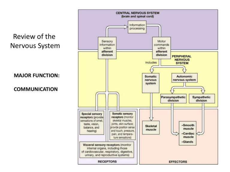
Review of the Nervous System MAJOR FUNCTION: COMMUNICATION Organization of the Nervous System Neuron Anatomy Neuron Gross Anatomy • Neurons are structurally different from other cells in the body. • Their unique structure belies their function. - Axon - Dentrites - Synapses Neuron Cell Body • Has pretty much the same organelles (ribosomes, RER, SER, Golgi apparatus, lysosomes, secretory vesicles, etc.) as those of other cells, with some minor differences: 1. Mitochondria present in processes, as well as the cell body itself, because of the energy requirements of maintaining the transmembrane ion potential (resting membrane potential); thus, usually more of them than in your average somatic cell. Neuron Structure Neuron Cell Body (cont’d) 2. Cytoskeleton – perhaps the most obvious structural difference between neurons and other cells, obviously because of the formers’ asymmetrical nature. a. Microfilaments [actin (growth cones) and myosin (growth cones, skeletal mus)]. b. Neurofilaments (intermediate in size between microfilaments and microtubules; implicated in tangles in AD). c. Microtubules – part of the cytoskeleton, act in axonal transport of vesicles, and act as molecular motors to move organelles (e.g., mitochondria) along. Axonal Transport of Membranous Organelles by Microtubules Motor Proteins • Kinesin – carries organelles in the (+)-end direction. • Dynein – carries organelles in the (-)-end direction. • See Figs. 1.10 and 1.11. Cytoskeleton Motor Proteins Kinesin and Dynein Myosin: A 3rd Motor Protein The headgroup of mysosin walks toward the head group of the actin filament (microfilament) Your Typical Presynaptic and Postsynaptic Neurons Axon • Thin, tube-like process that emanates from the cell body at a cone-shaped thickening, called the ‘axon hillock’. • Length can range from microns to meters. • Diameter remains relatively constant over its length. • Shape is maintained by the cytoskeleton. • Carries nerve impulses (information) to the soma Dendrite(s) • Neuronal processes that tend to be thicker and much shorter than axons. • Highly branched dendritic tree or arborization. • Unlike axon, dendrites can occur just about anywhere on the neuron. • Can grow small finger-like projections from the main dendritic shaft, called spines. • These spines are the synaptic input sites where the neuron receives information from other cells. • However, some dendrites also transmit electrical signals. Dendrites (cont’d) • Dendritic structure is not fixed and immutable. - Under certain physiological conditions, such as learning, stress, growth, disease, or aging (to name just a few), the size and shape of dendritic spines can dramatically change on a rapid time scale (a few minutes). • Carries nerve impulses (information) away from the soma. - These are the so-called plastic changes – more later in the quarter. Neuronal Polarity Dendrites Axons • • • • • • • • • • • Uniform calibre Few branches Lack polysomes Little, if any, protein synthesis Fast growth Neurofilament abundant Uniform polarity of microtubles Narrow spacing between microtubules Abundance of tau protein Presence of αγ spectrin Highly phosphorylated NF-M and NF-H • • • • • • • • Tapered morphology Highly branched Presence of polysomes Some protein synthesis Slow growth Abundance of microtubles Mixed polarity of microtubules Wide spacing between microtubules • Presence of MAP2A, B • Presence of αβ spectrin • Nonphosphorylated NF-M and NF-H Classification of Neurons Sensory Neurons • afferent • carry impulse to CNS • most are unipolar • some are bipolar Interneurons • link neurons • multipolar • in CNS Motor Neurons • multipolar • carry impulses away from CNS • carry impulses to effectors Synapse (= Connect) (Greek) • First discovered by Charles Sherrington near the end of the 19th century. • Studied reflexes, which do not require the brain at all. • Form the basis of intercellular communication. • But synapses, like dendrites, are not fixed. - Rather, there is a thing, called synaptic strength or strength of synaptic transmission. Synapses Nerve impulses pass from neuron to neuron at synapses Synaptic Transmission Neurotransmitters are released when impulse reaches synaptic knob Synapses (cont’d) • Synaptic strength can be modified according to the physiological state of the animal. - Is the animal learning? - Is the animal suffering from some disease? (Does not necessarily have to be a neurally-oriented disease). - Is the animal thriving or deprived? - Is the animal aging? - Has the animal been taking drugs (abuse or therapeutic)? Synapses (cont’d) Two Kinds of Synapses 1. Chemical 2. Electrical • Both types of synapses relay information, but do so by very different mechanisms. • Much more is known about chemical than about electrical synapses. - Information gleaned from NMJ in frog leg (sciatic n. – gastrocnemius m.). - However, this is n-m, rather than n-n. - n-m relay is much faster than n-n. Axon-dendrite Axo-axonic Axon-soma Chemical Synapses • Asymmetric morphology with distinct features found in the pre- and postsynaptic parts. • Synaptic cleft is ~ 200-300 angstroms wide. • CHO moities intersperse the synapse. • Most presynaptic endings are axon terminals. • Most postsynaptic elements in the CNS are dendrites. • There are all the combinations of synapses: axosomatic, axodendritic, axoaxonic. Chemical Synapses (cont’d) • Convergence. • Divergence. • Presynaptic ending: - swelling of the axon terminal. - mitochondria. - a variety of vesicular structures, clustered at/near the very edge of the axon terminal. Convergence • neuron receives input from several neurons • incoming impulses represent information from different types of sensory receptors • allows nervous system to collect, process, and respond to information • makes it possible for a neuron to sum impulses from different sources Divergence • one neuron sends impulses to several neurons • can amplify an impulse • impulse from a single neuron in CNS may be amplified to activate enough motor units needed for muscle contraction Chemical Synapses (cont’d) • Postsynaptic element - comprised largely of an electron-dense structure, called the postsynaptic density (PSD). Function of PSD? - Anchor receptors for nts in the postsynaptic membrane. - Involved in the conversion of chemical signal into an electrical one = transduction. Chemical Synapses (cont’d) • Associated with the morphological asymmetry is that chemical synapses are, for the most part, unidirectional. • There is a delay of a msec or more between the arrival of information at the presynaptic terminal and its transfer to the postsynaptic cell. This delay may reflect the several steps required for the release and action of the chemical neurotransmitter. The response of the postsynaptic neuron may be sustained (long-lasting), much longer than the presynaptic signal the evoked it. This may reflect long-lasting changes in the target (receiving) cell. • More on Chemical Synapses later in the course. Electrical Synapses • Symmetrical morphology. • Bidirectional transfer of information. • Pre- and postsynaptic cell membranes are in close apposition to each other, separated only by gap junctions. - Ions can flow through these gap junctions, providing low-resistance pathway for ion flow between cells without leakage to the extracellular space. - Instantaneous, fast transfer from 1 cell to the next, unlike the delay seen with chemical synapses. Electrical Synapses (cont’) Putative Functions • Synchronization of the electrical activity of large populations of neurons; - e.g., the large populations of neurosecretory neurons that synthesize and release biologically active peptide neurotransmitters and hormones are extensively connected by electrical synapses. - e.g., Synchronization may be required for neuronal development, including the development of chemical synapses. - e.g., Synchronization may be important in functions that require instantaneous responses, such as reflexes. • More on Electrical Synapses later in the course. Neuroglia Anatomy Types of Neuroglial Cells Types of Neuroglia Cells Schwann Cells • PNS • myelinating cell Oligodendrocytes • CNS • myelinating cell Microglia • CNS • phagocytic cell • respond to injury, infection or disease Astrocytes • CNS • scar tissue (“nerve glue”) • clean up • mop up excess ions (K+), nts, etc. • induce synapse formation • connect neurons to blood vessels Ependyma • CNS • ciliated • line central canal of spinal cord • line ventricles of brain Glial Functions • Provide myelin [Oligodendrocytes (CNS); Schwann cells (PNS)] • Act as scaffolding for neuronal migration and axon outgrowth. • Participate in the uptake and metabolism of the neurotransmitters that neurons use for intercellular communication. • Take up and buffer ions from the extracellular environments. • Act as scavengers to remove debris produced by dying neurons. • Segregate groups of neurons from each other and act as electrical insulators between neurons. • Provide structural support for neurons (c.f., role fulfilled by connective tissue cells in other organs). • Nurturant role: Supply metabolic components and even certain proteins necessary for neuronal function. • Participate in intercellular signaling; perhaps playing a role in information handling and memory storage. Glial Cell Functions Myelination of Axons • As per the previous slide, certainly not the only function, but arguably the most important function, whose malfunction is the basis for several wellknown, highly debilitating diseases. White Matter • contains myelinated axons Gray Matter • contains unmyelinated structures • cell bodies, dendrites OLIGODENDROCYTES (CNS) & SCHWANN CELLS (PNS) Processes of both types wrap around axon(s), forming an insulating sheath called myelin. Oligodendrocyte: its processes form multiple internodes on different axons and its cell body is located between the different axons. Schwann cell: its process forms only one internode, and its cell body is located on the axon. Astrocytes buffer the Environment Against an Accumulation of K+ • Astrocytes take up K+ ions from the extra-cellular space. • This occurs through K+ channels located on astrocyte cell bodies. • K+ is then released back into the extracellular space through their endfeet in areas that are relatively low in [K+] (running down the conc gradient). • Particularly important at the Nodes of Ranvier. • Why might such K+ buffering be important anyway? •Astrocytes take up K+ ions from the extracellular space. •K+ is released back into the extracellular space through their endfeet in areas that are relatively low in [K+] (running down the concentration gradient). Astrocytes Regulate Neuronal Cytosolic [Ca2+]s • Astrocytes release chemicals called “gliotransmitters”, glutamate, ATP, D-serine, and TNFα. • This suggests that astrocytes are excitable cells. • Yet, their excitability is not based on Vm as it is in neurons, but rather, on changes in [Ca2+]i. • Astrocytes, connected together by gap junctions, have been shown to signal each other (and neurons) via intercellular Ca2+ pulses. Astrocytes Regulate Neuronal Cytosolic [Ca2+]s (cont’d) • The activity-driven response in astrocytes is extremely sensitive to the level of neuronal activity: e.g., A 1% increase in [isofluorane] causes a 16% decrease in neuronal response to visual stimulation, but a 77% decrease in the astrocytic response to the same visual stimulus. This suggests that astrocytes’ role may be in finetuning the stimulation-induced responses (e.g., orientation and spatial frequency) of neurons. Astrocytes Regulate Neuronal Cytosolic [Ca2+]s (cont’d) • Such studies have shown that stimulation in rodents (e.g., whisker, limb, odor) releases Ca2+ from astrocytes. • Astrocytic Ca2+ release is largely mediated by the activation of metabotropic glu receptors (mGluRs). • Stimulation of mGluRs modulates arteriole diameter (bp) and are dependent on Ca2+ signaling, involving multiple glial cells. Astrocytes are Neuroprotective • Secrete neurotrophins. • Main defense against brain glutamate excitotoxicity. • K+ buffering. • Astrocytes express high amounts of connexins, the main protein of gap junctions. Through these gap junctions, glucose and metabolites are allowed to pass, thereby strengthening the astrocytic network, which, in turn, strengthens the neuronal network. Bidirectional Communication between Neurons and Astrocytes (b) Hippocampal activity is increased, but is scaled back and fine-tuned by astrocytereleased TNFα Astrocytes Participate in the Neurotransmitter Cycle • Astrocytes help take up neurotransmiters in the synaptic cleft so that they do not continuously bind their receptors on the postsynaptic cell. • This supplements the uptake mechanisms and degradation mechanisms that neurons already have in place. • Such supplementation may be particularly important for high [neurotransmitters]s (e.g., glutamate, whether naturally or drug-induced). Life cycle of a neurotransmitter An excitatory (glutamatergic) synapse A synapse using g-aminobutyric acid (GABA) A synapse that uses acetylcholine (ACh) Astrocytes Modify Synaptic Function in Time and Space SPACE • Astrocytes release gliotransmitters not only at processes of the astrocyte contacting the synapse: - Transmitter release may occur close to the synapse. - Transmitter release may occur into the synapse. e.g., release of D-ser modifies synaptic NMDA receptors. Astrocytes Modify Synaptic Function in Time and Space (cont’d) TIME • Both neuronal and astrocytic input determine the outcome of synaptic plasticity. • Timing is critical for determining extent of synaptic plasticity. e.g., Astrocytes release ATP both tonically and in an activity-dependent manner: Constant release of ATP that is rapidly degraded to adenosine provides a tonic suppression at hippocampal synapses. [recall tonic Ca2+ release for cell-cell communication]. But activity-mediated ATP release from astrocytes is responsible for heterosynaptic depression. Astrocytes can provide Neurons with ATP both Directly and Indirectly Directly • Some of the adenosine released by astrocytes are taken up by neurons to be used to manufacture ATP. • Astrocytes can provide neurons with glucose. Indirectly • Astrocytes numerous radiating processes that cover neurons and nearby capillaries support and brace the neurons and anchor them to their nutrient supply lines (capillaries). Astrocyte Astrocytes Help Form the Blood Brain Barrier • Basal lamina of the astrocytes + the astrocytic endfeet produce help maintain the BBB. • Notice how astrocytes send processes to the external surface of the CNS where the endfeet form the glia limitans externa, which separate the pia mater from the nervous tissue. • Gap junctions and desmosomes join the endfeet to form a space between neurons and vascular endothelial cells (Fig. 2.2). Ependymal Cells • Line the walls of the ventricles of the brain and central canal of the spinal cord. • Produce CSF. • Have cilia on their apical ends, which beat and help distribute the CSF that help cushion the brain and spinal cord (refer to preceding slide). Microglia • Branches touch nearby neurons to monitor the latters’ health. • If there is neuronal ill-health or injury, microglia mobilize and migrate towards them. • If invading microorganisms or dead neurons are present, microglia transform into a microphage, which then phagocytises the debris. • Critically important, given that the immune system cells have no access to the CNS. Microglia
