Chapter 27
Chapter 27
The Male Reproductive System
• Sexual reproduction
• Sex determination & development
• Male reproductive anatomy
• Puberty and climacteric
• Spermatogenesis, spermatozoa
& sperm
• Male sexual response
The Essence of Sex
• Reproduction is one property of a living thing
– great variety of methods
• Sexual reproduction means each offspring has 2 parents and receives genetic material from both
– provides genetic diversity & is considered the foundation for the survival and evolution of species
The Two Sexes
• Male and female gametes (sex cells) combine their genes to form a fertilized egg (zygote)
– one gamete has motility (sperm)
• the parent producing sperm is considered the male
• has Y chromosome
– other gamete (egg or ovum) contains most of the nutrients for the developing zygote
• the parent producing eggs is considered the female
• in mammals the female also provides shelter for developing fetus (uterus and placenta)
Overview of the Reproductive System
• Primary sex organs
– organs that produce the gametes (testes or ovaries)
• Secondary sex organs (essential for reproduction)
– male is ducts, glands and the penis that deliver the sperm cells
– female is uterine tubes, uterus & vagina that receive the sperm & nourish the developing fetus
• Secondary sex characteristics
– features that develop at puberty to attract a mate
• pubic, axillary & facial hair, scent glands, body morphology and low-pitched voice in males
Role of the Sex Chromosomes
• Our cells contain 23 pairs of chromosomes
– 22 pairs of autosomes
– 1 pair of sex chromosomes (XY males: XX females)
• males produce 50% Y carrying sperm and 50% X carrying
• all eggs carry the X chromosome
• Sex of the child is determined by the type of sperm that fertilizes the mother’s egg
Hormones and Sex Differentiation
• Gonads begin to develop at 6 weeks as gonadal ridges
– near the primitive kidneys (mesonephros)
• 2 sets of ducts exist at that time
– mesonephric ducts develop into male reproductive system
(paramesonephric ducts degenerate)
– paramesonephric ducts ( müllerian ducts) develop into female reproductive tract (mesonephric ducts degenerate)
• SRY gene ( S ex-determining R egion of Y gene)
– in males , codes for a protein that causes development of testes
• secrete testosterone
• secrete müllerian -inhibiting factor
degenerates paramesonephric ducts
• Female development occurs in the absence of male or female hormones
Embryonic Development
Embryonic Development
Embryonic Development
Male Female
Androgen-Insensitivity Syndrome
• Genetically male (XY)
• Testosterone is secreted
• Target cells lack receptors for the hormone
• No masculizing effects occur
Development of External Genitalia
• All 8 week old fetuses have same 3 structures
– by end of week 9, begin to show sexual differentiation
– distinctly male or female by end of week 12
Development of External Genitalia
10 week 10 week
Male
Female
12 week 12 week
Descent of the Testes
• Begin development near the kidney
– gubernaculum (cordlike structure containing muscle) extends from the gonad to the abdominopelvic floor
– it shortens and guides the testes to the scrotum
• passes through abdominal wall via the inguinal canal
• accompanied by testicular nerve, artery & vein
• Descent begins in weeks 6-10 & is finished by 28
– 3% of boys are born with an undescended testes
(cryptorchidism)
• Location outside the pelvic cavity is essential for low temperatures needed for sperm production
Descent of Testis
Boundaries of Male Perineum
Male Reproductive System
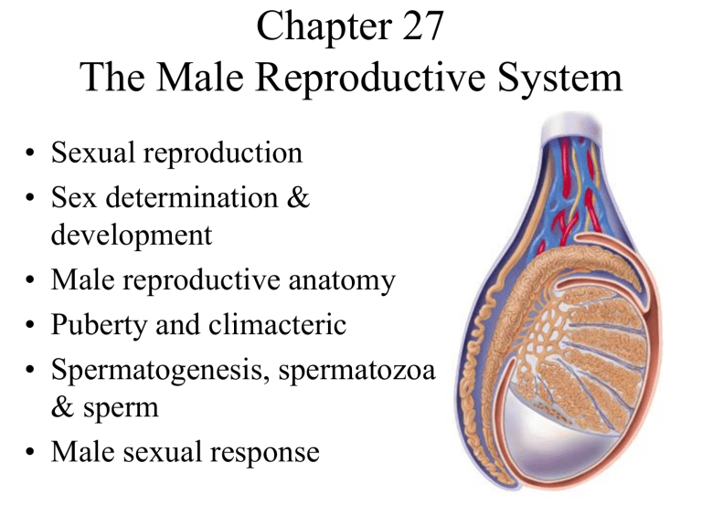
Testes
• Oval organ, 4 cm long x 2.5 cm in diameter
• Covered anteriorly by a saclike extension of the peritoneum (tunica vaginalis) that descended into the scrotum with the testes
• Tunica albuginea = white fibrous capsule
– septa divide the organ into compartments containing seminiferous tubules where sperm are produced
• each tubule is lined with a thick germinal epithelium composed of germ cells in the process of becoming sperm
– sustentacular cells promote sperm cell development
• blood-testis barrier is formed by tight junctions between sustentacular cells -- separating sperm from immune system
Testis and Associated Structures
• Seminiferous tubules drain into network called rete testis
• Low BP of testicular artery results in poor O
2 supply
– sperm develop very large mitochondria helping them survive the hypoxic environment of the female reproductive tract
• Testicular veins drain to the inferior vena cava
Histology of the Testis
Male Inguinal & Scrotal Region
Scrotum
• Pendulous pouch holding the testes
– divided into 2 compartments by median septum
• Spermatic cord travels up from the scrotum to pass through the abdominal wall (inguinal canal)
– contains testicular artery, vein, nerve & lymphatics
• Testicular thermoregulation is necessary since sperm are not produced at core body temperature
– cremaster muscle = segments of internal oblique muscle that pull testes closer to body
– dartos muscle = smooth muscle wrinkles skin reducing surface area of scrotum & lifting it upwards
– pampiniform plexus = veins ascending near testicular artery
• countercurrent heat exchanger cools arterial blood entering the testis
Heat Exchange of Pampiniform Plexus
Spermatic Ducts
• Efferent ductules
– 12 small ciliated ducts collecting sperm from the rete testes and transporting it to the epididymis
• Epididymis (head, body & tail)
– 6 m long coiled duct adhering to the posterior of testis
– site of sperm maturation & storage (fertile for 60 days)
• Ductus deferens (peristalsis during orgasm)
– muscular tube 45 cm long passing up from scrotum through inguinal canal to posterior surface of bladder
• Ejaculatory duct
– 2 cm duct formed from ductus deferens & seminal vesicle & passing through prostate to empty into urethra
Male Duct System
Male Urethra
• Regions of male urethra: prostatic, membranous and penile --- totals 20 cm long
Accessory Glands
• Seminal vesicles
– posterior to bladder
– empty into ejaculatory duct
• Prostate gland
– below bladder, surrounds urethra and ejaculatory duct
– 2 x 4 x 3 cm
• Bulbourethral glands
– near bulb of penis
– empty into penile urethra
– lubricating fluid
Penis
• Internal root and visible shaft and glans
– external portion is 4 in. long when flaccid
– skin over shaft is loosely attached allowing expansion
• extends over glans as prepuce or foreskin
• Consists of 3 cylindrical bodies of erectile tissue
– single corpus spongiosum along ventral side of penis
• encloses penile urethra
• ends as a dilated bulb ensheathed by bulbospongiosus muscle
– paired corpora cavernosa
• diverge like arms of a Y
• each crus attaches to pubic arch & is covered with ischiocavernosus muscle
Anatomy of the Penis
Puberty and Climacteric
• Reproductive system remains dormant for years after birth
– surge of pituitary gonadotropins begins development
• 10-12 in most boys; 8-10 in most girls
• Puberty = period from onset of gonadotropin secretion until first menstrual period or first ejaculation of viable sperm
• Adolescence = ends when person attains full adult height
Brain-Testicular Axis
• Mature hypothalamus produces GnRH
• Stimulation of gonadotrope cells in anterior pituitary causes secretion of FSH & LH
– LH stimulates interstitial cells to produce testosterone
– FSH stimulates sustentacular cells to secrete androgen-binding protein that interacts with testosterone to stimulate spermatogenesis
• Other effects of testosterone
– enlargement secondary sexual organs
• penis, testes, scrotum, ducts, glands and muscle mass enlarge
• hair, scent and sebaceous glands develop
• stimulates erythropoiesis and libido
• During adulthood, testosterone sustains libido, spermatogenesis and reproductive tract
Hormones & Brain-Testicular Axis
Aging and Sexual Function
• Decline in testosterone secretion
– peak secretion at 7 mg/day at age 20
– declines to 1/5 of that by age 80
• Rise in FSH and LH secretion after age 50 produces male climacteric (menopause)
– mood changes, hot flashes & “illusions of suffocation”
• Impotence (erectile dysfunction)
– 20% of those in 60s and 50% of those in 80s
– 0ver 90% of impotent men remain able to ejaculate
Mitosis and Meiosis
• Mitosis produces 2 genetically identical daughter cells (occurs in tissue repair & embryonic growth)
• Meiosis produces gametes haploid cells required for sexual reproduction
– 2 cell divisions (after only one replication of DNA)
• meiosis I separates homologous chromosome pairs
2 haploid cells
• meiosis II separates duplicated sister chromatids
4 haploid cells
– meiosis keeps chromosome number constant from generation to generation after fertilization
– occurs in seminiferous tubules of males
Meiosis I
Meiosis I
Meiosis II
Meiosis II
Spermatogenesis
• Spermatogonia produce 2 kinds of daughter cells
– type A remain outside blood-testis barrier & produce more daughter cells until death
– type B differentiate into primary spermatocytes
• cells must pass through
BTB to move inward toward lumen - new tight junctions form behind these cells
• meiosis I
2 secondary spermatocytes
• meiosis II
4 spermatids
• Spermiogenesis is transformation into spermatozoon
– sprouts tail and discards cytoplasm to become lighter
Spermatogenesis & Sustentacular Cells
• Blood-testis barrier is formed by tight junctions between and basement membrane under sustentacular cells.
Spermiogenesis
• Changes that transform spermatids into spermatozoa
– discarding excess cytoplasm & growing tails
The Spermatozoon
• Head is pear-shaped front end
– 4 to 5 microns long structure containing the nucleus, acrosome and basal body of the tail flagella
• nucleus contains haploid set of chromosomes
• acrosome contains enzymes that penetrate the egg
• basal body
• Tail is divided into 3 regions
– midpiece contains mitochondria around axoneme of the flagella (produce ATP for flagellar movement)
– principal piece is axoneme surrounded by fibers
– endpiece is very narrow tip of flagella
Spermatozoon
Semen or Seminal Fluid
• 2-5 mL of fluid expelled during orgasm
– 60% seminal vesicle fluid, 30% prostatic & 10% sperm
• normal sperm count is 50-120 million/mL (< 25 million/mL is associated with infertility)
• Other components of semen
– fructose provide energy for sperm motility
– fibrinogen causes clotting
• enzymes convert fibrinogen to fibrin
– fibrinolysin liquefies semen within 30 minutes
– prostaglandins stimulate female peristaltic contractions
– spermine is a base stabilizing sperm pH at 7.2 to 7.6
Male Sexual Response -- Anatomy
• Arteries of the penis
– dorsal & deep arteries(brs. of the internal pudendal)
– deep artery supplies lacunae of corpora cavernosa
• dilation fills the lacunae causing an erection
– normal penile blood supply comes from dorsal a.
• Nerves of the penis
– abundance of tactile, pressure & temperature receptors
– dorsal nerve of the penis and internal pudendal nerves lead to integrating center in sacral spinal cord
– both autonomic and somatic motor fibers carry impulses from integrating center to penis & other pelvic organs
Excitement and Plateau
• Excitement is characterized by vasocongestion of genitals, myotonia, and increases in heart rate, BP,
& pulmonary ventilation
• Initiated by many different erotic stimuli
• Erection of penis is due to parasympathetic triggering of nitric oxide (NO) secretion
– dilation of deep arteries & filling of lacunae with blood
• corpora spongiosum not nearly as hardened
– enlarged elevated penis is ready for intromission
• Erection is maintained during plateau phase
Parasympathetic Signals & Sexual Response
• Parasympathetic signals produce an erection with direct stimulation of the penis and other perineal organs
Orgasm and Ejaculation
• Climax (orgasm) is 15 second reaction that includes the discharge of semen (ejaculation)
• Ejaculation
– emission = sympathetic nervous system propels sperm through ducts as glandular secretions are added
– expulsion = semen in urethra activates muscular contractions that lead to expulsion
• Ejaculation and orgasm are not the same
– can occur separately
Orgasm - Emission
Orgasm - Ejaculation
Resolution
• Sympathetic signals constrict the internal pudendal artery & reduce blood flow to penis
– penis becomes soft & flaccid (detumescence)
• Cardiovascular & respiratory responses return to normal
• Refractory period (10 minutes to few hours)
– impossible to attain another erection and orgasm

