Essential Question 2 - Monmouth Regional High School
advertisement

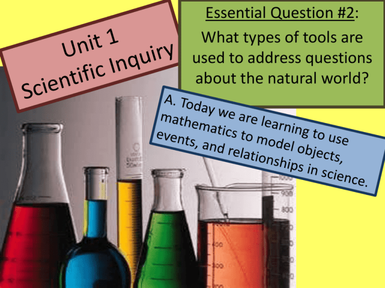
Essential Question #2: What types of tools are used to address questions about the natural world? Textbook References: Unit 1 Miller & Levine: Ch. 1-The Science of Biology 1.1 What is Science (Pgs. 4-9) 1.2 Science in Context (Pgs. 10-16) Campbell: Ch. 1- Biology: Exploring Life 1.7 & 1.8 The Process of Science (Pgs. 9-10) 1.9 Biology and Everyday Life (Pg. 12) SAFETY FIRST! Ruler Tape measure Meter stick A Typical Metric Ruler - shows distance in centimeters. - the other side of the ruler shows inches. 1 inch = 2.54 cm cm inches According to the ruler: How many mm are in 1 cm? _______ 2 inches can be converted into ______ cm. Triple Beam Balance Digital Scale Beaker Flask Graduated Cylinder The Metric System – It is used by scientists when collecting data. – It is a universal system of measurement. – It consists of standard base units. – Other units are obtained by multiplying and dividing by powers of 10, or simply moving the decimal. -Metric SystemBase Units Measurement Base Unit • • • • • Meter Gram Liter Second Celsius Length Mass Volume Time Temperature Unit Conversions Unit Conversions • To convert length from a large unit into a small unit, multiply by the relevant power of 10. • OR simply move the decimal to the right. • To convert length from a small unit into a large unit, divide by the relevant power of 10. • OR simply move the decimal to the left Practice CIRCLE ONE 1 m = ____________ mm Length Volume Mass 25g = ____________ kg Length Volume Mass 400 ml = __________ L Length Volume Mass 12 cm = ____________ m Length Volume Mass Make up your own problem in the space below. Ask your neighbor to try and solve it. _________________________________________________ Microscopy Essential Question #2: What types of tools are used to address questions about the natural world? Today we will describe how the different types of microscopes are used and how they have impacted the study of biology. MicroAngela is a creation of Tina (Weatherby) Carvalho of the Biological Electron Microscope Facility, (BEMF), part of the Pacific Biomedical Research Center at the University of Hawaii at Manoa. http://www5.pbrc.hawaii.edu/microangela/methods.htm Types of Microscopes • Light Microscope – the models found in most schools – use compound lenses and light to magnify objects. – The lenses bend or refract the light, which makes the object beneath them appear closer. Stereoscope • this microscope allows for binocular viewing of larger specimens. (Binocular means 2 eyes) Scanning Electron Microscope (SEMs) - allow scientists to view a universe too small to be seen with a light microscope. - they use electrons (instead of light waves) to magnify objects up to two million times!! Transmission Electron Microscope Also uses electrons, but instead of scanning the surface (as with SEM's) electrons are passed through very thin specimens. Parts of a Microscope Light Microscope Magnifications • Your microscope has three magnifications: – Scanning – Low – High • Each objective will have the magnification number engraved on the side. • The ocular lens (eyepiece) has a magnification of 10x • Total magnification = ocular x objective Calculating Total Magnification Objective Magnification Ocular lens Magnification Total Magnification Scanning 4x 10x 40x Low Power 10x 10x 100x High Power 40x 10x 400x Power General Procedures to Start 1. Make sure all backpacks and junk are out of the aisles. 2. Always carry microscopes by the arm and base. 3. Set the microscope flat on your desk and not at the edge. 4. Plug your microscope into the floor outlet under your desk 5. Always start with the Scanning Objective and increase magnification as needed. Making a Wet Mount Gather a thin slice/piece of your specimen. If your specimen is too thick, then the coverslip will wobble on top of the sample like a see-saw. Place ONE drop of water directly over the specimen. Too much water makes the the coverslip float on top of the water so it is hard to find the specimen through the microscope lens. Place the cover slip at a 45° angle with one edge touching the water drop and then gently let go. Focusing Specimens 1. Always start with the scanning objective. – Use the Coarse Knob to focus. • The image may be small, but you won't be able to find it on the higher powers without this first step. – Try moving the slide around until you find something. SCANNING POWER 2. Now switch to Low Power. – Use the coarse adjustment knob to bring the specimen back into focus. – Use the fine adjustment knob to increase the sharpness – Again, if you haven't focused on this level, you will not be able to move to the next level. LOW POWER (40X) 3. Now switch to High Power. – Use only the fine adjustment knob. – Do not use the coarse adjustment knob or you may crack the slide. HIGH POWER (100x) Troubleshooting Here are some common problems & solutions. Image is too dark or light! – Adjust the diaphragm, make sure your light is on. There's a spot in my viewing field, even when I move the slide the spot stays in the same place! – Your lens is dirty. – Use lens paper, and only lens paper to carefully clean the objective and ocular lens. I can't see anything under high power! – Remember the steps, if you can't focus on scanning and then low power, you won't be able to focus anything on high power. Scanning Low Power High Power Only half of my viewing field is lit, it looks like there's a half-moon in there! – You probably don't have your objective fully clicked into place. I see a line going through my viewing field. Try twisting the eyepiece and the line should move. This needle is used for pointing out things to your lab partner or teacher. SEE I’M POINTING OUT THE POINTER THAT’S POINTING OUT THE CELL! GET IT ? Drawing Specimens Use pencil - you can erase and shade areas Drawings should be labeled with the specimen name and magnification. All drawings should include clear and proper labels (and be large enough to view details). Labels should be written on the outside of the circle. o The circle indicates the viewing field as seen through the eyepiece Specimens should be drawn to scale. o What does drawing to scale mean? o For instance, if your specimen takes up the whole viewing field, make sure your drawing reflects that. CHECKLIST: Used Pencil Shaded areas, colored Name of Specimen Total Magnification Clear, proper labels Lines do not crossover Labels are outside circle Drawn to scale Sample Drawing General Procedures to End 1. Do not remove slides while on High Power Objective is in place- this will scratch the lens! 2. Wash slides in the sink and dry them, placing them back in the slide boxes 3. Throw cover slips away. 4. Microscopes should be stored with the Scanning Objective clicked into place. 5. Always wrap electric cords and place the cover over the microscope before returning them to the cabinet. Essential Question #2: What types of tools are used to address questions about the natural world? Measurement and observation tools are used to categorize, represent, and interpret the natural world.


