Anatomy - Internal Features of Heart
advertisement
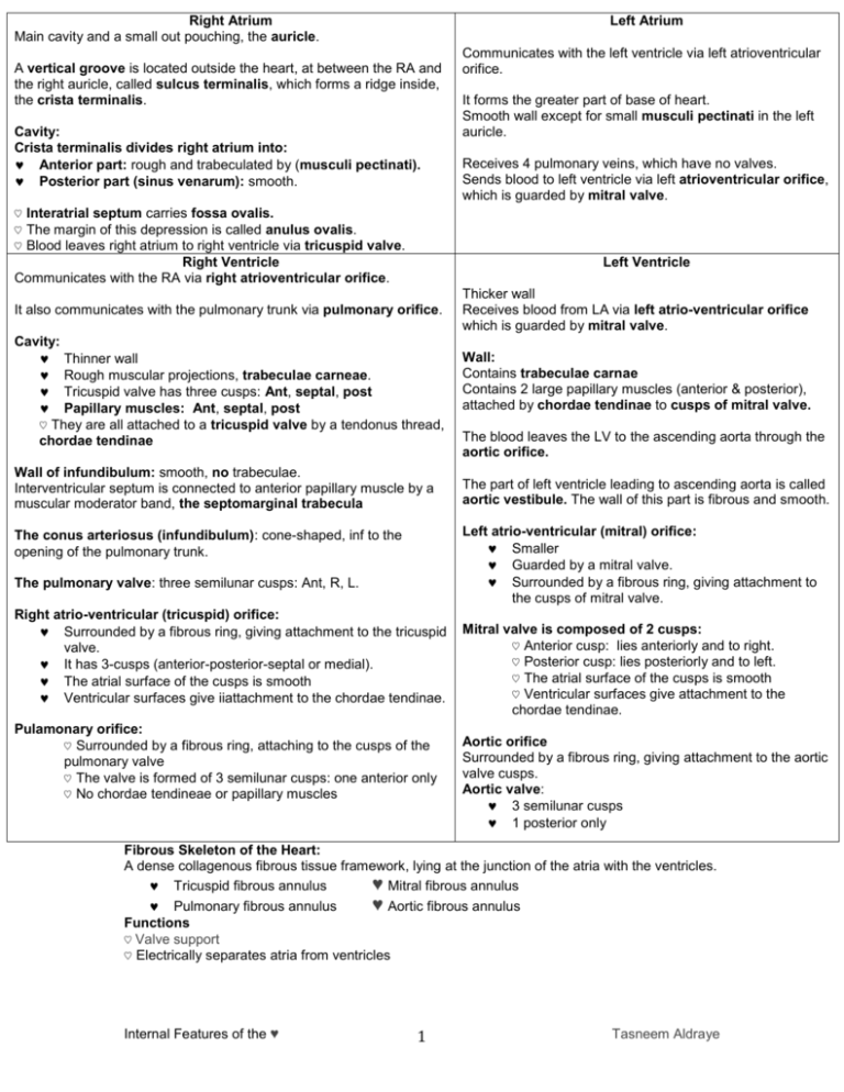
Right Atrium Main cavity and a small out pouching, the auricle. Left Atrium A vertical groove is located outside the heart, at between the RA and the right auricle, called sulcus terminalis, which forms a ridge inside, the crista terminalis. Cavity: Crista terminalis divides right atrium into: Anterior part: rough and trabeculated by (musculi pectinati). Posterior part (sinus venarum): smooth. Communicates with the left ventricle via left atrioventricular orifice. It forms the greater part of base of heart. Smooth wall except for small musculi pectinati in the left auricle. Receives 4 pulmonary veins, which have no valves. Sends blood to left ventricle via left atrioventricular orifice, which is guarded by mitral valve. ♡ Interatrial septum carries fossa ovalis. ♡ The margin of this depression is called anulus ovalis. ♡ Blood leaves right atrium to right ventricle via tricuspid valve. Right Ventricle Communicates with the RA via right atrioventricular orifice. Left Ventricle It also communicates with the pulmonary trunk via pulmonary orifice. Cavity: Thinner wall Rough muscular projections, trabeculae carneae. Tricuspid valve has three cusps: Ant, septal, post Papillary muscles: Ant, septal, post ♡ They are all attached to a tricuspid valve by a tendonus thread, chordae tendinae Wall of infundibulum: smooth, no trabeculae. Interventricular septum is connected to anterior papillary muscle by a muscular moderator band, the septomarginal trabecula Thicker wall Receives blood from LA via left atrio-ventricular orifice which is guarded by mitral valve. Wall: Contains trabeculae carnae Contains 2 large papillary muscles (anterior & posterior), attached by chordae tendinae to cusps of mitral valve. The blood leaves the LV to the ascending aorta through the aortic orifice. The part of left ventricle leading to ascending aorta is called aortic vestibule. The wall of this part is fibrous and smooth. Left atrio-ventricular (mitral) orifice: Smaller Guarded by a mitral valve. Surrounded by a fibrous ring, giving attachment to the cusps of mitral valve. The conus arteriosus (infundibulum): cone-shaped, inf to the opening of the pulmonary trunk. The pulmonary valve: three semilunar cusps: Ant, R, L. Right atrio-ventricular (tricuspid) orifice: Surrounded by a fibrous ring, giving attachment to the tricuspid valve. It has 3-cusps (anterior-posterior-septal or medial). The atrial surface of the cusps is smooth Ventricular surfaces give iiattachment to the chordae tendinae. Pulamonary orifice: ♡ Surrounded by a fibrous ring, attaching to the cusps of the pulmonary valve ♡ The valve is formed of 3 semilunar cusps: one anterior only ♡ No chordae tendineae or papillary muscles Mitral valve is composed of 2 cusps: ♡ Anterior cusp: lies anteriorly and to right. ♡ Posterior cusp: lies posteriorly and to left. ♡ The atrial surface of the cusps is smooth ♡ Ventricular surfaces give attachment to the chordae tendinae. Aortic orifice Surrounded by a fibrous ring, giving attachment to the aortic valve cusps. Aortic valve: 3 semilunar cusps 1 posterior only Fibrous Skeleton of the Heart: A dense collagenous fibrous tissue framework, lying at the junction of the atria with the ventricles. Tricuspid fibrous annulus ♥ Mitral fibrous annulus Pulmonary fibrous annulus ♥ Aortic fibrous annulus Functions ♡ Valve support ♡ Electrically separates atria from ventricles Internal Features of the ♥ 1 Tasneem Aldraye
