6.5 Nerves Hormones and Homeostasis - IBDPBiology-Dnl
advertisement
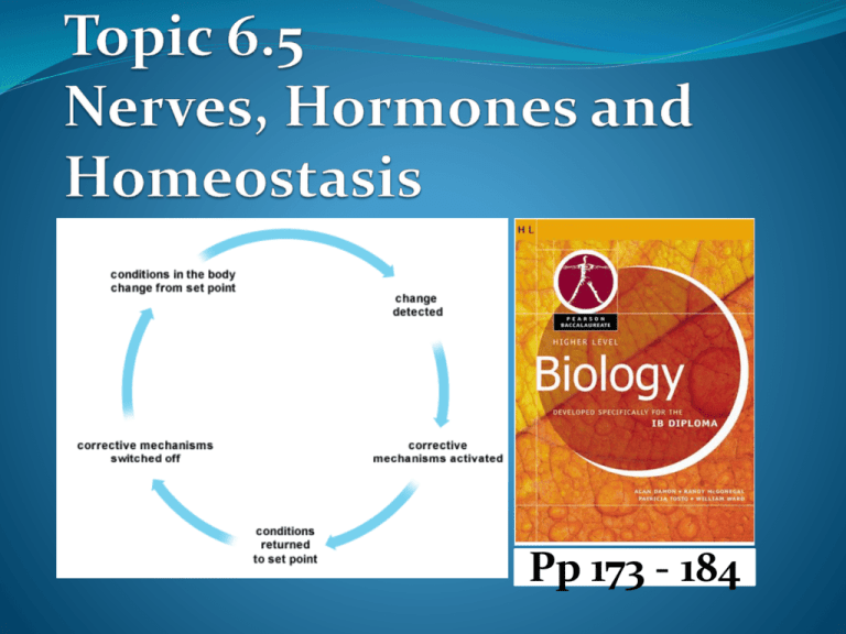
Pp 173 - 184 Nervous System Nervous system consists of two parts: Central Nervous System (CNS); brain and spinal cord relays messages, processes and analyzes information Peripheral Nervous System (PNS) peripheral nerves: cranial and spinal (outside CNS) connects sense organs to CNS connects CNS to muscles and glands Central Nervous System • The central nervous system (CNS) consists of the brain and spinal cord. • Both structures receive sensory information from receptors all over the body and they interpret the information, process it and decide if a response is required. • A response by the brain or spinal cord is known as a motor response. Peripheral Nervous System • The peripheral nervous system includes all other nerves that connect to the CNS. • Peripheral nerves are composed of sensory neurons and motor neurons. The peripheral system has two categories of peripheral nerves. • There are 31 pairs of spinal nerves which emerge directly from the spinal cord and are a mix of sensory neurons and motor neurons. It also has 12 pairs of cranial nerves which emerge from the brain stem. Neurons • The nervous system is made up of special nerve cells called neurons. • These cells are very different in shape from other eukaryotic cells and they transmit messages in the form of electrical impulses at incredible speed. • Many neurons grouped together form a nerve. 3 main types of nerve cells sensory neurone relay neurone motor neurone Sensory neurons Conducts impulses from receptors e.g. pain receptors in skin to the CNS( brain or spinal cord) Relay neuron Conducts impulses within the CNS, from sensory neurons to motor neurons. Motor neuron Conducts impulses from CNS to effectors e.g. muscle to bring about movement or gland to bring about secretion of hormone Draw a labelled diagram of a motor neurone Parts of a Motor Neuron • Cell body: Contains the nucleus, rough ER and other • • • • • organelles. Axon: a very long and thin extension of cytoplasm from the cell body. Carries impulses away from the cell body. Dendrites: much smaller cytoplasm extensions which carry impulses to the cell body. Myelin Sheath: a type of insulation that is wrapped around the axons of neurons; composed of Schwann cells. Helps reduce signal loss. Schwann Cells: special type of glial cells that produce the myelin sheath Nodes of Ranvier: regularly occurring gaps between sections of myelin sheath where the axon is “naked”. Reflex How the impulse is transmitted Impulse begins when a neuron is stimulated by another neuron or by the environment Electrical impulse moves in one direction: Dendrites → Cell Body → Axon Synapse: gap between 2 neurons Neurotransmitters send the signal to the following neuron No myelin = 5-25m/s With myelin = 10-120m/s Resting potential and Action potential (depolarization and repolarization). • Resting potential can be defined as the electrical potential across the plasma membrane of a cell that is not sending an impulse. The resting potential of all neurons depends on the ionic gradients that exist across the plasma membrane of the cell. • Action potential can be defined as the reversal and restoration of the electrical potential across the plasma membrane of a cell as an electrical impulse passes along it. It is also measured in mV. Nerve Impulse – Animated Tutorial Action Potential – Animated Tutorial Resting Potential All cells have an electrical potential difference (voltage) across their plasma membrane that is measured in mV and the difference is known as the membrane potential. Neurons typically have a membrane potential between -60 and -80 mV (millivolts) when the cell is not transmitting a signal and this is called the resting potential • Neurons use active transport to maintain a balance of ions across their membranes. Sodium ions are pumped out and potassium ions are pumped in. • There are chloride ions and other negatively charged ions inside the neuron that are fairly large and have a tendency to stay inside which creates a net negative charge inside the neuron as compared with the more positive charge outside the neuron. • This creates the resting potential and the membrane is said to be polarized. NOTE: Sodium ions are highly concentrated outside of the cell and they have a tendency to diffuse inside the cell. Potassium ions diffuse out of the cell as sodium diffuses in but the membrane is about 50 times more permeable to potassium than sodium so the movement is unequal. The sodium-potassium pumps use active transport to control the movement of these ions. When an impulse is passing along a neuron the potassium and sodium ions are allowed to diffuse (passive transport) across the membrane through proteins known as voltagegated ion channels. This reverses the electrical potential of the neuron but it is quickly restored and this is the action potential. The neuron is depolarized and then quickly repolarized. When there is a stimulus... Na+ gates open = Na + enter the cell Electrical potential of the cell changes depolarization (normal charge is reversed) = +30mV Action potential is recorded Na + channels close K + channels open repolarization occurs (charges back to normal) K + channels stay open longer hyperpolarization = -85mV (refractory period = prevents one impulse to catch up with another) Stimulus = self-propagating http://outreach.mcb.harvard.edu/animations/actionpotential.swf Depolarization and Repolarization Depolarization is the diffusion of sodium ions into the nerve cell resulting in a charge reversal. Repolarization is the process of restoring the original polarity of the nerve membrane. How a nerve impulse passes along a non-myelinated neuron. • Understanding of how an action potential works is the key to understanding how a nerve impulse passes along the axon of a neuron. • An action potential in one part of a neuron will cause the development of an action potential in the next section of the neuron. • This can occur because sodium ions flow from a region with an action potential to a region with a resting potential. • As the ions move the resting potential is reduced which results in the opening of the voltage-gated channels. • When a neuron is excited the membrane becomes more permeable to sodium than potassium. • Scientists believe this occurs because sodium gates open while potassium gates close. The membrane potential is reduced and more sodium channels open. • As sodium ions flow into the neuron via diffusion and charge attraction the inside of the membrane becomes positive (charge reversal) and depolarization occurs. The membrane potential has been reversed. • Once depolarization occurs the sodium gates are closed and the potassium channels open. Potassium diffuses out of the neuron in the direction of the concentration gradient. • The loss of the positive potassium ions causes the internal environment of the neuron to become negative once again and the potential across the membrane is restored. • This is known as repolarization; the return to the original polarity of the nerve membrane. • Sodium-potassium pumps are used to restore the concentration gradients of the ions via active transport. Sodium is pumped back out of the neuron while potassium is pumped back in. • The resting potential of the neuron is now restored and can now conduct another impulse. • The action potential moves along the membrane of the neuron creating a wave of depolarization and repolarization. As the impulse moves along the axon of the neuron it moves from a depolarized region and initiates depolarization in the next region. A synapse is a junction between two neurons • At the end of the axons of neurons there are swollen • • membranous areas called terminal buttons. Inside the terminal buttons are small vesicles filled with chemicals known as neurotransmitters. These chemicals are used for synaptic transmission and they always pass in the same direction; from the presynaptic neuron to the postsynaptic neuron. A synapse is a junction between two neurons and the plasma membranes of those neurons are separated by a narrow fluid-filled gap known as the synaptic cleft. Synaptic transmission is the transmission of an action potential across the synapse from the pre-synaptic neuron to the postsynaptic neuron. Steps in Synaptic Transmission 1. The nerve impulse (action potential) arrives at the end of the presynaptic neuron. 2. Depolarization of the membrane occurs and the voltage gated calcium channels open allowing calcium ions diffuse into the terminal button. 3. The movement of the calcium ions in causes the vesicles containing the neurotransmitter such as acetylcholine to fuse with the plasma membrane of the neuron and release the neurotransmitter into the synaptic cleft. 4. The neurotransmitter diffuses across the cleft from the presynaptic neuron and binds to receptors (transmittergated ion channels) on the postsynaptic neuron. 5. The binding of the neurotransmitter results in the opening of the Na+ ion channels and sodium ions along with other positively charged ions diffuse into the postsynaptic neuron. 6. The movement of the ions causes the depolarization of the postsynaptic membrane and this initiates the movement of the action potential down the neuron. 7. The neurotransmitter in the cleft is quickly broken down by enzymes to prevent continuous synaptic transmission and the calcium ions are pumped out of the presynaptic neuron and into the synaptic cleft. The ions channels close to sodium ions. 8. The fragments of the broken down neurotransmitters diffuse back across the synaptic cleft and into the vesicles where they can be reassembled. 9. Ca2+ ions are pumped back into the synaptic cleft from presynaptic neuron Endocrine System The endocrine system The endocrine system consist of glands in various locations around the body. Each gland secretes one or more hormone into the bloodstream to be transported to target organ(s) i.e. a specific organ of the body where hormones initiates a specific response. Hormones are chemical messengers made of proteins and secreted from the cells of ductless glands. Hormones help to control and coordinate body activities then they are broken down in the liver. Homeostasis maintaining the internal environment constant between narrow limits Example of conditions that need to be kept constant: blood sugar, blood pH, oxygen & carbon dioxide concentrations, body temperature and water balance Homeostasis involves negative feedback Homeostasis:- monitoring levels of variables and correcting changes in levels by negative feedback mechanisms change in internal environment is detected response to bring the system back to set point / within narrow limits occurs when the set point is reached, the response is stopped to prevent over reaction internal environment fluctuates around the set point (norm) Negative feedback loop Receptor Coordination Centre Stimuli Effector Response - ve feedback Control of body temperature; - (thermoregulation) Humans are endothermic (i.e. they are able to regulate their body temperature) they maintain a constant body temperature at approximately 37 ˚C. Body temperature is regulated by a negative feedback mechanism Thermoreceptors in thermoregulatory centre (hypothalamus) in brain detect temperature change The message is carried by neurons to various parts of the body which responds by either increasing or decreasing the output of heat depending on the situation. warming the body actions: cooling the body actions: Shivering/ increased vasodilation of skin arterioles metabolism to produce heat through respiration no release of sweat/ sweating behaviours such as increased motion, huddling, reduction of exposed body surfaces vasoconstriction of skin arterioles thus less blood flow near the skin surface to reduce heat loss leading to retention of heat thus more blood flow near the skin surface increasing loss of heat through radiation Increased sweating/ perspiration accompanied by evaporative cooling reduction of metabolic activity, relaxation of body muscles resulting in lower rate of respiration thus less heat is produced behavioural responses such as panting, licking of fur resulting in cooling Control of blood glucose concentration homeostasis maintains the internal blood glucose levels between narrow limits i.e. 70-110 mg glucose 100cm −3 of blood homeostasis involves both nervous and endocrine systems blood glucose level is maintained constant by negative feedback mechanism islets cells in the pancreas monitor blood glucose levels High blood glucose level after a carbohydrate meal blood glucose increases high blood glucose level stimulates release of insulin by β -islet cells in pancreas insulin causes muscles, adipose tissue and liver cells to take up more glucose as cells become more permeable to glucose glucose is converted to glycogen for storage in muscle & liver cells storage of glucose in form of glycogen lowers blood glucose levels low blood glucose level if blood glucose levels drops after starvation, glucagon is produced by α -islet cells in pancreas glucagon causes liver to break down glycogen to glucose tissue cells become less permeable to glucose glycogen breakdown into glucose causes blood glucose level increase Diabetes In both types of diabetes, there is: a build up of glucose in the There are two types of diabetes: type I diabetes type II diabetes blood stream and it will then subsequently appear in urine high concentrations of blood glucose (hyperglycaemia) results in the movement of water from body cells by osmosis into blood this extra fluid in the blood results in production of larger quantities of urine lack of glucose in cells means that fats then proteins have to be metabolised in respiration breakdown of protein for energy creates organ damage. By use of a table, distinguish between type I and type II diabetes. Type I diabetes autoimmune disease i.e. the immune system attacks and destroys its own beta (β) - islet cells so that little or no insulin is produced usually caused by body producing antibodies against insulin and (or) β cells in islet of Langerhans it is treated by using insulin injections to control blood sugar levels usually the age of onset is around the age of 14, majority of sufferers are diagnosed before their twentieth birthday Type II diabetes insulin is produced but the body cells (target cells) do not respond to insulin (insulin resistance) associated with genetic history, obesity, lack of exercise, advanced age, and certain ethnic groups it can be controlled by adopting a low carbohydrate diet usually the age of onset is 40+ years with the majority being diagnosed between the ages of 50 and 60 Side Effects of Diabetes Regardless of the type of diabetes, if it is not controlled it can lead to many serious side effects such as: Retina damage leading to blindness Kidney failure Nerve damage Increased risk of cardiovascular disease Poor healing of wounds which can result in gangrene and amputation.


