click - Uplift Education
advertisement
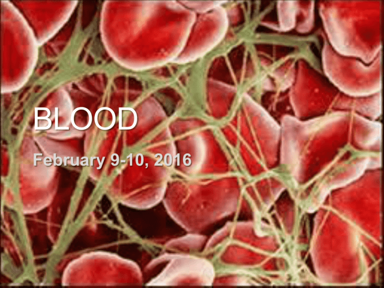
BLOOD February 9-10, 2016 What is the function of blood? Transports material throughout the body, including: • • • • • • • • Water Oxygen Carbon dioxide Nutrients – glucose, fatty acids, amino acids, vitamins) Electrolytes Hormones Immune cells HEAT!! Why can do we you need die oxygen? from blood loss? Lack We need of oxygen oxygen in to heart do cellular and brain respiration – to convert glucose into ATP What is blood made of? plasma ~55%, platelets & white blood cells ~1%, red blood cells ~44% Blood Components Plasma (~55% TOTAL VOLUME) • ~92% water • ~7% proteins • • • • Albumin (osmotic balance) Fibrinogen (clotting) Antibodies (immune) Hormones (regulation) • ~1% other solutes • Electrolytes (osmotic and pH balance, regulating membrane permeability) • Nutrients (glucose, fatty acids, amino acids) • Oxygen • Carbon dioxide Blood Components Erythocytes (~44% TOTAL VOLUME) • aka red blood cells • tiny! • lack a nucleus, have few organelles • contain hemoglobin – an iron- containing protein that reversibly binds to oxygen (and a small amount of CO2) How does the structure of erythrocytes facilitate their function? - very small to fit through small capillaries - small size and lack of nucleus, most organelles designed to maximize oxygen transport Blood Components Leukocytes (< 1% TOTAL VOLUME) • aka white blood cells Blood is typically stained with chemicals that bind to certain proteins to aid in identification of cells • fight pathogens (bacteria, viruses, parasites) and cancer • can leave the blood stream to go to infected tissue – diapedesis • summoned to damaged areas by chemotaxis, move by ameboid motion • Blood Components Leukocyte Types • Granulocytes – contain granules in cytoplasm and unusually shaped nuclei a) Neutrophils – most numerous leukocyte; phagocytic; abundant during bacterial infection b) Eosinophils – kill parasitic worms and increase during allergy attacks c) Basophils assist in inflammatory response Blood Components Leukocyte Types • Agranulocytes – lack granules in cytoplasm and have normal nuclei a) Lymphocytes – 2nd most numerous; include B and T cells; produce antibodies and attack infected cells b) Monocytes – engulf and destroy pathogens Blood Components Platelets (<1% TOTAL VOLUME) • cell fragments • involved in blood clotting Review 1: What is blood made of? Identify each component, and justify your response! A B C D Review 2: Structure & Function Identify the component of blood that transports each material, and justify your response! • • • • • • • • Water Oxygen Carbon dioxide Nutrients – glucose, fatty acids, amino acids, vitamins) Electrolytes Hormones Immune cells HEAT!! Review 3: Compare & Contrast Turn & Talk – 2 min Compare & contrast the structure and function of erythrocytes and leukocytes Hemostasis Hemostasis is the process of blood clotting • Occurs when small blood vessel (capillary) is damaged • Clot seals the blood vessel until the it regenerates • Occurs in just 3-6 minutes Process of Hemostasis Three major events occur, all beginning the moment the vessel is damaged: 1) Vasoconstriction 2) Platelet plug formation 3) Coagulation of blood Coagulation takes longer, and is completed after vasoconstriction and platelet plug formation occur Process of Hemostasis Watch me! This is a simplified overview of the clotting cascade Hemostasis: Review & Connections What is the recommended way to treat a bleeding wound (until you see a doctor)? Why? Gauze and pressure The gauze acts much like collagen fibers – provide a rough surface that helps activate platelets. Pressure manually constricts blood vessels and also increases the release of thromboplastin, which helps initiate coagulation. Never remove gauze or a bandage from an actively bleeding wound. Why? Removing the bandage will remove both clotting factors and the beginnings of a platelet plug or blood clot, causes an increase in bleeding. Hemostasis: Review & Connections How is blood clotting an example of positive feedback? Which part(s) of the process best exemplify positive feedback? (Turn & Talk – 2 min) Platelet plug formation Activated platelets release chemicals that cause more platelets to activate, until a large number of platelets clump together forming a plug. Hemostatic Disorders – Blood Clots A thrombus is a blood clot that forms in an unbroken vessel. A large thrombus may block blood flow, causing tissue death. An embolus is a blood clot that forms then breaks away and floats freely in the blood vessels. An embolus may then lodge in a capillary and block blood flow. coronary thrombosis cerebral embolism – pulmonary embolism – Hemostatic Disorders – Blood Clots A thrombus is a blood clot that forms in an unbroken vessel. A large thrombus may block blood flow, causing tissue death. An embolus is a blood clot that forms then breaks away and floats freely in the blood vessels. An embolus may then lodge in a capillary and block blood flow. coronary thrombosis – in heart cerebral embolism – in brain pulmonary embolism – in lungs Hemostatic Disorders – Blood Clots Causes of thrombus • Injury to blood vessel or build-up of fatty plaques Both create rough surfaces inside vessel, which may activate platelets • Poor blood circulation Clotting factors may accumulate Immobility increases the risk of deep vein thrombus in legs! Hemostatic Disorders – Blood Clots Blood thinners (such as warfarin, aspirin, and heparin) can be used to prevent thrombus Aspirin – blocks thromboxane reduces formation of platelet plug Wafarin – blocks production of certain clotting factors reduces coagulation interrupting clotting cascade Heparin – helps inactivate thrombin reduces coagulation by preventing conversion of fibrinogen to fibrin Review your notes – how exactly does each of these reduce clotting? Hemostatic Disorders - Hemophilia Causes • lack of one or more clotting factors • Recessive sex-linked trait (more common in men) Symptoms • • • • Prolonged bleeding even from minor injuries Excessive bruising Bruised and swollen joints Excessive clumsiness and falling Treatment • Intravenous injection of clotting factors • Donated plasma • Synthetic clotting factors Hematopoiesis Hematopoiesis is the process of blood formation. • Occurs in the red bone marrow Where is this found? In babies, nearly all bones have red marrow In adults, just the flat bones and epiphyses • All blood cells and platelets derive from hemocytoblast stem cells Erythrocyte life cycle and production Develop in red marrow (for 3-5 days) Eject nucleus, then enter blood stream. Red blood cells life for 3-4 months Digested by phagocytes Production is controlled by hormone erythropoietin. Erythropoietin release is stimulated by low levels of O2 in blood. Erythrocyte life cycle and production Develop in red marrow (for 3-5 days) Eject nucleus, then enter blood stream. Red blood cells life for 3-4 months Digested by phagocytes Why is there no hormone to decrease RBC production? - High numbers of RBCs don’t cause major problems - High levels are temporary. RBC levels will decline due to death of cells. Erythrocyte life cycle and production Develop in red marrow (for 3-5 days) Eject nucleus, then enter blood stream. Red blood cells life for 3-4 months Digested by phagocytes Why do world-class athletes train at high altitude before major competitions? - High altitude has lower oxygen levels, which stimulates the production of erythrocytes - The high levels of erythrocytes will persist for a while after leaving high altitude Closure • What were our objectives, and what did you learn about them? • What was our learner profile trait and how did we exemplify it? • How does what we did today address our unit question? Exit Ticket Make mini posters illustrating the following terms. The posters should • be in color • prominently feature the term • have both a picture • a definition / explanation of the term Exit Ticket Neutrophil hemostasis Fibrin hemophilia Basophil Serotonin fibrinogen agglutination Eosinophil Platelet plug Platelet factor 3 Albumin Lymphocyte Thromboplastin coagulation Antibody Monocyte Prothrombin Clotting factor Antigen platelet thrombin vasoconstriction Plasma hemocytoblast erythropoietin Thrombus Erythrocyte hemotopoeisis Prothrombin activator embolus leukocyte
