The Neuron - Central Web Server 9
advertisement
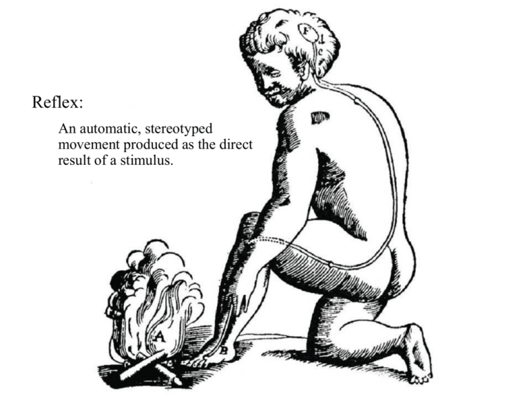
Reflex: An automatic, stereotyped movement produced as the direct result of a stimulus. The Neuron number: 10 billion to a trillion 10,000 connections each parts: dendrites cell body (or "soma") axon terminal endings (or terminal buttons) Questions… 1) how does a neuron "fire"? (what is the nerve impulse?) 2) how does it cause the next neuron to fire? (how does it communicate?) nerve impulse = ACTION POTENTIAL: 1) start with electrical RESTING POTENTIAL: inside of cell is 70 mV more negative than outside due to Cl- ions inside and Na + ions outside (so RESTING POTENTIAL is -70 mV). 2) stimulation of neuron lets in Na+ ions, which makes the inside more positive: -70,-69,-68,-67... ACTION POTENTIAL (continued)… 3) when enough Na+ ions get in for the potential to be reduced to -55 mV, suddenly the doors (ion gates) to the cell membrane are flung open allowing Na+ to rush in. 4) so much Na+ enters that the potential doesn't just go to 0 -- it shoots all the way up to +40 mV, so the inside is now positive relative to the outside (the ACTION POTENTIAL) Action potential (conclusion) 5) ion pumps work to reduce potential back to -70 mV by pushing positive ions out (actually K+ because Na+ goes out slower; then ANOTHER pump takes Na+ back out and puts K+ back in) ACTION POTENTIAL (continued)... • note that -55mV is a threshold: below that voltage there is no action potential - firing is "all-or-none" • more intense stimulation doesn't cause a more intense action potential -- just more frequent ones (up to 1000/sec!), and in more neurons ACTION POTENTIAL (continued)… • action potential travels down length of axon by depolarizing neighboring areas • travels NOT at speed of electrical current in wire, but rather at about 50 to 100 m/sec communication across the synapse: NEUROTRANSMITTERS 1) synapse is gap between two neurons (the presynaptic and the postsynaptic neurons); terminal endings of presynaptic neuron relay impulse to dendrites of postsynaptic neuron NEUROTRANSMITTERS (continued) 2) terminal buttons contain little sacs ("vesicles") of chemicals ("neurotransmitters"); at action potential, vesicles burst and release neurotransmitters into synapse 3) receptor molecules on membrane of dendrite are like little locks to be opened: neurotransmitters are the keys, and this is what opens ion gates to allow Na+ inside in the first place NEUROTRANSMITTERS (continued…) 4a) neurotransmitters may open a gate to let Na+ inside: excitatory (more likely to fire) because potential is getting smaller, toward -55 4b) or they may open a gate that pushes positive K+ ions out: inhibitory (less likely to fire) because potential is getting larger (e.g., -70, -71, -72...) Reciprocal Inhibition NERVOUS SYSTEM ("NS") central - “center” peripheral - “outside of center” somatic - “body” autonomic - “self rule” sympathetic - excited states parasympathetic - vegetative, calm states central NS (brain, spinal cord) peripheral NS (everything else) somatic NS (muscles, senses) autonomic NS (vital functions: heart rate, breathing, digestion, reproduction) sympathetic NS - arousal: mobilizes for emergency (speeds heart and lungs, inhibits digestion and sexual function) parasympathetic NS - calm: conserves energy (slows heart and lungs, etc.) Organization of the Nervous System BRAIN: bottom to top (=inside to outside=old to new) hindbrain: medulla - breathing, heartbeat, blood circulation pons - arousal and attention cerebellum - integration of muscles to perform fine movements, but no coordination / direction of these movements; balance cat transected above hindbrain: can move but not act midbrain: forms movements into acts; controls whole body responses to visual and auditory stimuli cat transected above midbrain can act, but without regard to environment: without purpose forebrain… BRAIN (continued) forebrain: thalamus - sensory and motor relay center (to various cerebral lobes) hypothalamus - controls responses to basic needs (food, temperature, sex) basal ganglia - regulates muscle contractions for smooth movements limbic system - memory (hippocampus) and emotion (amygdala) cerebral cortex (or “neocortex”) - four lobes (frontal, parietal, occipital, temporal); seat of "higher" intellectual functions cat transected above limbic system: acts normal, with purpose - but clumsy CEREBRAL HEMISPHERES (or CEREBRUM): corpus callosum: connects hemispheres each hemisphere controls OPPOSITE SIDE of body cerebral cortex (= skin or bark): 1 to 3 mm thick; 2 or 3 ft square if flattened out higher motor, sensory, and intellectual functions Corpus Callosum large band of neural fibers largest "commissure" (or pathway between hemispheres) of the brain • but not the ONLY one! FOUR LOBES of cortex: frontal lobe: planning; social behavior; motor control front of brain parietal lobe: somatosensory (sense of touch) on top and toward back of brain occipital lobe: vision back of brain temporal lobe: hearing; memory side of brain The Cerebral Cortex Motor and Somatosensory Cortex TWO GENERAL RULES of cortical function: 1. Left Hemisphere: Right Hemisphere: language spatial abilities 2. Front: expression / actions / plans Back: reception / perceptions / interpretations DAMAGE TO NON-PRIMARY ("ASSOCIATION") CORTEX: pre-frontal lesions: loss of planning, moral reasoning, sensitivity to social context or... loss of initiation of action, deliberation apraxia ("no doing"): failure in sequencing components of actions; inability to organize movements FRONTAL - lesions just forward of motor cortex NOT paralysis, as from motor cortex lesion agnosia ("no knowing"): - deficit in interpreting, categorizing, labeling, knowing OCCIPITAL (visual) or TEMPORAL (auditory) lesions sensory systems themselves (e.g., eyes) are okay DAMAGE TO NON-PRIMARY ("ASSOCIATION") CORTEX (continued): neglect: RIGHT hemisphere (PARIETAL) damage causes inattention to whole left side aphasia: LEFT hemisphere (FRONTAL or TEMPORAL) damage causes deficits in language function... APHASIA: disorder of language left hemisphere brain lesions essentially apraxia (from frontal lesion) or agnosia (from back lesion) of language expressive aphasia: cannot produce speech lesion to BROCA'S AREA (frontal assoc. area) receptive aphasia: cannot understand speech - and consequently cannot produce speech! lesion to WERNICKE'S AREA (temporal assoc. area) SPLIT BRAIN STUDIES sever corpus callosum to reduce severity of seizures leaves patient mostly normal, but with left and right brain independent in subtle ways note visual pathways: left side of each eye sends info to left hem. right side of each eye sends info to right hem. result: left visual field goes to right hem. right visual field goes to left hem. SPLIT BRAIN STUDIES (continued) experiment on split brain patient: patient looks straight ahead; picture flashed quicker than eyes can move; ask "what did you see?" picture of cup on right: LH says "cup" picture of spoon to left: LH says"nothing" BUT when told to reach for that object with the left hand, RH grabs spoon ask "what is it?" and LH guesses "pencil" (and RH may frown at that) Apparatus for Studying Split-Brain Patients
