10-2 Cell Division - Lincoln Park High School
advertisement

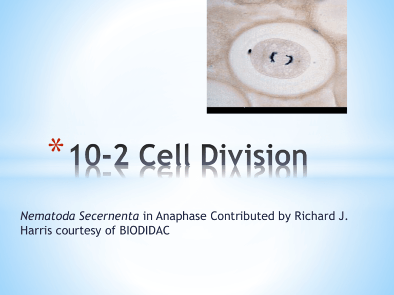
* Nematoda Secernenta in Anaphase Contributed by Richard J. Harris courtesy of BIODIDAC 1. 2. Mitosis- division of the cell nucleus Cytokinesis- division of the cytoplasm * SEM image of Chromosomes I. 1. Chromosomes- contain DNA (genetic info) which is passed from parent to offspring. 2. Before a cell divides each chromosome is copied 3. Structure of the chromosome: * Two sister chromatids; are identical * Centromere- holds the sister chromatids together resulting in an x-shaped c’some Sister chromatids Centromere * When the cell divides, the chromatids separate. * Each new cell gets one chromatid. Cell division Micrograph II. 1. Cell Cycle- the series of events that takes place in a cell leading to its division & replication 2. During the cell cycle: * a cell grows * prepares for division 3. Divides to form two daughter cells, each of which begins the cycle again 4. The cell cycle consists of four phases: *G1 (First Gap Phase) *S Phase (Synthesis) *G2 (Second Gap Phase) *M Phase (Mitosis) III. 1. Interphase- the first stage of the cell cycle. During this phase the cell: * Grows * Makes new proteins & organelles * C’somes are replicated * DNA is made 2. Interphase has 3 phases: G1, S, & G2 3. During G1, the cell: * increases in size * synthesizes new proteins and organelles 4. During the S phase: * chromosomes are replicated * DNA synthesis takes place 5. Once a cell enters the S phase, it usually completes the rest of the cell cycle 6. The G2 Phase (Second Gap Phase) * organelles & molecules required for cell division are produced * Once G2 is complete, the cell is ready to start the M phase— Mitosis IV. 1. Mitosis has 4 phases: *Prophase *Metaphase *Anaphase *Telophase 2. Centrioles- barrel shaped structures that are made of groupings of microtubules 3. Spindle- a fanlike microtubule structure that helps separate the chromosomes. 4. Chromatin- a combination of DNA and proteins that make up c’somes 5. Prophase *The 1st & longest phase of mitosis *The centrioles separate & end up on opposite sides of the nucleus in an area called the centromere A cell in prophase of mitosis. (An epithelial cell in prophase of mit treated with the anti-cancer drug taxol. Chromosomes are labeled Taricha granulosa (newt) lung.) *Chromatin condenses into chromosomes. *The nuclear envelope breaks down. Observing Mitosis with Fluorescence Microscopy. Prophase. © 1995-2013 by Michael W. Davidson and The Florida State University. 6. Metaphase *2nd phase of mitosis *The chromosomes line up across the center of the cell *Microtubules connect the centromere of each chromosome to the poles of the spindle. Observing Mitosis with Fluorescence Microscopy. Metaphase. © 1995-2013 by Michael W. Davidson and The Florida State University. 7. Anaphase *3rd phase of mitosis *the duplicated centromeres of each pair of sister chromatids separate *the now-daughter chromosomes begin moving toward opposite poles until they have separated into two groups. Observing Mitosis with Fluorescence Microscopy. Anaphase. © 1995-2013 by Michael W. Davidson and The Florida State University 8. Telophase *The last phase of mitosis *Chromosomes gather at opposite ends of the cell *A new nuclear envelope forms around each bunch of chromosomes. Observing Mitosis with Fluorescence Microscopy. Telophase. © 1995-2013 by Michael W. Davidson and The Florida State Universi 9. Cytokinesis *the cytoplasm pinches in half. *Each daughter cell has an identical set of duplicate chromosomes. Actin (blue) and microtubules (orange) at the end of cytokinesis in a gr urchin zygote. University of Washington Center for Cell Dynamics 10. *structure known as the cell plate forms midway between the divided nuclei *The cell plate gradually develops into a separating membrane. *A cell wall then begins to appear in the cell plate. Cell plate Cell wall 11. *A cleavage furrow forms-An indention formed by microfilaments *The pulling of the filaments separates the cytoplasm of the daughter cells 12. *95% of the cell cycle is spent in interphase *A typical rapidly dividing human cell with a total cycle time of 24 hours: *the G1 phase might last about 11 hours *S phase about 8 hours *G2 about 4 hours *and M about 1 hour. * *Page 249 “Analyzing DataThe Life Span of Human Cells” *Read the paragraph and answer questions 1-4 *


