Unit 2 Phylum Porifera PPT
advertisement
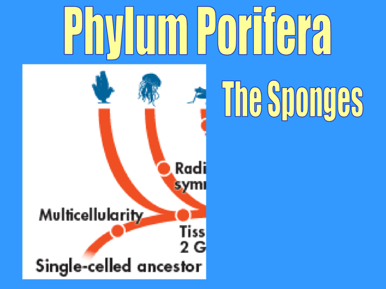
The Cladogram of Animals Main Topics I. II. III. IV. General Characteristics Sponge Anatomy- The Basics Feeding and Reproduction Types of Sponges I. General Characteristics • Porifera- Latin for “pore-bearing;” contain pores all over their body • Simplest multicellular animals (most primitive) • Invertebrates General Characteristics (continued) Levels of Organization Specialized cells, no true tissues Body Symmetry Asymmetrical Germ Layers Absent Body Cavity - Embryological Development - Segmentation Absent Cephalization Absent General Characteristics (continued) • Can be better described as a colony of single celled organisms living together • Over 10,000 species; all aquatic and 99% marine • All are filter feedersfilter small particles of food from water that passes through the organism General Characteristics • Some defend themselves with chemicals and are focus of biotech research companies • Adults are sessile- live permanently attached to a substrate (base material like a reef or a crab) and are not able to move on their own Current Event Homework • Find an article about how Poriferans (sponges) are used in biomedical research. • Due Friday Requirements: 1. Print a copy of the article 2. On a separate paper, write a summary of the article. 3. Counts as a double Homework assignment II. Sponge Anatomy- The Basics • “Vaselike” general shape • Osculum- one large excurrent pore where water flows out of • Ostium- many tiny, incurrent pores where water flows in (plural Ostia); found throughout the body • Atrium- inner filter chamber Oscula Holdfast- root-like structure that anchors aquatic sessile organisms, such as seaweed and sponges II. Sponge Anatomy- The Basics • Wall of the body is made of 3 cell layers (remembernot true germ layers) Outer Layer • Lined with Pinacocytesflattened cells packed tightly together that forms the outer boundary Sponge Anatomy- The Basics (continued) Gelatinous layer • Middle layer that forms the skeleton and support for the sponges shape • Amebocytes- cells carry food and oxygen to cells of each layer • Spicules- needle-like structures made of silica or calcium • Spongin- tough protein fibers Sponge Anatomy- The Basics (continued) Inner Layer • Lined with choanocytes (collar cells)- specialized cell that has a flagella. These cells help control water flow into and out of the cell. • The flagella beats to create water current and the collar traps food particles. III. Feeding and Reproduction • Mostly feed on plankton and organic molecules Feeding and Reproduction (continued) 1. Water enters through ostia. 2. Water is pumped into the atrium. 3. Collar cells trap food particles with flagella. 4. Amebocytes take food to other cells within the sponge. 5. Water leaves through the osculum. Figure 12.07 Figure 12.09 Feeding • http://www.youtube.com/watch?feature=pl ayer_detailpage&list=PL6174CA65320048 CE&v=RmPTM965-1c Feeding and Reproduction (continued) Sponges reproduce both asexually and sexually. Asexual Branches or buds break off & grow into separate sponges (budding) Sexual Sponges produce both male & female gametes to produce larva. Larvae floats off and establishes itself on a new base. Asexual Repro Budding • http://www.youtube.com/watch?v=4JIytOLQ18&feature=player_detailpage IV. Types of Sponges • There are 3 major Classes of sponge. • Sponges are classified mainly based on what their skeleton is made of in the gelatinous layer. • Common names are mainly based on their shapes • Non-synthetic cleaning sponges have spongin skeletons; Synthetic sponges are made based off spongin skeletons
