46.5: Interplay of tropic and sex hormones regulates - APBio10-11
advertisement
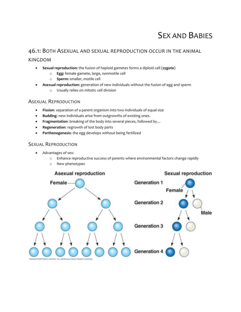
SEX AND BABIES 46.1: BOTH ASEXUAL AND SEXUAL REPRODUCTION OCCUR IN THE ANIMAL KINGDOM Sexual reproduction: the fusion of haploid gametes forms a diploid cell (zygote) o Egg: female gamete, large, nonmotile cell o Sperm: smaller, motile cell Asexual reproduction: generation of new individuals without the fusion of egg and sperm o Usually relies on mitotic cell division ASEXUAL REPRODUCTION Fission: separation of a parent organism into two individuals of equal size Budding: new individuals arise from outgrowths of existing ones. Fragmentation: breaking of the body into several pieces, followed by… Regeneration: regrowth of lost body parts Parthenogenesis: the egg develops without being fertilized SEXUAL REPRODUCTION Advantages of sex: o Enhance reproductive success of parents where environmental factors change rapidly o New phenotypes REPRODUCTIVE CYCLES AND PATTERNS Ovulation: release of mature eggs Hermaphroditism: each individual has both male and female reproductive systems 46.2: FERTILIZATION DEPENDS ON MECHANISMS THAT BRING TOGETHER SPERM AND EGGS OF THE SAME SPECIES Fertilization: union of sperm and egg o External: female releases eggs into the environment where the male fertilizes them Allows mate selection, triggering release of both sperm and eggs, increase probability of fertilization o Internal: sperm deposited in or near the female reproductive tract Enables sperm to reach egg effectively Copulation, sophisticated and compatible reproductive systems o Spawning: individuals in the same area release their games into the water at the same time ENSURING THE SURVIVAL OF OFFSPRING Species generally make more than necessary for survival o External fert: produce large # of gametes small fraction of survivors o Internal fert: produce fewer, protect and care for young. Marsupials: mammary glands Eutherian mammals: fetuses remain in the uterus, nourished by mother’s blood supply through the placenta Offspring are fed by adult birds and mammals Parental care important to survival GAMETE PRODUCTION AND DELIVERY Gonads: organs that produce gametes in most animals Spermatheca: sac in which sperm may be stored for extended periods Cloaca: reproductive systems have a common opening to the outside 46.3 REPRODUCTIVE ORGANS PRODUCE AND TRANSPORT GAMETES FEMALE REPRODUCTIVE ANATOMY OVARIES Follicles: made up of oocytes, partially developed eggs Oogenesis: formation and development of an ovum Corpus luteum: residual follicular tissue that grows within the ovary, secretes additional estrogen, as well as progesterone OVIDUCTS AND UTERUS Oviduct: fallopian tube, extends from uterus to each ovary Uterus: womb, thick muscular organ that can expand during pregnancy to accommodate for big fetus Endometrium: lining of the uterus Cervix: opening into the vagina VAGINA AND VULVA Vagina: muscular but elastic chamber that is the site for insertion of the penis and deposition of sperm Vulva: external female genitals Labia majora: fatty ridges that protects the vulva Labia minora: slender skin folds that border the urethra and the vaginal opening Hymen: covers the vagina opening at birth until sex/physical activity breaks it Clitoris: short shaft supported by the glans, covered by a small hood of skin, the prepuce o During arousal, these, the vagina and the labia minora all engorge with blood MAMMARY GLANDS Mammary glands: present in both sexes, but normally produce milk in only females o Important to reproduction MALE REPRODUCTIVE ANATOMY TESTES Testes: consist of many highly coiled tubes surrounded by several layers of connective tissue Seminiferous tubules: where sperm form, make up the testes Leydig cells: scattered between the seminiferous tubules, produce testosterone Scrotum: fold of the body wall that maintains the testis temperature DUCTS Epididymis: where the sperm goes after the seminiferous tubes, 6-ft long Ejaculation: sperm are propelled from each epididymis through the muscular duct, vans deferens, which extends around and behind the bladder, where it joins a duct from the seminal vesicle, forming a short ejaculatory duct. Urethra: outlet tube for both excretory system and reproductive system ACCESSORY GLANDS Semen: fluid that’s ejaculated Seminal vesicles: contribute 60% of the semen Prostate gland: secretes products directly into the urethra through several small ducts, source of some of the most common medical problems of men over 40 Bulbourethral glands: small glands along the urethra below the prostate, makes precum PENIS Penis: contains the urethra, three cylinders of spongy, erectile tissue HUMAN SEXUAL RESPONSE Vasocongestion: filling of tissue with blood Myotonia: increased muscle tension Coitus: sexual intercourse The Stages of Sex o Excitement: vasocongestion, myotonia o More excitement: plateau phase: continue as a result of direct stimulation of the genitals o Orgasm: rhythmic, involuntary contractions of reproductive structures in both sexes o Resolution: reverses responses of earlier stages 46.4: TIMING AND PATTERN OF MEIOSIS DIFFERS FOR MALES/FEMALES Gametogenesis: production of gametes o Sperm are motile and small o Eggs are large and stay stationary, have to provide initial food stores for the embryo Spermatogenesis: formation and development of sperm, continuous and prolific in adult males o Cell division and maturation occurs throughout the seminiferous tubules coiled within testes Oogenesis: development of mature oocytes, prolonged process in female o Immature eggs form in the ovary, but do not complete their development until later Differences o Spermatogenesis: all four products form mature gametes Oogenesis: cytokinesis during meiosis is unequal, almost all cytoplasm is segregated to a single daughter (secondary oocyte) polar bodies degenerate o Spermatogenesis: occurs throughout adolescence and adulthood Oogenesis: mitotic divisions are thought to be complete before birth, production of mature gametes ceases at age 50 46.5: INTERPLAY OF TROPIC AND SEX HORMONES REGULATES MAMMALIAN REPRODUCTION Principle sex hormones are steroid hormones o Males: androgens (testosterone) o Females: estrogens and progesterones (estradiol) Sex hormones serve many functions o Responsible for male vocalizations o Development of primary sex characteristics of males, structures directly involved in reproduction (seminal vesicles and other ducts) o Males: voices deepen, facial and pubic hair to grow, mucsles to grow Promote sex drive and general aggressiveness o Females: influences female sexual behavior, induces fat (breasts and hips), increases water retention and alters calcium metabolism HORMONAL CONTROL OF THE MALE REPRODUCTIVE SYSTEM GnRH from Hypothalamus makes FSH (Sertoli Cells) and LH (Leydig cells) o FSH Spermatogenesis (inhibin) o LH testosterone Spermatogenesis REPRODUCTIVE CYCLE OF FEMALES Menstruation: cyclic shedding of endometrium from uterus Menstrual cycle (uterine cycle): changes in the uterus Ovarian cycle: changes in the ovaries OVARIAN CYCLE 1. 2. 3. 4. 5. 6. 7. 8. Release of GnRH from hypothalamus Stimulates anterior pituitary to secrete FSH and LH FSH stimulates follicle growth Cells of growing follicles start to make estradiol follicular phase, follicles grow and oocytes mature Estradiol levels rise b/c excreted by growing follicle FSH and LH levels increase, LH triggers ovulation LH surge causes rupture of the follicle wall and release of the secondary oocyte Luteal phase: follows ovulation: LH stimulate follicular tissue left behind to transform into corpus luteum secretes progesterone and estradiol promote thickening of the endometrium a. Corpus luteum degenerates, sharp decrease in estradiol and progesterone concentrations UTERINE (MENSTRUAL) C YCLE 9. Proliferative phase: coordinated with follicular phase, estradiol and progesterone secreted by corpus luteum stimulate continued development and maintenance of uterine lining a. Secretory phase: coordinated w/ luteal phase 10. Menstrual flow phase: disintegration of the corpus luteum, estradiol and progesterone levels drop and blood Endometriosis: disorder: some of the cells of the uterine lining migrate to an abnormal location (ectopic- abnormal location) ectopic tissue swells and breaks down each ovarian cycle, resulting random bleeding and other shitty shit MENOPAUSE Menopause: cessation of ovulation and menstruation- ovaries lose responsiveness to FSH and LS, resulting in a decline in estradiol production Why do we have it? Nobody knows for sure. Awesome, bio book. Thanks for wasting 30 seconds of my life. MENSTRUAL VS ESTROUS C YCLES Estrous cycles: in the absence of pregnancy, the uterus reabsorbs the endometrium (FUCK YOU WHY DON’T WE HAVE THAT?!) 46.6: IN PLACENTAL MAMMALS, AN EMBRYO DEVELOPS FULLY WITHIN A MOTHER’S UTERUS CONCEPTION, EMBRYONIC DEVELOPMENT AND BIRTH Conception: Your bed is the scene of the crime: when sperm fuses with an egg in the oviduct o Cleavage: 24 hours, zygote divides o Blastocyst: sphere of cells surrounding central cavity 1 weekish o + a few days: embryo implants into the endometrium, starts developing into a fetus o Human chorionic gonadotropin (hCG): acts like LH and maintains secretion of progesterone and estrogens by corpus luteum through the first months of pregnancy o Pregnancy/gestation: condition of carrying one+ embryos in the uterus, averages 266 days for humans Sometimes pregnancies spontaneously terminate o Chromosomal/developmental abnormalities o Ectopic pregnancy (egg lodges in the oviduct, may rupture the oviduct and result in internal bleeding, etc) FIRST TRIMESTER Weeks 2-4: Embryo obtains nutrients from the endometrium o Trophoblast: outer layer of the blastocyst, grows outward and mingles with endometrium in this period o Placenta: disk-shaped organ, containing embryonic and maternal blood cells Supplies nutrients, provides immune protection, exchanges respiratory gases, disposes of wastes for the embryo, blood travel o Monozygotic (one egg) twins – identical o Dizygotic (two eggs) twins – fraternal o Organogenesis: development of body organs 8 weeks: all major structures are present, embryo is now a fetus Heart beats by 4th week, heartbeat detected at 8-10 weeks Mother is… o Makin’ more progesterone cervix plus with mucus, maternal placenta grows, uterus gets larger and ovulation/menstruation stops o Momma is getting sick, getting bigger and having larger boobs SECOND TRIMESTER Pregnancy is obvious, baby starts moving hCG levels declining, corpus luteum and placenta takes over production of progesterone THIRD TRIMESTER Fetus grows, mother feels the pain Labor: process by which childbirth occurs; series of strong, rhythmic uterine contractions during the three stages of labor bring about birth (parturition) o Dilation, opening up and thinning of the cervix o Expulsion/delivery of the baby (ow ow ow ow) o Delivery of the placenta Hormones o Regulators (prostaglandins) and hormones (estradiol and oxytocin) Lactation: hypothalamus signals anterior pituitary to secrete prolactin, which stimulates mammary glands to produce milk o Suckling also triggers release of oxytocin, which triggers release of milk CONTRACEPTION AND ABORTION Contraception: deliberate prevention of pregnancy o Rhythm method: temporary abstinence o Natural family planning: refraining from intercourse with conception is most likely o Coitus interruptus: withdrawal method o Barrier methods: condoms, diaphragms, female condom, cervical cap o Sterilization: tubal ligation (females) cauterizing/tying oviducts Vasectomy: cutting/tying off a small section of each vas deferens o IUD: implanted, interferes with fertilization and implantation o Hormonal contraceptives: birth control pills Synthetic estrogen and progestin Mimics negative feedback of the ovarian cycle, stopping the release of GnRH and FSH and LH blocks ovulation Progestin: causes thickening of woman’s cervical mucus so that it blocks sperm from entering the uterus, decreases frequency of ovulation and changes endometrium o Abortion: termination of pregnancy in process mifepristone: drug that allows a woman to terminate pregnancy nonsurgically MODERN REPRODUCTIVE TECHNOLOGIES Ultrasound, amniocentesis and chorionic villus sampling Fetal blood cells genetic disorders TREATING INFERTILITY Assisted reproductive technologies: procedures that involve surgically removing eggs, stimulating them with hormones and returning them to her body In vitro fertilization: oocytes are mixed with sperm in culture dishes and fertilized eggs are incubated until they have formed at least 8 cells uterus Intracytoplasmic sperm injection: head of spermatid/sperm is drawn into a needle and injected directly into an oocyte. CHAPTER 47: PREVIEW Cytoplasmic determinants: molecules that are placed into the egg by the mother, as the zygote divides, differences arise between early embryonic cells due to uneven distribution of cytoplasmic determinants Cell differentiation: process of cell specialization in structure and function Morphogenesis: process by which an organism takes shape and differentiated cells occupy their appropriate locations Model organism: species that lends itself to the study of a particular question, that is representative of a larger group and is easy to grow in a lab 47.1: AFTER FERTILIZATION , EMBRYONIC DEVELOPMENT PROCEEDS THROUGH CLEAVAGE, GASTRULATION AND ORGANOGENESIS Cleavage: cell division creates a blastula Gastrulation: rearranges the blastula into a three-layered embryo, the gastrula Organogenesis: create rudimentary organs FERTILIZATION Model: Sea urchins ACROSOMAL REACTION Eggs are fertilized externally; the egg is coated with jelly that exudes soluble molecules that attract the sperm Acrosomal reaction: begins when a specialized vesicle at the tip of the sperm, the acrosome, discharges hydrolytic enzymes o The enzymes digest the jelly coat, enabling a sperm structure to elongate, penetrating the coat o Molecules of the protein on the tip of the acrosomal process adhere to specific sperm receptor proteins that extend from the egg plasma membrane through the surrounding meshwork of extracellular matrix (vitelline layer) lock and key: recognition of molecules ensures that eggs will be fertilized only by sperm of the same species o Contact of tip of the acrosomal process with the egg membrane leads to fusion of sperm and egg plasma membranes Sperm then enters the egg cytoplasm Change in membrane potential fast block to polyspermy, only one sperm can fertilize the egg CORTICAL REACTION b/c membrane depolarization only lasts for a short time cortex: rim of cytoplasm just beneath the plasma membrane cortical granules: vesicles that fuse with egg plasma membrane, initiating cortical reaction o contain a lot of molecules that are secreted into the perivitelline space (between the plasma membrane and the vitelline layer) o o o the enzymes and other macromolecules push the vitelline layer away from the egg and hardens the layer, creating a protective fertilization envelope that resists the entry of additional sperm slow block to polyspermy: takes longer, but is a longer-term ordeal due to high concentration of Ca2+ ions ACTIVATION OF THE EGG egg activation: increase in rates of cellular respiration and protein synthesis by the egg, caused by sharp rise in Ca2+ concentration FERTILIZATION IN MAMMALS we have internal fertilization the egg is cloaked by follicle cells released along w/ the egg during ovulation, the sperm must travel through this layer of follicle cells before it reaches the zona pellucida, the extracellular matrix of the egg o zona pellucida functions as a sperm receptor CLEAVAGE cleavage: rapid cell division, cells carry out the synthesis and mitosis phases of the cell cycle, but skip the growth phases and little protein synthesis blastomeres: smaller cells that make up the blastula blastocoel: fluid-filled cavity of the blastula blastula: hollow ball of cells some animals have definite polarity, established as the egg developed within the mother during oogenesis o yolk: stored nutrients, influences the pattern of cleavage vegetal pole: more concentration of yolk animal pole: yolk concentration decreases towards this end, the polar bodies of oogenesis bud from the cell animal hemisphere is dark gray because it has dark-colored melanin granules o determines anterior/posterior (head-tail) of the embryo lack of melanin granules in the vegetal hemisphere allows the yellow color of the yolk to be visible cortical rotation: movement where the plasma membrane and associated cortex rotate with repsect to eh inner cytoplasm o animal hem cortex moves toward the vegetal inner cytoplasm on the side where the sperm entered o allows molecules in the unpigmented vegetal cortex on the side opposite sperm entry to interact with molecules in the inner cytoplasm of the animal hemisphere, which activates previous inactive proteins of the vegetal cortex which leads to formation of cytoplasmic determinants that will affect gene expressions in the cell gray crescent: light gray region of cytoplasm that was previously covered by the pigmented animal cortex near the equator of the egg serves a marker for future dorsal side of the embryo holoblastic cleavage: complete cleavage, cleavage furrow passes all the way through the cells meroblastic cleavage: incomplete division of yolk-rich egg cells (birds, reptiles, fishes and insects) GASTRULATION gastrulation: morphogenetic process, taking up new locations that will allow the later formation of tissues and organs gastrula: embryo is called this, two-layered body plan three layered embryo with primitive digestive tube germ layers: collective name for three layers produced by gastrulation ectoderm: outer layer endoderm: lines embryonic digestive tract mesoderm: fills space between endoderm and ectoderm S EA URCHIN 1. 2. 3. 4. 5. blastula gastrulation at vegetal pole (invagination) archenteron (inside the gastrula) blastopore: open end of the archenteron produces an embryo with a primitive digestive tube with three germ layers blastula consists of single-layered ciliated cells surrounding blastocoel, gastrulation begins with migration of mesenchyme cells from vegetal pole into the blastocoel vegetal pole invaginates, mesenchyme cells migrate throughout the blastocoel endoderm cells form the archeteron, new mesenchyme cells at the tip of the tube begin to send out thin extensions (filopodia) towards the blastocoel wall filopodia then contract, dragging the archenteron across the blastocoel fusion of archenteron with the blastocoel wall completes formation of the digestive tube F ROG 1. 2. 3. Gastrulation begins when a small indented crease, the blastopore, appears on the dorsal side of the late blastula. The crease is formed by cells changing shape and pushing inward from the surface. Sheets of outer cells then roll inward over the dorsal lip (involution) and move into the interior, where they will form endoderm and mesoderm. Meanwhile, cells at the animal pole, the future ectoderm, change shape and begin spreading over the outer surface Blastopore extends around both sides of the embryo, as more cells invaginate. When the ends finally meat on the other side, the blastopore forms a circle that becomes smaller as ectoderm spreads downward over the surface. Internally, continued involution expands the endoderm and mesoderm, and the archenteron begins to form; as a result, the blastocoel becomes smaller Late in gastrulation, the endoderm-lined archenteron has completely replaced the blastocoel and the three germ layers are in place. The circular blastopore surrounds a plug of yolk-filled eggs Primitive streak: is like the blastopore, but it runs along the embryo’s anterior-posterior axis ORGANOGENESIS Organogenesis: creation of organs Notochord: skeletal rod characteristic of all chordate embryos, formed from dorsal mesoderm that condenses when the cells associate tightly as a group just above the archenteron Neural tube: runs along the anterior-posterior axis of the embryo, will become the animal’s central nervous system, brain and spinal chord Neural crest cells: migrate to various parts of the embryo, forming peripheral nerves Somites: separated blocks of the notochord, arranged serially on both sides along the length of the notochord, dissociate into mesenchyme cells, which migrate to new locations. o Some mesenchyme cells gather around the notochord and forme the vertebrae, others persist as inner portions of vertebral disks o Other somite cells can form muscles associated with vertebral column and ribs The three germ layers DEVELOPMENTAL ADAPTATIONS OF AMNIOTES Amniotes: animals that have their embryos surrounded by fluid within a sac, the membrane is called the amnion Extraembryonic membranes: membranes located outside the embryo o Chorion: completely around the embryo and the yolk, serves as gas exchange o Amnion: closes embryo in a protective fluid-filled amniotic cavity o Yolk sac: encloses the yolk, which provides nutrients until the time of hatching o Allantois: disposes of waste MAMMALIAN DEVELOPMENT Eggs are typically quite small, most fertilization occurs in the oviduct, and the earliest stages of development occur while the embryo completes its journey down to the uterus 1. blastocyst: embryonic version of a blastula. Clustered at one end of the blastocyst cavity is a group of cells called the inner cell mass, which will develop into the embryo proper and form/contribute to all extraembryonic membranes 2. trophoblast: outer layer of the blastocyst, does not contribute to the embryo itself but provides support services a. Initates implantation b. Extends fingerlike projections into surrdounding maternal titssue 3. as implantation is completed, gastrulation begins: cells move inwards from the epiblast through a primitive streak and form the mesoderm and endoderm, placenta starts forming 4. Formation of germ layers 47.2 MORPHOGENESIS IN ANIMALS INVOLVES SPECIFIC CHANGES IN CELL SHAPE, POSITION AND ADHESION CYTOSKELETON , CELL MOTILITY , AND CONVERGENT EXTENSION 1. Cuboidal ectodermal cells form a continuous sheet 2. Microtubules help elongate the cells of the neural plate. 3. Actin filaments at the dorsal end of the cells may then contract, forming the cells into wedge shapes 4. Cell wedging in the opposite direction causes the ectoderm to form a dinge 5. Pinching off of the neural plate forms the neural tube Cells “crawl” within the embryo by using cytoskeletal fibers to extend and retract cellular protrusions During gastrulation in some organisms, invagination begins with cuboidal cells on the surface of the blastula become wedge shaped Convergent extension: involves cell crawling, it’s a type of morphogenetic movement in which the cells of a tissue layer rearrange themselves so that the sheet becomes narrower while it becomes longer ROLE OF CELL ADHESION MOLECULES AND THE EXTRACELLULAR MATRIX Cell adhesion molecules (CAMs): key group of proteins that contribute to cell migration and stable tissue structure, glycoproteins o Transmembrane cell-surface proteins that bind to CAMs on other cells o Cadherins: require calcium ions outside the cell for proper function, there are many of these o Process of cell migration and tissue organization involves ECM, meshwork of secreted glycoproteins and other macromolecules lying outside the plasma membranes of cells Helps to guide cells in many types of morphogenetic movements, such as migration of individual cells/shape changes 47.3: DEVELOPMENTAL FATE OF CELLS DEPEND ON THEIR HISTORY AND ON INDUCTIVE SIGNALS 1. During early cleavage divisions, embryonic cells must somehow become different form one another: differences in cells’ cytoplasmic composition help specify the body axes and influence expression of genes that affect the cells’ developmental fate 2. Once initial cell asymmetries are set up, subsequent interactions among the embryonic cells influence their fate, usually by causing changes in gene expression—induction: eventually brings about differentiation, may be mediated by diffusible signally molecules FATE MAPPING Fate maps: territorial diagrams of embryonic development o 1920s: Walther Vogt: charted fate maps for different regions of early amphibian embryos o Sydney Brenner, Robert Horvitz and Jon Sulston o Every adult hermaphrodite of the worm Caenorhabditis elegans has 959 somatic cells ESTABLISHING CELLULAR ASYMMETRIES AXES OF BASIC BODY PLAN Nonamniote vertebrates: established during oogenesis/fertilization: locations of melanin and yolk in the unfertilized egg definite the animal and vegetal poles o Animal-vegetal axis indirectly determines the anterior-posterior body axis, fertilization triggers cortical rotation, establishes dorsal-ventral axis – gray crescent (dorsal side) Amniotes: body axes are not established until later o Chicks: gravity, pH differences RESTRICTION OF DEVELOPMENTAL POTENTIAL OF CELLS Totipotent: only the zygote is capable of developing into all the different cell types of the species o First cleavage is asymmetrical, two blastomeres receiving different cytoplasmic determinants o Fates of embryonic cells can be affected not only by distribution of cytoplasmic determinants, but also by how distribution relates to the zygotes characteristic pattern of cleavage Mammalian embryos: remain totipotent until the morula stage General feature of development in all animals is the progressive restriction of developmental potential CELL FATE DETERMINATION AND PATTERN FORMATION BY INDUCTIVE SIGNALS Spemann’s organizer/gastrula organizer/organizer: scientists are using this method to figure out how to identify the molecular basis of induction Ex: induction by the BMP-4 of lateral and ventral structures the organizer inactivates BMP on the dorsal side of the embryo by binding proteins to BMP-4, rendering it unable to signal FORMATION OF THE VERTEBRATE LIMB Pattern formation: development of an animal’s spatial organization, arrangement of organs and tissues in their characteristic places in 3D space Positional information: molecular cues that control pattern formation, tell a cell where it is in respect to the animals’ body axes and help determine how the cell and its descendants will respond to molecular signaling a. Organizer regions: vertebrae limbs develop from protrusions called limb buds, each consisting of mesoderm cells covered by a layer of ectoderm. Two regions in each limb bud, the AER and the ZPA play key roles as organizers in limb pattern formation b. Wing of chick embryo: as the bud develops into a limb, specific patterns of tissues develop: chick wing: digits are always present in the arrangement. Pattern formation requires the embryonic cell to receive positional info indicating location along the three axes of the limb. Apical ectodermal ridge (AER): thickened area of ectoderm at the tip of a bud removal blocks outgrowth of the limb along the proximal-distal axis Zone of polarizing activity (ZPA): block of mesodermal tissue located underneath the ectoderm where the posterior side of the bud is attached to the body, necessary for proper pattern formation along the anterior-posterior axis of the limb Hox genes: important to limb pattern formation
