FETAL PIG DISSECTION
advertisement
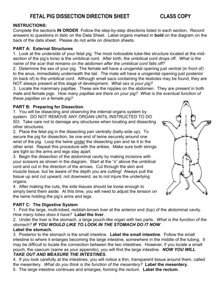
FETAL PIG DISSECTION DIRECTION SHEET CLASS COPY INSTRUCTIONS: Complete the sections IN ORDER. Follow the step-by-step directions listed in each section. Record answers to questions in italic on the Data Sheet. Label organs marked in bold on the diagram on the back of the data sheet. Please do not write on direction sheets. PART A: External Structures 1. Look at the underside of your fetal pig. The most noticeable tube-like structure located at the midsection of the pig’s torso is the umbilical cord. After birth, the umbilical cord drops off. What is the name of the scar that remains on the abdomen after the umbilical cord falls off? 2. Determine the sex of your pig. The female will have a urogenital opening just ventral (in front of) to the anus, immediately underneath the tail. The male will have a urogenital opening just posterior (in back of) to the umbilical cord. Although small sacs containing the testicles may be found, they are NOT always present at this stage of development. What sex is your pig? 3. Locate the mammary papillae. These are the nipples on the abdomen. They are present in both male and female pigs. How many papillae are there on your pig? What is the eventual function of these papillae on a female pig? PART B: Preparing for Dissection 1. You will be dissecting and observing the internal organs system by system. DO NOT REMOVE ANY ORGAN UNTIL INSTRUCTED TO DO SO. Take care not to damage any structures when locating and dissecting other structures. 2. Place the fetal pig in the dissecting pan ventrally (belly-side up). To secure the pig for dissection, tie one end of twine securely around one wrist of the pig. Loop the twine under the dissecting pan and tie it to the other wrist. Repeat this procedure with the ankles. Make sure both strings are tight so the arms and legs stay apart. 3. Begin the dissection of the abdominal cavity by making incisions with your scissors as shown in the diagram. Start at the “x” above the umbilical cord and cut in the direction of the arrows. Cut through the skin and muscle tissue, but be aware of the depth you are cutting! Always pull the tissue up and cut upward, not downward, as to not injure the underlying organs. 4. After making the cuts, the side tissues should be loose enough to simply bend them aside. At this time, you will need to adjust the tension on the twine holding the pig’s arms and legs. PART C: The Digestive System 1. Find the large, multi-lobed, reddish-brown liver at the anterior end (top) of the abdominal cavity. How many lobes does it have? Label the liver. 2. Under the liver is the stomach, a large pouch-like organ with two parts. What is the function of the stomach? IF YOU WOULD LIKE TO LOOK IN THE STOMACH DO IT NOW Label the stomach. 3. Posterior to the stomach is the small intestine. Label the small intestine. Follow the small intestine to where it enlarges becoming the large intestine, somewhere in the middle of the tubing. It may be difficult to locate the connection between the two intestines. However, if you locate a small pouch, the caecum (same as your appendix), you will find the large intestine. NOW YOU WILL TAKE OUT AND MEASURE THE INTESTINES. 4. If you look carefully at the intestines, you will notice a thin, transparent tissue around them, called the mesentery. What do you think is the function of the mesentery? Label the mesentery. 5. The large intestine continues and enlarges, forming the rectum. Label the rectum. 6. The point at which the rectum opens to the outside of the body is called the anus. What’s the function of the anus? Label the anus. 7. Relocate the liver. Lift up and trace the stomach anteriorly (upward) to the esophagus. What’s another name for the esophagus? Label the esophagus. 8. Observe the dome-shaped, muscular wall just above the liver. This is the diaphragm. To what system does the diaphragm belong? Label the diaphragm. 9. Embedded in the underside of the right central lobe of the liver is the greenish gall bladder. What is the function of the gall bladder? Label the gall bladder. 10. Directly below the gall bladder on the underside of the stomach, you will find the pancreas, a granular-looking organ. What is its function? Label the pancreas. 11. Attached to the left edge of the stomach is the spleen. What is its function? Label the spleen. PART D: The Respiratory and Circulatory Systems 1. Open the thoracic cavity (chest) using scissors to cut through the bone. The bones are very soft;, therefore, when cutting them be careful not to cut too deeply or you may damage the heart and lungs. Carefully cut the diaphragm on either side of the body and fold it back over the liver. 2. Beginning at the top of the stomach, trace the esophagus as it passes through the diaphragm and under the heart. At the anterior end of the esophagus, on the ventral (belly) side is the trachea. What is another name for the trachea? Label the trachea. 3. Notice the small “rings” around the trachea; these are made of cartilage. These rings give the trachea the appearance of a vacuum cleaner hose. What is the function of these cartilage rings? 4. Trace the trachea to the point where it branches into each lung. What is the function of the lung? Label the lung. 5. Continue the dissection of the neck anteriorly (toward the head). Locate an enlarged area of cartilage, the larynx. What is another name for the larynx? Label the larynx. 6. Locate the “roundish” structure between the lungs. This is the heart. Label the heart. 7. Notice that it is covered by a translucent tissue called the pericardium. It encloses the heart and keeps it free from all organs around it. Label the pericardium. Remove the pericardium to take a closer look at the heart. You can see two lighter-colored structures that look like little ears on the upper part of the heart. These chambers are the atria. The lower, more pointed chambers are the ventricles. NOW YOU MAY TAKE OUT THE LUNGS AND HEART. FEEL FREE TO CUT THE HEART IN HALF TO LOOK AT THE CHAMBERS MORE CLOSELY. PART E: The Urogenital System 1. Carefully push aside the digestive organs in the abdominal cavity. Along the backside behind the intestines is a pair of dark, bean-shaped organs, the kidneys; they are covered by a translucent connective tissue called the peritoneum. TAKE ONE KIDNEY OUT AND CUT IT IN HALF TO LOOK AT THE MEDULLA AND CORTEX. What is the function of the kidneys? 2. If you have a male pig, you may find the oval-shaped testes in the abdominal cavity. If you have a female pig, you may find the ovaries near the rear of the abdominal cavity.
