Handout G: The Eye and How We See
Prevent Blindness America. (2003c). The eye and how we see. Retrieved
July 31, 2003, from http://www.preventblindness.org/resources/howwesee.html
Your eyes are wonderful sensory organs. They help you learn about the
world in which you live.
Your eyes see all sorts of things—big or small, near or far, smooth or
textured, and even colors and dimensions.
Your eyes constantly send information by way of the optic nerve to your
brain. Your brain processes the date in a fraction of a second, and the
result is that you see something.
It requires many parts for your eyes to see properly. And your eyes don't
work alone.
Light and Dark
To control the amount of light entering the eye, the pupil changes in size.
It shrinks in bright light since your eye doesn't need as much light to see in
these conditions (1). Your pupil enlarges in dim light because it needs as
Visual Conditions Module 06/04/04
The Eye and How We See
S3 Handout A
Page 1 of 5
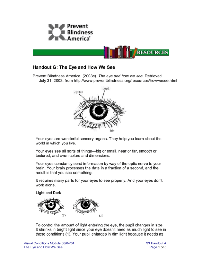
much of the available light as possible to see (2). The pupil is only an
opening in the iris; it looks black because the eye is dark inside. The
whole front of the eye, including the pupil, is covered with a protective
layer called the cornea.
Color
Rods and cones in the retina line the back of the eye and work together to
help you see well. Cones allow you to see colors, fine detail and function
best in bright light. Rods function best in dim light and are important for
side (peripheral) vision.
Near and Far
Muscles around the eye adjust the shape of the lens to focus on an object
either nearby or far away. The lens gets thicker when focusing for near
objects (3), and thinner for distant objects (4). The size of the image
reflected on the retina changes, too.
Visual Conditions Module 06/04/04
The Eye and How We See
S3 Handout A
Page 2 of 5
Upside Down
Light reflected from an object passes through the cornea (5), moves
through the lens, which focuses it (6), and then reaches the retina at the
very back (7), where it meets with a thin layer of color-sensitive cells
called the rods and cones. Because the light criss-crosses while going
through the cornea, your retina "sees" the image upside down. But your
brain "reads" it right side up.
Inside the Eye
Think of the eye in the shape of a ping-pong ball. The outside of the ball
represents the outer globe of the eyeball or the outer protective layer. This
outside layer is covered in sclera. The eyeball sits inside of the eye socket
and a thick membrane lining called the conjunctiva covers the inner
surface of the eyelid and part of the outer surface of the eyeball. At the
top and bottom of the eyeball are muscles that hold the eyeball and move
it inside the socket. At the front of the outside layer is the cornea where
light enters the eye. Just past the cornea is a space called the anterior
chamber of the eye. The back of anterior chamber is formed by the iris,
the colored part of the eye, that admits light through an opening called the
pupil. In the space between the cornea and iris is a liquid called the
aqueous humor. Directly behind the iris is a biconvex structure called the
lens. The center of the inside of the eye, the space between the lens and
Visual Conditions Module 06/04/04
The Eye and How We See
S3 Handout A
Page 3 of 5
the retina, or the inside of the ping pong ball, is the vitreous. The vitreous
maintains the shape of the eyeball. The posterior two thirds of the eye is
lined with the retina. Between the retina and the sclera is a vascular layer
called the choroid. The very back of the eye has a small nerve similar to a
chord, called the optic nerve.
Many different parts of the eye must work together to see properly. Also,
muscles are attached to the outer walls of the eyeball to hold it in place.
The picture above shows these main parts. If anything goes wrong, such
as an eye disease or eye injury, you might not be able to see well again
even after your eye heals.
Vision Vocabulary
Aqueos Humor: A clear, watery fluid that fills the front part of the eye between the
cornea, lens and iris.
Choroid: The middle layer of the eyeball, which contains veins and arteries that furnish
nourishment to the eye, especially the retina.
Conjunctiva: A mucous membrane that lines the eyelids and covers the front part of the
eyeball.
Cornea: The transparent outer portion of the eyeball that transmits light to the retina.
Iris: The colored, circular part of the eye in front of the lens. It controls the size of the
pupil.
Lens: The transparent disc in the middle of the eye behind the pupil that brings rays of
light into focus on the retina.
Optic Nerve: The important nerve that carries messages from the retina to the brain.
Pupil: The circular opening at the center of the iris that controls the amount of light
allowed into the eye.
Retina: The inner layer of the eye containing light-sensitive cells that connect with the
brain through the optic nerve.
Sclera: The white part of the eye that is a tough coating which, along with the cornea,
forms the external protective coat of the eye.
Vitreous Body: A colorless mass of soft, gelatin-like material that fills the eyeball behind
the lens.
Visual Conditions Module 06/04/04
The Eye and How We See
S3 Handout A
Page 4 of 5
For more information about your eyes, contact Prevent Blindness America
or the Prevent Blindness affiliate near you.
Copyright © 1998-2000 Prevent Blindness America ® | Disclaimer
URL: http://www.preventblindness.org/resources/howwesee.html
graphics/images courtesy of ClickArt®
© 1996 BrØderbund Software, Inc.
All rights reserved. Used by permission.
ClickArt and BrØderbund are registered trademarks of BrØderbund Software, Inc.
Reprinted with permission from PREVENT BLINDNESS AMERICA. Copyright 2003.
Visual Conditions Module 06/04/04
The Eye and How We See
S3 Handout A
Page 5 of 5

