Paint_Frontal_Lobe_3-21-05 - CCB
Painting Frontal Lobe Gyri on the Brain Surface
Using BrainImageJava (BIJ)
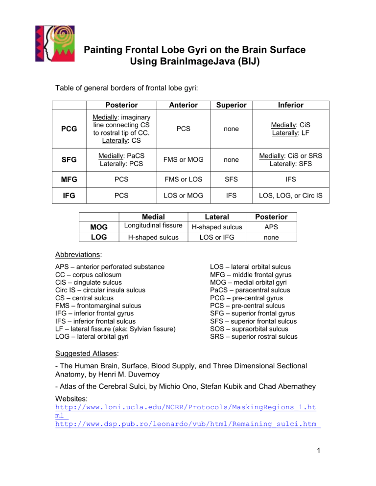
Table of general borders of frontal lobe gyri:
Posterior Anterior Superior Inferior
PCG
Medially: imaginary line connecting CS to rostral tip of CC.
Laterally: CS
PCS none
Medially: CiS
Laterally: LF
SFG
Medially: PaCS
Laterally: PCS
FMS or MOG none
Medially: CiS or SRS
Laterally: SFS
MFG PCS FMS or LOS SFS IFS
IFG PCS LOS or MOG IFS LOS, LOG, or Circ IS
MOG
LOG
Abbreviations:
Medial
Longitudinal fissure
H-shaped sulcus
APS – anterior perforated substance
CC – corpus callosum
CiS – cingulate sulcus
Circ IS – circular insula sulcus
CS – central sulcus
FMS – frontomarginal sulcus
IFG
– inferior frontal gyrus
IFS – inferior frontal sulcus
LF
– lateral fissure (aka: Sylvian fissure)
LOG – lateral orbital gyri
Suggested Atlases:
Lateral
H-shaped sulcus
LOS or IFG
Posterior
APS none
LOS – lateral orbital sulcus
MFG – middle frontal gyrus
MOG – medial orbital gyri
PaCS – paracentral sulcus
PCG – pre-central gyrus
PCS – pre-central sulcus
SFG
– superior frontal gyrus
SFS – superior frontal sulcus
SOS
– supraorbital sulcus
SRS – superior rostral sulcus
- The Human Brain, Surface, Blood Supply, and Three Dimensional Sectional
Anatomy, by Henri M. Duvernoy
- Atlas of the Cerebral Sulci, by Michio Ono, Stefan Kubik and Chad Abernathey
Websites: http://www.loni.ucla.edu/NCRR/Protocols/MaskingRegions_1.ht
ml http://www.dsp.pub.ro/leonardo/vub/html/Remaining_sulci.htm
1
http://www9.biostr.washington.edu/cgi- bin/DA/PageMaster?atlas:Neuroanatomy+ffpathIndex:Splash^Page+2
2
Painting the Precentral Gyrus
Notice that the pcg is continuous with the postcentral gyrus on the medial plane, and the inferior frontal gyrus and superior frontal gyrus o\in the lateral view.
Painting the surface helps to delineate these borders for full parcellation on the coronal plane.
3
Adding to the Pre-Central Gyrus (PCG)
Medial view of the central sulcus cingulate sulcus
Midsagittal view of the brain, showing the medial surface.
The area within the oval is the paracentral lobule and includes medial cortex of both the pre- and post-central gyri. The central sulcus extends posteriorly from the top of the brain. Imagine a line connecting the tip of this sulcus to the caudal border of the corpus callosum, shown here by a dotted line – this line delineates the border between the pre- and post-central gyri on the medial surface. The cingulate sulcus delineates the inferior border of the medial pcg.
When drawn, the sagittal view of the
PCG will look similar to this:
4
Adding to the Pre-Central Gyrus (PCG)
1. The anterior PCG arises from the posterior extreme of the IFG.
2. It soon includes neighboring gyri dorsal to the anterior portion.
3. Due to its sloped-upward shape, and depending on the brain, some coronal slices may include small separated
PCG gyri. Viewing the painted surface view may help in defining the PCG here.
4. The PCG widens as it reaches the dorsal aspect of the brain, often including several gyri on the coronal view.
5. A few coronal slices will include both lateral and medial surfaces in the PCG
6. The posterior extreme of the PCG is located on the medial surface, with the posterior central gyrus located dorsolateral to it.
5
Painting the Superior, Middle and Inferior Frontal Gyri
(SFG, MFG, IFG)
The posterior borders of the three gyri are easy to locate due to the pre-central sulcus.
They encompass the lateral frontal surface except for the pre-central gyrus and lateral orbital gyrus. They are separated from each other by prominent sulci: the SFS and IFS.
Even with these clear landmarks, it is sometimes difficult to separate small portions due to discontinuity of the sulci, creating continuous segments with neighboring gyri.
SFG : The SFG is often continuous with the MFG, pre-central gyrus and orbital gyri due to discontinuous sulci. When painting, connect these sulci with a straight line.
MFG : Likewise, the MFG is often continuous with the SFG, IFG, pre-central gyrus and orbital gyri due to discontinuous sulci. When painting, connect these sulci with a straight line.
IFG : The IFG is usually continuous with the MFG, pre-central gyrus and lateral orbital gyrus.
Be sure to exclude the orbital gyri from your surface painting (use frontomarginal sulcus as the inferior boundary for the SFG). Usually one layer of gyrus on the inferior part of the brain is excluded (See figure below, with orbital gyri in purple and bright green)
6
ROIs: It is important when drawing coronal ROIs to refer to the behavior of the gyri on the surface . This is important in determining how IFG, MFG, and SFG should begin and end (see BIJ movies for examples of this). Also refer to the X-axis or Sagital
View, this will also help in determining where jumps in your ROIs should occur.
Adding to the Superior Frontal Gyrus (SFG)
7
1. The medial aspect of the SFG extends from the pre-central sulcus to the frontal pole, is further delineated by the cingulate and inferior rostral sulci. Refer to the X-axis
(Sagital view) as you draw.
2. The anterior SFG begins at the tip of the frontal pole, with the orbital gyri and MFG soon appearing on the same plane. Note the internal snakelike frontomarginal sulcus.
3. When the frontomarginal sulcus disappears, connect tips of sulci as shown here. The bilateral cingulate sulci are usually aligned and symmetrical.
4. The appearance of the cingulate gyrus marks the end of the most inferior medial portion of the SFG.
5. Note the double gyri of the cingulate cortex; the general rule is to use the deepest sulcus to delineate cingulate gyrus from SFG.
6. As the posterior extreme of the SFG approaches, the lateral extent narrows.
Adding to the Middle Frontal Gyrus (MFG)
8
1. The anterior MFG begins to appear as a separate section sandwiched between the SFG and the lateral orbital gyrus.
2. The MFG is separated from the SFG by the snake-like supramarginal gyrus, and is continuous with the lateral orbital gyrus until the IFG appears.
3. As the IFG takes shape, the MFG is situated between the SFG and IFG.
4. Shallow sulci often split the MFG into multiple gyri as you move posteriorly.
5. In general, deeper sulci should be used to define the MFG, as shown here.
6. The posterior MFG ends at the precentral gyrus a few slices beyond this one.
9
Adding to the Inferior Frontal Gyrus (IFG)
1. The anterior IFG begins to appear as a separate section wedged between the
MFG and the lateral orbital gyrus.
2. The IFG becomes continuous with the lateral orbital gyrus.
3. By referring to previous and upcoming slices, separate the IFG from the lateral orbital gyrus similar to this.
4. The IFG separates from neighboring gyri as it continues posteriorly.
5. In general, deep sulci should be used to help define the IFG, as shown here.
6. The extreme posterior IFG blends into the pre-central gyrus near here.
10
Locating the posterior IFG border
The IFG is contiguous with the precentral gyrus (pcg), so finding the inferior/anterior border of the pcg is helpful in finding the posterior edge of the IFG. The following are two examples of the inferior/anterior border of the pcg: inf/ant pre central gyrus
Depending on the angle of the coronal sections:
The coronal slice that begins the posterior border of the IFG will be slightly anterior to the anterior commissure. The optic chiasm may be visible in this slice, as it is in the figure below, or it may be posterior to the slice. Caudate, putamen, internal capsule and the temporal pole should also be visible in the slice. The third ventricle will be posterior to this slice. Often, the IFG is visible in one hemisphere before the other. In the following figure, the IFG has just emerged in the left hemisphere, but not the right: posterior
IFG optic chiasm
Typically the IFG will gradually fade into PCG, as Par Opercularis disappears under this “L” shaped structure. For this reason, remember to continue including Pars
Opercularis until it ends, because your surface drawing typically will not “bleed” into the coronal view to include the last few ROIs needed to delineate IFG.
11
Painting the Medial and Lateral Orbital Gyri (MOG and LOG)
Specific gyri of the orbital frontal gyri are hard to visualize from the surface view using
BIJ. The H-shaped sulci separating the medial orbital gyrus from the anterior and posterior orbital gyri can usually be seen well enough so that the medial orbital gyri and the lateral orbital gyri can be discerned. For the purpose of this protocol, the medial orbital gyri includes the gyrus rectus and medial orbital gyrus, while the lateral orbital gyri include the anterior and posterior orbital gyri, and the lateral orbital gyrus. It is helpful to view this ventral picture of the brain when painting these gyri in BIJ: lateral orbital gyri (red) medial orbital gyri (blue)
12
Adding to the Medial Orbital Gyri (MOG)
1. The MOG begin at the frontal pole, with both the gyrus rectus and the medial orbital gyrus quickly taking shape.
2. The gyrus above the supra-orbital sulcus begins to be included in the
MOG as the frontomarginal sulcus tip rises. Include medial tissue that would not be part of SFG or cingulate gyrus.
3. The MOG widens as you move caudally through the slices.
4. As the genu of the corpus callosum appears, the MOG remains basically the same.
5. As the posterior extreme of the MOG approaches, the sulci depths decrease substantially.
6. The MOG ends when sulci are no longer visible, just beyond this slice, and the anterior perforated substance begins.
13
Adding to the Lateral Orbital Gyri (LOG)
1. The LOG are continuous with the MOG in the frontal pole region.
2. The LOG are continuous with the MFG at this level, but separated from the
MOG by a shallow sulcus.
3. The IFG is just beginning to appear and is continuous with the LOG, so it must be separated by referring to nearby slices.
,
4. The IFG is now separate from the
LOG.
5. As the temporal poles appear, the surface of the LOG will no longer be painted, as this area is not visible on the surface view.
6. As the posterior extreme of the LOG approaches, only a small part of the posterior orbital gyrus remains.
14
