IB Biology – Cell Test Review Guide
advertisement
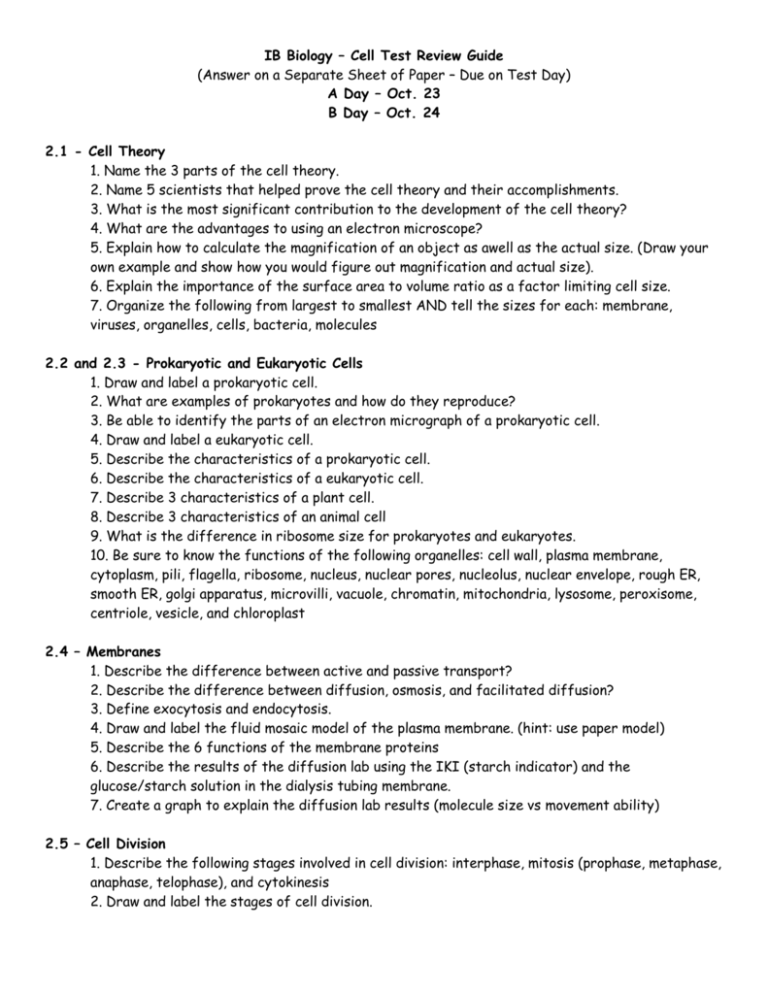
IB Biology – Cell Test Review Guide (Answer on a Separate Sheet of Paper – Due on Test Day) A Day – Oct. 23 B Day – Oct. 24 2.1 - Cell Theory 1. Name the 3 parts of the cell theory. 2. Name 5 scientists that helped prove the cell theory and their accomplishments. 3. What is the most significant contribution to the development of the cell theory? 4. What are the advantages to using an electron microscope? 5. Explain how to calculate the magnification of an object as awell as the actual size. (Draw your own example and show how you would figure out magnification and actual size). 6. Explain the importance of the surface area to volume ratio as a factor limiting cell size. 7. Organize the following from largest to smallest AND tell the sizes for each: membrane, viruses, organelles, cells, bacteria, molecules 2.2 and 2.3 - Prokaryotic and Eukaryotic Cells 1. Draw and label a prokaryotic cell. 2. What are examples of prokaryotes and how do they reproduce? 3. Be able to identify the parts of an electron micrograph of a prokaryotic cell. 4. Draw and label a eukaryotic cell. 5. Describe the characteristics of a prokaryotic cell. 6. Describe the characteristics of a eukaryotic cell. 7. Describe 3 characteristics of a plant cell. 8. Describe 3 characteristics of an animal cell 9. What is the difference in ribosome size for prokaryotes and eukaryotes. 10. Be sure to know the functions of the following organelles: cell wall, plasma membrane, cytoplasm, pili, flagella, ribosome, nucleus, nuclear pores, nucleolus, nuclear envelope, rough ER, smooth ER, golgi apparatus, microvilli, vacuole, chromatin, mitochondria, lysosome, peroxisome, centriole, vesicle, and chloroplast 2.4 – Membranes 1. Describe the difference between active and passive transport? 2. Describe the difference between diffusion, osmosis, and facilitated diffusion? 3. Define exocytosis and endocytosis. 4. Draw and label the fluid mosaic model of the plasma membrane. (hint: use paper model) 5. Describe the 6 functions of the membrane proteins 6. Describe the results of the diffusion lab using the IKI (starch indicator) and the glucose/starch solution in the dialysis tubing membrane. 7. Create a graph to explain the diffusion lab results (molecule size vs movement ability) 2.5 – Cell Division 1. Describe the following stages involved in cell division: interphase, mitosis (prophase, metaphase, anaphase, telophase), and cytokinesis 2. Draw and label the stages of cell division. 3. Be sure to know the following terms: centriole, chromosomes, centromere, sister chromatids, cell plate, cleavage furrow
