outline27956
advertisement
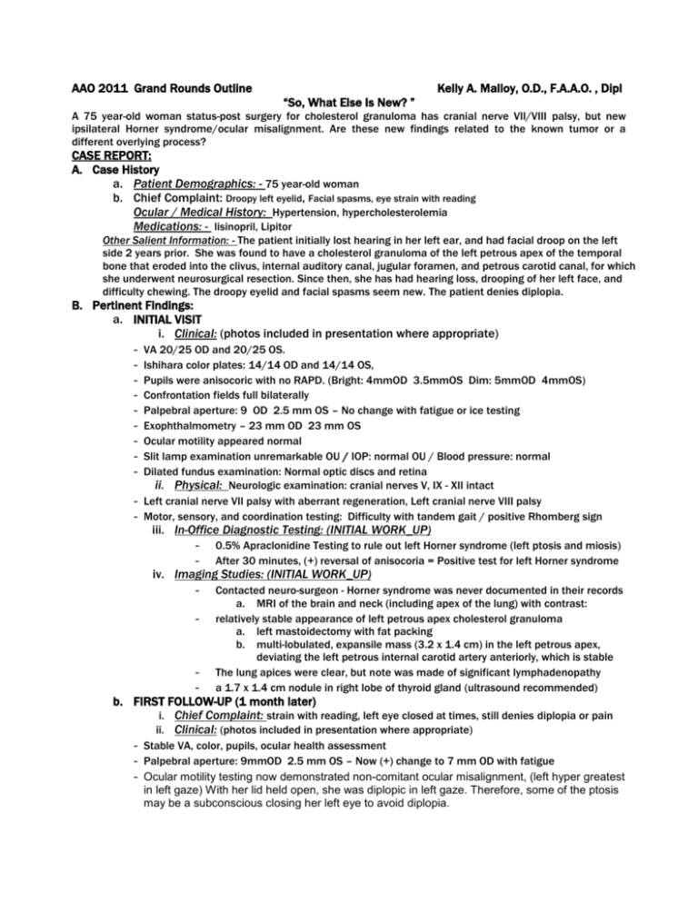
AAO 2011 Grand Rounds Outline Kelly A. Malloy, O.D., F.A.A.O. , Dipl “So, What Else Is New? ” A 75 year-old woman status-post surgery for cholesterol granuloma has cranial nerve VII/VIII palsy, but new ipsilateral Horner syndrome/ocular misalignment. Are these new findings related to the known tumor or a different overlying process? CASE REPORT: A. Case History a. Patient Demographics: - 75 year-old woman b. Chief Complaint: Droopy left eyelid, Facial spasms, eye strain with reading Ocular / Medical History: Hypertension, hypercholesterolemia Medications: - lisinopril, Lipitor Other Salient Information: - The patient initially lost hearing in her left ear, and had facial droop on the left side 2 years prior. She was found to have a cholesterol granuloma of the left petrous apex of the temporal bone that eroded into the clivus, internal auditory canal, jugular foramen, and petrous carotid canal, for which she underwent neurosurgical resection. Since then, she has had hearing loss, drooping of her left face, and difficulty chewing. The droopy eyelid and facial spasms seem new. The patient denies diplopia. B. Pertinent Findings: a. INITIAL VISIT i. Clinical: (photos included in presentation where appropriate) - VA 20/25 OD and 20/25 OS. Ishihara color plates: 14/14 OD and 14/14 OS, Pupils were anisocoric with no RAPD. (Bright: 4mmOD 3.5mmOS Dim: 5mmOD 4mmOS) Confrontation fields full bilaterally Palpebral aperture: 9 OD 2.5 mm OS – No change with fatigue or ice testing Exophthalmometry – 23 mm OD 23 mm OS Ocular motility appeared normal Slit lamp examination unremarkable OU / IOP: normal OU / Blood pressure: normal Dilated fundus examination: Normal optic discs and retina ii. Physical: Neurologic examination: cranial nerves V, IX - XII intact Left cranial nerve VII palsy with aberrant regeneration, Left cranial nerve VIII palsy Motor, sensory, and coordination testing: Difficulty with tandem gait / positive Rhomberg sign iii. In-Office Diagnostic Testing: (INITIAL WORK_UP) - 0.5% Apraclonidine Testing to rule out left Horner syndrome (left ptosis and miosis) After 30 minutes, (+) reversal of anisocoria = Positive test for left Horner syndrome - Contacted neuro-surgeon - Horner syndrome was never documented in their records a. MRI of the brain and neck (including apex of the lung) with contrast: relatively stable appearance of left petrous apex cholesterol granuloma a. left mastoidectomy with fat packing b. multi-lobulated, expansile mass (3.2 x 1.4 cm) in the left petrous apex, deviating the left petrous internal carotid artery anteriorly, which is stable The lung apices were clear, but note was made of significant lymphadenopathy a 1.7 x 1.4 cm nodule in right lobe of thyroid gland (ultrasound recommended) iv. Imaging Studies: (INITIAL WORK_UP) - - b. FIRST FOLLOW-UP (1 month later) i. Chief Complaint: strain with reading, left eye closed at times, still denies diplopia or pain ii. Clinical: (photos included in presentation where appropriate) - Stable VA, color, pupils, ocular health assessment - Palpebral aperture: 9mmOD 2.5 mm OS – Now (+) change to 7 mm OD with fatigue - Ocular motility testing now demonstrated non-comitant ocular misalignment, (left hyper greatest in left gaze) With her lid held open, she was diplopic in left gaze. Therefore, some of the ptosis may be a subconscious closing her left eye to avoid diplopia. iii. Additional Laboratory Studies due to diplopia / ocular misalignment: - - CBC with differential, platelet count, ESR, C-reactive protein, Lyme titer, ANA, RPR, FTA-ABS, ACE, TSH, T4, T3, thyroperoxidase antibody, thyroglobulin antibody, thyroid stimulating immunoglobulin, and acetylcholine receptor antibodies (binding, blocking, and modulating). Results remarkable for significantly low TSH level - MRI of orbits to look for enlarged extra-ocular muscles Thyroid ultrasound iv. Additional Imaging Studies: v. Additional Consults: endocrinology c. (ALL ADDITIONAL FOLLOW-UP VISITS WILL BE INCLUDED) C. Differential Diagnoses: - For Horner syndrome (cholesterol granuloma, thyroid nodule?, other) For additional ptosis (cholesterol granuloma, myasthenia gravis, lid closure to avoid diplopia) For diplopia (cholesterol granuloma (skew deviation), thyroid, myasthenia gravis) For facial spasms (cholesterol granuloma, aberrant regeneration of CN VII, other) D. Diagnosis and Discussion: a. Cholesterol granuloma b. c. i. rare, benign cysts that can occur at the tip of the petrous apex ii. expanding masses that contain fluids, lipids, and cholesterol crystals surrounded by fibrous lining iii. air cells in the petrous apex are obstructed, creating a vacuum that causes blood to be drawn into air cells. As red blood cells break down, cholesterol is released. The immune system reacts to cholesterol as a foreign body, producing an inflammatory response. Associated small blood vessels rupture as a result of the inflammation. Recurrent hemorrhaging makes the mass expand. iv. Permanent hearing loss, nerve damage, and bone destruction can occur if mass is untreated v. Symptoms may include: hearing loss in one ear, tinnitus, facial twitching ,vertigo, facial numbness Thyroid nodule i. More common in women, may be asymptomatic ii. May be benign or malignant (features more associated with malignancy: <age 20 or > age 70, hard nodule, radiation exposure to head/neck, male gender, associated enlarged lymph nodes) iii. Can be associated with hyperthyroidism - Thyroid orbitopahty (diplopia, proptosis, lid edema etc all possible) Horner Syndrome –have been reports of Horner syndrome as the presenting feature of thyroid malignancy E. Treatment / Management: a. Treatment / Response: i. Cholesterol granuloma: surgery (translabrynthine/infralabrynthine/endoscopic endonasal approach) - Monitor by neuro-surgery ii. Thyroid issues – ultrasound, labs, possible fine needle biopsy, endocrinology consult, medical treatment of hyperthyroidism) iii. Facial spasms – Botox (but not if the lid closure is eliminating the diplopia) b. F. Bibliography / Literature Review: i. Conclusion: a. b. c. ii. iii. Almog Y, Gepstein R Kesler A. Diagnostic value of imaging in Horner syndrome in adults. J Neuroophthalmol. 2010 Mar;30(1):7-11. Broome JT Gauger PG Miller BS Doherty GM. Anaplastic thyroid cancer manifesting as new-onset Horner syndrome. Royer MC Pensak ML. Cholesterol granulomas. Curr Opin Otolaryngol Head Neck Surgery. 2007 Oct;15(5):319-22. Just because a patient has a known neurologic problem, all subsequent neurologic signs/symptoms cannot be unequivocally attributed to the initial problem. There may be another overlying problem. Horner syndrome can be the initial manifestation of thyroid malignancy. Work-up is crucial. We must work together with many different specialties to fully evaluate and treat the patient appropriately.
