USMLE Step 1 Web Prep — Electrical Activity of the Heart 152500
advertisement
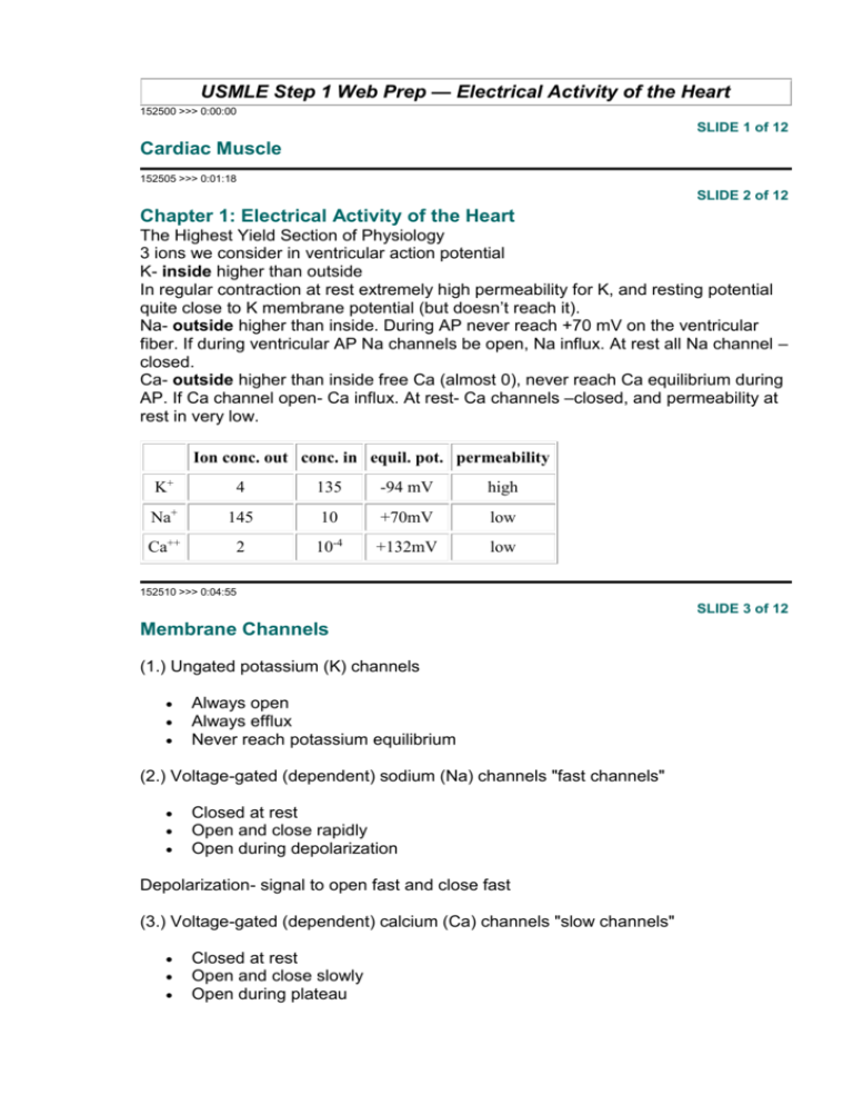
USMLE Step 1 Web Prep — Electrical Activity of the Heart 152500 >>> 0:00:00 SLIDE 1 of 12 Cardiac Muscle 152505 >>> 0:01:18 SLIDE 2 of 12 Chapter 1: Electrical Activity of the Heart The Highest Yield Section of Physiology 3 ions we consider in ventricular action potential K- inside higher than outside In regular contraction at rest extremely high permeability for K, and resting potential quite close to K membrane potential (but doesn’t reach it). Na- outside higher than inside. During AP never reach +70 mV on the ventricular fiber. If during ventricular AP Na channels be open, Na influx. At rest all Na channel – closed. Ca- outside higher than inside free Ca (almost 0), never reach Ca equilibrium during AP. If Ca channel open- Ca influx. At rest- Ca channels –closed, and permeability at rest in very low. Ion conc. out conc. in equil. pot. permeability K+ 4 135 -94 mV high Na+ 145 10 +70mV low Ca++ 2 10-4 +132mV low 152510 >>> 0:04:55 SLIDE 3 of 12 Membrane Channels (1.) Ungated potassium (K) channels Always open Always efflux Never reach potassium equilibrium (2.) Voltage-gated (dependent) sodium (Na) channels "fast channels" Closed at rest Open and close rapidly Open during depolarization Depolarization- signal to open fast and close fast (3.) Voltage-gated (dependent) calcium (Ca) channels "slow channels" Closed at rest Open and close slowly Open during plateau Depolarization is also signal to open and close slowly (4.) Voltage-gated potassium (K) channels – different form neuron K Open at rest – (unlike to neuron) – both types of K channel are open- very high K permeability- very close to K equilibrium, but not reach it Begin closing during depolarization – signal to closing voltage –gated K channel Closed during plateau Reopen with repolarization 152515 >>> 0:10:58 SLIDE 4 of 12 ACTION POTENTIAL OF A VENTRICULAR FIBER (FAST RESPONSE) Phase 0 - depolarization Increase sodium (Na) conductance - influx Fast channels open during depolarization allowing sodium to enter Phase 1 - Slight Repolarization Due to special potassium channel efflux through voltage gated potassium channels (the new details) – not important for step 1 Phase 2 - Plateau phase – very important Slow channels open (voltage- gated calcium channels) increasing calcium conductance Calcium influx occurs o Participate in contraction o Release additional calcium from sarcoplasmic reticulum o Very low potassium (K) conductance resulting in delayed repolarization and stable plateau Not only participate in contraction, BUT RELEASE Ca from SR – voltage -gated channel closed, but ungated K channel – open; so we have K influx through ungated channel (always open). They balance each other in the plateau and we have stable plateau. WE wouldn’t have stable plateau if we have voltagegated channel open. Phase 3 - Repolarization o o o Voltage gated calcium channels close Voltage gated potassium (K) channels reopen increasing potassium conductance Massive potassium efflux Can be tested: If Drug in ventricular muscle (regular contracted) prevented opening voltage – gated K channel – in repolarization you will not have massive K efflux, but still have small efflux of K through ungated K channel, but you still have slow repolarization. If in neuron drug voltage –gated Na- block – no AP, but in ventricular muscle- block of fast channel- you don’t block AP, because slow Ca can replace fast channel and will have a slow depolarization Phase 4 - Rest - only in regular contracting fiber of the heart have a stable resting potential (very long plateau phase) 152520 >>> 0:25:58 SLIDE 5 of 12 ELECTRICAL VERSUS MECHANICAL EVENTS One electrical event per mechanical event Due to very long absolute refractory period in ventricular muscle – impossible to saturate the interior with free Ca Impossible to tetanize heart muscle Electrical event in cardiac muscle – longer than skeletal muscle. In skeletal – we have several electrical event for every mechanical event because the electrical event is short we have tetanization. 152525 >>> 0:28:04 SLIDE 6 of 12 ACTION POTENTIAL CHARACTERISTICS OF SPECIALIZED CELLS SA node Contain few fast channels Depolarization due to slow calcium channels, not fast sodium channels Repolarization due to potassium efflux Innervated by sympathetic and parasympathetic neurons Will be tested: Do not have very many fast channels Repolarization- K efflux Unstable Phase 4 in all specialized cell – gradual depolarization (Pacemakers potential or prepotential) toward threshold Gradual depolarization is probably due to decrease K conductance, or influx of Na or Ca, or due to all 3 mechanisms, So it won’t be tested on step 1 AV node Purkinje fibers 152530 >>> 0:32:43 SLIDE 7 of 12 Effect of Sympathetics on SA node Increase slope of prepotential Reach threshold sooner Intrinsic firing rate increased 152535 >>> 0:33:27 SLIDE 8 of 12 Effect of Parasympathetics on SA node Hyperpolarization of Phase 4 Take longer to reach threshold (decreased slope) Intrinsic rate slows down 152540 >>> 0:34:30 SLIDE 9 of 12 Conduction pathways and velocity of conduction SA node atrial muscle AV node (delay) Purkinje fibers ventricular muscle Fastest conducting fiber = Purkinje cell Slowest conducting fiber = AV node Automaticity 1. SA nodal cells - Highest intrinsic rate – primary pacemaker 2. AV nodal cell - Second highest intrinsic rate- secondary pacemaker 3. Purkinje cells - Slowest intrinsic rate If heart rate - 100 beats/min and it’s normal heart, - firing rate of AV nodal cellalso 100 beat/min, and all cells beat at 100 beat/min If heart have AV rhythms (SA knocked out), and beats 60 then every cell will beat at 60 beat/min. 152545 >>> 0:38:21 SLIDE 10 of 12 ELECTROCARDIOGRAPHY- supplemental topic 152550 >>> 0:38:44 SLIDE 11 of 12 HEART BLOCK 1. Partial (first-degree) block Slowed conduction through the AV node P - R interval is increased Every impulse will take longer to convey Parasympathetic input (large)- could induce 1-st degree heart block 2. Second-degree block – most tested Some impulses are not transmitted through the AV node 2 subcategories – not HY if you have ECG- then it is 2-nd degree. Every 4 or 3 will not be followed by QRS complex 3. Complete (third-degree block) No impulses conducted from the atria to ventricles They beating independently We don’t need to know ECG, but may be asked. 1. atria and ventricles beating independently- no correlation btw P waves and QRS complex 2. pacemakers tissue in atria have higher intrinsic rated than ventricle tissuethe frequency of P wave is greater of QRS complex 152555 >>> 0:43:02 SLIDE 12 of 12 EINTHOVEN’S TRIANGLE – supplemental topic If you have left axis deviation- left ventricular hypertrophy If you have right axis deviation- right ventricular hypertrophy
![Cardio Review 4 Quince [CAPT],Joan,Juliet](http://s2.studylib.net/store/data/005719604_1-e21fbd83f7c61c5668353826e4debbb3-300x300.png)
