Fingerprints NZAHT Nov/Dec 2010 - New Zealand Association of
advertisement
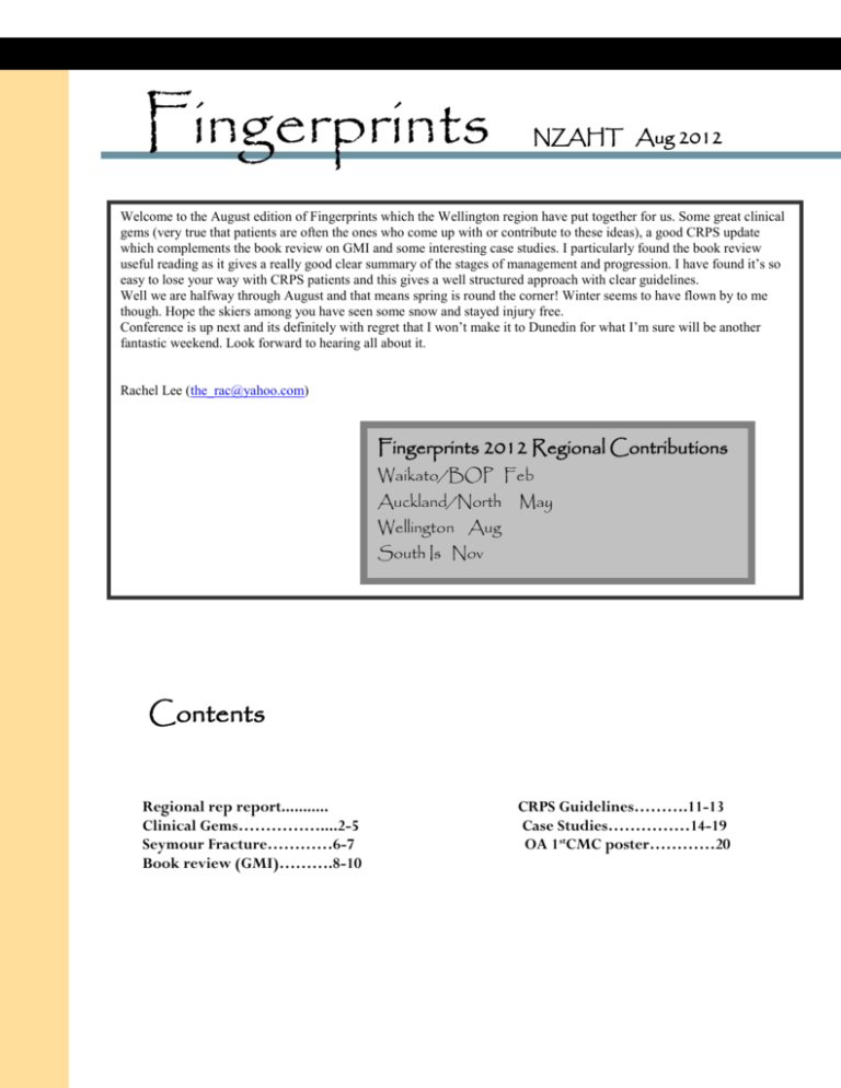
Fingerprints NZAHT Aug 2012 Welcome to the August edition of Fingerprints which the Wellington region have put together for us. Some great clinical gems (very true that patients are often the ones who come up with or contribute to these ideas), a good CRPS update which complements the book review on GMI and some interesting case studies. I particularly found the book review useful reading as it gives a really good clear summary of the stages of management and progression. I have found it’s so easy to lose your way with CRPS patients and this gives a well structured approach with clear guidelines. Well we are halfway through August and that means spring is round the corner! Winter seems to have flown by to me though. Hope the skiers among you have seen some snow and stayed injury free. Conference is up next and its definitely with regret that I won’t make it to Dunedin for what I’m sure will be another fantastic weekend. Look forward to hearing all about it. Rachel Lee (the_rac@yahoo.com) Fingerprints 2012 Regional Contributions Waikato/BOP Feb Auckland/North May Wellington Aug South Is Nov Contents Regional rep report........... Clinical Gems……………....2-5 Seymour Fracture…………6-7 Book review (GMI)……….8-10 CRPS Guidelines……….11-13 Case Studies……………14-19 OA 1stCMC poster…………20 Fingerprints (Aug 2012) Regional Rep Report The Wellington Group has met on three occasions so far this year. At our first meeting in February, we were fortunate to have Philippa Grimes (Occupational Health Physiotherapist) come and speak to us about her work and provide us with some good tidbits to apply in our practice. In April, Doug led us in a discussion over the latest management approach for extensor tendon repairs from Hutt Hospital; this generated discussion over how we should be standardizing our measurements such as ROM in our region to make this more reliable/relevant between clinics. In June, Caroline fed back highlights of Lynn’s workshop from last years’ conference while Gail lead the 2nd part of the evening covering dynamic taping. For our next meeting coming up later this month, we decided to follow on from our discussion in April and actually have a practical workshop to review our assessment/recording methods and hopefully manage to make this more uniform within our region and in line with what is being taught at AUT. We are hoping to clarify questions such as when measuring IPJ motion, whether we should measure the joints in a composite flexion or extension range or have these done in isolation and so forth. We have also been continuing on with our attempts at videoconferencing with the other members outside of Wellington. This year we have been trialing “teamviewer” as this has allowed an unlimited number of people to join our meetings unlike skype which only allowed one user when viewing (not just listening to) the presenter. There have been limitations with this; such as difficulty hearing when there are multiple discussions occurring at the same time, not to mention the skill of the person operating the app (which is mainly me, the self-confessed techno phobe!) However, it does operate very closely to real time and is overall showing some good potential. (I would like to – at this point - send my ongoing “thank you”s to all those members who have been soooo patient with me with all my glitches as we attempt to fine-tune this process+++.) So, our next meeting on Assessment of the Hand is scheduled for 24th August at 7.30pm. Anyone who is interested (and I can actually put this invite out to everyone), please feel free to contact me, so I can direct you on setting up your computer. E: workinghands@paradise.net.nz or M: 021 76 4263 Finally, I would like to send another big thank you to all members again for contributing to this edition. We hope you’ll enjoy reading our edition of fingerprints. Look forward to catching up with everyone at conference. Amanda Johnson, Wellington Regional Rep Clinical Gems Modifications to the Neoprene Wrist and Thumb Wrap (Ross Simmons, Focus on Hands) I use a few neoprene wrist and thumb wrap supports from time to time, typically as clients are weaning out of a thermoplastic splint and progressing their strengthening. I currently use an off the shelf wrist and thumb wrap from USL but find that it needs a few tweaks before I give it out, as it has a few design faults. Standard wrist / thumb neoprene support Placing the splint on, Fault 1- the tongue of the splint migrates out from under the wrist strap after 5 mins of use, when carrying loads, or moderate amounts of ulnar/radial deviation with functional activities. Fault 2- Velcro strap never makes it across all the hook section of the Velcro, and with larger clients, it struggles to make it to the hook portion of the Velcro. This leads to exposed hook, which in turn then gets messy. Parts required for modification. 1- Off cut of 50mm loop Velcro, 2- Small length of adhesive backed loop If I have time, (which is not often…) then I sew the extra length of loop onto the end of the strap, otherwise I just place the off cut of Velcro on the exposed hook, and it remains there while the client only removes the strap that is sewn onto the neoprene. Splint with modifications. Splint with modifications, which stays in place with improved compliance, and less complaints. 2 Issues with normal Scissors’??? Another little gem that I have had some positive feedback from, were these little spring loaded thread scissors that I source from the 1-2-3$ shop locally. They have been well received by my elderly clients who sew or knit, but who struggle with standard scissors, They only require a relatively closed fist to grip, and some light thumb flexion to operate with ease. Or they can be used without the thumb at all, just by clenching a fist. Because they are designed like shears, the blades always run together, so little threads etc that are often not cut with blunt scissors, or with poor scissors control, are now cut with ease. I expect most 2$ shops may stock these, especially if they have a Chinese supplier. Microbreaks (Matt Beal, Hand Rehab) For those patients who need to do micro breaks or regular exercises at their work station, a free seven day trial of a program which blacks your screen to reminds you can be downloaded. Try googling www.eyeprotectorpro.com; it also has lots of good information regarding work stations. 3 Patient’s Clinical Gems (Amanda Johnson, Working Hands) Patients can be very good sources of clinical gems describing their own discoveries or refinements on advice given to them. Below are a few that I have come across so far this year: 1. Scar Massage In the past, I had advised those patients wanting to simulate the mini vibrator effect at home to use the handle of an electric toothbrush - until a patient went one up on that idea by placing a pencil rubber on the tip of it … this is so much better! 2. Hand Warmers These are merino gloves that can be sourced from “Kilt” a NZ made designer clothing outlet for $49 a pair. These gloves can be purchased from their stores in Napier, Palmerston North, Wellington, Nelson, New Plymouth, Tauranga and Ponsonby. Much nicer to wear than tubigrips or bulky woollen gloves!! 4 3. Funky Splint Designs This previously white polyflex splint was painted by a patient’s son who used model making paint. It was then covered further with nail polish varnish to prevent it from chipping – ingenious! 5 Seymour Fracture – Distal Phalanx Fracture with a Difference (Laura-Kate Matheson, Hutt Hospital) Have you seen an XR like this before? Figure 1. Lateral View Figure 2. PA View Definition A Seymour fracture is a paediatric fracture. It is classified as a Salter Harris I/II open fracture or fracture dislocation of the distal phalanx epiphyseal plate (growth plate) with associated avulsion of the proximal edge of the nail from the eponychial fold (Yeh, P. & Dodds, S., 2009). 6 Clinical Appearance - Flexion (mallet) deformity of DIPJ - Exposed proximal nail plate - Signs of trace bleeding around the nail bed and/or the visible nail Surgical management may appear too long - Irrigation/debridement XR: Dorsal epiphyseal widening - and Reduction is common flexionof ofthe thefractur distal segment Repair of nail bed and nail plate - Figure 3. Clinical Appearance of Seymour Fracture - Treatment Surgical management - Irrigation/debridement of the open fracture - Reduction of the fracture with k-wire (k-wire in situ for 4/52) - Repair of nail bed and nail plate Hand Therapy - Protective mallet splint pre and post k-wire removal - Wound/scar management - ROM exercises and splint weaning 7 Book Review (Caroline Durney, Wellhand) We have been using the GMI for a few years since Lorimer talked at our conference in 2005 but Noigroup publications 2012 have recently published a new book with more recent research and useful clinical tips. It is well worth reading. These are some notes and take home points from the book and a recent Noigroup email. THE GRADED MOTOR IMAGERY HANDBOOK 2012 G. Lorimer Moseley, David S. Butler, Timothy B. Beames, Thomas J. Giles Ramachandran "Phantoms in the brain" he was the first person to use mirror for phantom pain. Moseley tried using mirror for chronic pain esp. CRPS with some good and some terrible results. He then looked in to motor imagery and found an article about research on reduced reaction times for L/R hand judgement. NEUROTAGS - a network of interconnected neurones that are distributed throughout the brain. When a neurotag is activated it produces an output. The output defines the neurotag e.g. the neurotag for thumb pain refers to the network of brain cells that, when activated, produces thumb pain. The neurotag of the smell of bread is that network of brain cells that, when activated, produces the smell of bread (not the odour itself, nor does it detect it, rather it produces the EXPERIENCE of smelling the bread). Criteria to work: the neurons that make up the neurotag have to fire, but nearby brain cells provide inhibition. Neurotags have an activation threshold. DANGER messages come up the spinal cord. When pain persists there are brain changes and the pain neurotag becomes sensitised (increased excitability of member brain cells so they are more easily excited) so that even imagined movements can activate the neurotag. It also becomes disinhibited (a decrease in the inhibition of the non-member brain cells) which means the neurotag loses precision may manifest as spreading, moving or less precisely defined pain. This does not follow a peripheral nerve or nerve root distribution. Disinhibition results in loss of precision which can disrupt movement commands, sensory function, perception of the body part and anything else related to that body part. GRADED MOTOR IMAGERY IS DONE IN THREE CONSEQUATIVE STAGES: STAGE 1 LEFT/RIGHT JUDGEMENTS or IMPLICIT MOTOR IMAGERY This involves looking at pictures of hands and making a judgement whether it is a left or right hand. "Recognise" is the programme developed by Noigroup and has vanilla hands (photos of L/R hands in different positions), context hands (hands doing activities) and abstract (drawings, sculptures etc). Explicit motor imagery is when we are aware of the movement; implicit motor imagery is when we do it without having to imagine our hand in that position. Initially doing L/R judgements the symptoms may be aggravated due to sensitive neurotags and using explicit motor imagery but with practice this will become implicit and the pain is less likely to be aggravated. The test should be done as quickly as possible, almost guessing, so there is less activation of the movement areas of the brain. Interpreting the results - still being researched; slow response times >2.5s , side-to-side difference in response times >0.3s, reduced accuracy of L/R judgements <80% correct. There is discussion on possible reasons in the manual. More recent research has suggested that when doing L/R judgements the patient should be able to do the 8 following before moving on to imagined movements: Accuracy of 80% and above, but why not aim for more! Hands - average speed of 2 seconds +/- 0.5 seconds Accuracies and response times should be reasonably equal left and right The above figures should be quite stable, i.e. they don't fade out with stress and are consistent for at least a week. A judgement will also be needed on the personal relevancy of the responses. For example, minor left right discrimination changes may not be so relevant in a person who has a severe pain related incapacity, whereas they may be more relevant in a person with a much more minor problem. This is a clinical reasoning judgement. If there has been no improvement in the scores they suggest asking the following questions: Has it been done enough? (might need up to 2 hours a day) Has it been done for long enough? (might need weeks or months) Has it been done in different contexts and with different tools? (use the online program, flashcards, apps, magazines, photo albums in safe and non-safe places and in different moods) Is it being done properly? (a quick but relaxed and automatic decision) Has it been embedded in a favourable education context? (can they engage their brains or is it off limits?) Is there an existing brain injury? (perhaps it was never going to change much) Explain to the patient that in a chronic pain state their brain has had a functional change, but it is not damaged which means it can be retrained. However they will need patience and persistence, courage and commitment! Research indicates that if imagined movements or mirror therapy are started before L/R judgements the patients tended to get worse. The hypothesis is that they promote intracortical inhibition and therefore the precision of motor neurotags. STAGE 2 IMAGINED MOVEMENTS or EXPLICIT MOTOR IMAGERY It is essentially thinking about moving without actually moving and is excellent training without the risk of sensory feedback that can evoke pain. There is greater activation of the neurotag during explicit rather than implicit motor imagery. You can use the Recognise pictures - progress from vanilla images to context as soon as possible, but monitor symptoms. If vanilla is fine and context cause pain, find out which of the images are sufficiently threatening to activate the pain neurotag, which usually the patient can identify. Then break down the activity into different components and practice each one separately. You can also use flash cards, magazines or photographs. They can imagine moving like someone else or sit in a mall watching 9 people. Need to get a sense of "feeling" that they are doing it, not just watching. You can start in a body part away from the painful area so they can experience it e.g. start with other hand or at shoulder and work down. STAGE 3 MIRROR THERAPY This is the final component of GMI. It provides visual feedback of a healthy limb that looks well, moves well and is fully endorsed by the brain. It provides a step between explicit motor imagery and functional exposure as brain activation is less than actual movement. People that experience the greatest pain relief following mirror therapy are those who are able to imagine moving their affected limb. Need to use a good quality mirror so there is no distortion which can make pain worse. Remove jewellery, watches and cover tattoos. Watch the reflection. May need to start with both hands still; then progress to just moving the outside hand keeping inside one still; then progress to movements of both hands. Some suggested movements are slow fisting, wrist flex and extn, finger opposn and gentle pinch, rotation, tasks using hands. You can move the outside hand through full range whilst moving the affected hand through only comfortable range or less forceful grip within pain limits. The mirror may be useful as a first step in acute problems, but generally the full GMI sequence is recommended. Some of the GMI tools available as suggested by Noigroup There are always plenty of GMI tools at your fingertips. Run through your Facebook photos or dust off the albums and pick out all the left hands. Use a magazine to do the same. Try turning it upside down! We have also built some easy to use and accessible tools. Recognise is the computer program and anybody can sign up for a 5 login trial to test their LRD or do motor imagery exercises. Apps are available as 'Recognise hands' and 'Recognise feet' for iPhones and Androids. Go to the iTunes App store (iPhones) or Google Play (Androids) to get the App for an unusually low price over the next 5 days! This offer ends July 18, 2012. Flash cards are available as sets of 48 pictures (24 lefts and 24 rights) in Hands, Feet, Necks, Shoulders and Backs. Play games like 'fish', 'snap' or 'memory'. Build a fear hierarchy based on the different postures and work through them gradually. The NOI Mirror Box is also useful for progression through the stages in GMI. It is a good idea to sign up to their regular emails which have lots of interesting updates, ideas and clinical gems. http://www.noigroup.com/en/Home 10 CRPS Guidelines (Lyndall MacKenzie, CCDHB) The Royal College of Physicians released a 55 page document in May this year titled ‘Complex Regional Pain Syndrome in Adults: UK guidelines for diagnosis, referral and management in primary and secondary care.’ Knowing how much we as Hand Therapists manage CRPS on a regular basis, our Pain Management Team Occupational Therapist forwarded the document onto me. The mechanism and management for CRPS is still very much up for debate in literature today, (I know this due to the recent completion of an assignment on CRPS for a Pain Management paper) so to have something presented to me in an organised, well-compiled fashion felt revolutionary (and slightly frustrating post assignment hand-in) and so I thought I would share it. I have pulled out specific aspects from the document that I think are particularly interesting and useful for Hand Therapists when treating CRPS in Adults and for those who are particularly keen I have added an internet link below that will take you the document itself. The document was developed via a literature review sourced from July 2000 – April 2010 using Medline and SCOPUS databases, in conjunction with a panel consensus and expert opinion and finally circulated for consultation to various bodies including the: College of Occupational Therapists, Chartered Society of Physiotherapy, British Association of Hand Therapist etc. From this the following outcomes and recommendations were made; Diagnosis: The onset of symptoms of CRPS for the majority of people occur within one month of trauma or immobilisation of the limb, with 15% of people affected having continued pain and physical impairment two years after the onset. The document refers to the ‘Budapest Criteria’ for diagnosis of CRPS and presents a neat Diagnostic tick-box table to use with a patient suspected to be affect with CRPS. As below: is The diagnosis of CRPS Type 1 (absence of nerve lesion) and Type 2 (with lesion) is accepted, however, it acknowledged that this distinction has no relevance for management of the condition. 11 Treatment Approach: A model of care is introduced – this is called the ‘Four Pillars of Care’ and these are described as: Education: Patient information and education to support self-management. Pain Relief: Medication and procedures. Physical Rehabilitation: Physical and Vocational rehabilitation Psychological Intervention. These four pillars are all connected and should be seen to have equal importance in the holistic treatment of CRPS. Treatment Guidelines: The rest of the document then continues as ‘Speciality Guidelines’. It addresses the specific input for the identification and management of CRPS in the areas of: Primary Care, Occupational Therapy and Physiotherapy, Orthopaedic Practice, Rheumatology, Neurology and Neurosurgery, Dermatology, Pain Medicine, Rehabilitation Medicine and Long-term support in CRPS. In the interest of not writing my own 55 page summary of the information presented, I have pulled the most relevant information to Hand Therapy practise from the Occupational Therapy/ Physiotherapy and Rehabilitation Medicine sections. An algorithm based on expert opinion for the rehabilitation of both acute and more establish CRPS is presented. People with an established diagnosis of CRPS (referred from GP/Surgeon) enter at phase 3. It is acknowledged that experienced occupational therapists and physiotherapists may make a diagnosis of CRPS. People affected with a mild form of CRPS or who receive early intervention may be discharged without any formal confirmation of diagnosis by a doctor. 12 It is recommended the Budapest criteria be used in the diagnosis of CRPS and common differential diagnoses are listed e.g. orthopaedic malfixation, compartment syndrome, arterial insufficiency etc. Yellow flags should be acknowledged and are listed e.g. poor coping strategies, distress, anxiety/depression, passive in treatment sessions etc. Symptoms should be determined to be either mild/moderate: few signs of significant pain-related disability or distress and pain intensity can be adequately managed by either conventional or neuropathic drugs. Or moderate to severe: severe signs and symptoms, presence of dystonia, condition deteriorates or improvements are not sustained despite ongoing treatment. With the algorithm being followed accordingly. If treatment is initiated by the therapist under mild/moderate severity and a person affected by CRPS fails to respond to treatment within four weeks they should be considered as having moderate/severe symptoms and a referral to onwards service’s (GP, Consultant, Pain Clinic etc.) should be completed. It is recommended that once a Pain Clinic referral is made that the therapists continued to treat the person affected by CRPS until the initial Pain Clinic assessment is completed. Treatment modalities are listed including; patient education, desensitisation, GMI programme, laterality re-training, mirror visual feedback, principals of stress-loading, functional movement patterns and management of CRPS-related dystonia. Splinting is suggested as clinically appropriate but advised to limit its use to avoid long periods of immobilisation or covering up of the limb. Appropriate Treatment Providers: Examples are given for patients best managed under a MDT rehabilitation team/Pain management team rather than an individual treating therapist. They are people: - With CRPS-related severe complex disability - With CRPS presenting in the context of another existing condition e.g. stroke or severe multitrauma - With complex psychological or psychiatric co morbidities – either pre or postdating in the onset of CRPS - Who require specialist facilities, equipment or adaptations or review - Who are unable to work and require specialist vocational rehabilitation or support - Who have ongoing litigation and require support to facilitate an early conclusion. The conclusion of the document is made up of functional appendices that can be used as therapy tools including a: - Desensitisation education leaflet - Post fracture/operation patient information leaflet - CRPS patient education leaflet Overall I have found the guidelines useful to primarily affirm my current practise around the management of CRPS and suspect this will be the case for the majority of you (even if you are only reading my very abridged version). This is also why I have not described in any specific details to actual treatment modalities other than list them off, but there are some more details in the document if you are interested. To me though, the document has been particularly useful in furthering my own clarification around treatment timeframes prior to onwards referral, as well as determining the appropriate service provision for a person affected by CRPS. Here’s hoping you have found something useful too, 13 Kind Regards, Lyndall MacKenzie NZROT/NZAHT Assoc CCDHB http://www.rcplondon.ac.uk/resources/complex-regional-pain-syndrome-guidelines Case Studies CASE STUDY Keleigh Smith, Physiotherapist, Associate member of NZAHT Hawkes Bay DHB This is a simple case study of a patient that I found interesting. She was unfortunate to have a long road to a diagnosis and she happened to be seen at all three hand therapy clinics in Hawkes Bay so four out of five of us are familiar with her. Ross Simmons, her post-surgery therapist, has kindly taken some photos and provided additional information for this case study. December 2011: A 76 year old woman is referred by her GP and presents to the DHB Hand Therapy clinic with 16-month history of right hand weakness of grip, loss of dexterity, aching pain along ulnar border of hand. Can wake with pins and needles and/or numbness during night, often in morning, improves over the day. Feels like hand ‘doesn’t belong to me’. Symptoms have fluctuated since onset but now feels they are worsening. Very concerned re: prognosis. History of presenting condition: May 2007 R) Colles fracture with ORIF, and subsequent ulnar shortening and TFCC debridement in January 2008. September 2010 sudden onset of weakness and clumsiness R) hand. Diagnosis of small vessel CVA at hospital following CT scan and clinical assessment. April 2011 Ongoing problems with right hand. After referral by GP and time on waiting list, a nerve conduction study is performed in April. From nerve conduction study report: Significant abnormality in R) ulnar nerve. Amplitudes are very reduced and polyphasic compound action potentials are obtained (there are three distinct phases obtained). Together with this she shows prolonged motor distal latency and slow distal motor conduction velocity. No sensory action potentials were obtained for the nerve. Findings were out of proportion to findings in the other nerves tested in both hands. Some distal slowing was evident in the median nerves, right worse than left, which could be due to mild carpal tunnel syndrome but amplitudes are better in these nerves than in the ulnar. Results are not explained by CVA and are due to an acute onset of ulnar mononeuropathy. Pathology is often small vessel disease and CT scan of brain did show some evidence of this. Advice to GP: manage vascular risk factors and no further follow up with neurologist. August 2011 – while pruning, a branch came down across the right hand, instantly felt a ‘ping’ in the ulnar border of the hand, since then has felt a constant ‘toothache’ in ulnar side of hand. 14 November 2011 saw GP again complaining of clumsiness and pain in hand, and mild decreased sensation of 5th digit. X-ray ordered to check plate stability, referral to orthopaedic specialist who performed the ulna shortening operation and referral to hand therapy. X-ray report: Plate with seven screws in situ in distal ulna, where alignment is normal and any fracture is completely united – no evidence of loosening, infection or other complication. Minor modelling deformity of the distal radius, including minor dorsal angulation, consistent with the history of an old Colles fracture and a small ossicle is projected in close relation to the tip of the ulna styloid. The bones and joints are otherwise unremarkable. COMMENT: Consistent with old healed fractures. No complication about the plate. No convincing secondary OA. December 2011 Presented to hand therapy. On waiting list to see orthopaedic specialist. Medical History CVA at 14 years of age, only ongoing effect is well controlled epilepsy. COPD. Wrist fracture 2007 as detailed above. Non-smoker Medication: aspirin, dilantin, inhalers for COPD. . Social history: lives alone although frequently acts as caregiver for an elderly friend who comes to stay in her home. Occupation: retired orchard manager ADL: 1 ½ hours/week home help. Has difficulty with meal preparation, personal grooming Hobbies: painting, singing, floral art, gardening Functional limitations: grip strength, both whole hand and pinch ie. opening a peg, opening cans/jars. Loss of dexterity when putting on makeup, holding a paintbrush Right hand dominant Objective assessment Pain: Numerical pain rating scale 8/10 at worst and at assessment 0/10 Observation: Mild clawing ring finger and little finger Marked wasting of hypothenar eminence, 1st web space Froments sign positive AROM: Full composite flexion MCP extension MF +14° PIP extension MF -30° RF + 18° RF -35° LF + 15° LF - 15° Grip strength 14kg R) vs 19-20kg on L) Sensation tested with Semmes-Weinstein monofilaments: Normal ulnar border of hand Diminished light touch on volar surfaces of middle phalanx and distal phalanx in ring finger and little finger Ulnar nerve excursion testing: range OK but some soreness in palm immediately after testing Patient’s Goals: Improve functional grip and pinch – ability to paint, to use hair spray, apply make up, open jars, open pegs. To have a more full diagnosis, with a cause and a prognosis, as she is distressed by her loss of function in her dominant hand, and concerned about her future level of function. Analysis: distal ulnar nerve neuropathy. Cause unestablished but location of neuropathy appears to be 15 within Guyon’s canal as sensation is predominantly preserved and motor loss is severe. This would imply that the neuropathy is unrelated to her previous wrist fracture and surgery to the ulna and TFCC. However, anatomical variations of the nervous system must be considered. Awaiting consultation with hand specialist. Use splinting to aid in function and prevent deformity. Treatment: Made an aquatube figure 8 splint bringing ring finger and little finger into metacarpophalangeal flexion. Splint directs extension force to interphalangeal joints to improve function, and prevents hyperextension deformity of MCPs Education on prevention of deformity and maintaining passive ROM so her hand is in good condition to make use of any recovery of nerve function. Patient went for a second opinion to another hand therapy clinic. Therapist altered the splint a little and supported the referral to the specialist. On her next appointment she said the splint was tolerable but was not that happy with it. I made her a Tailor splint ulnar border splint which had larger pieces to apply pressure to the proximal phalanx of both LF and RF, and a strap through the 1st web space. This was able to worn more comfortably at night for deformity prevention. On review she was happy enough with both splints and used one or the other depending on tasks. Prefers the aquatube splint. Reports symptoms to be fluctuating in severity. Faces a three month wait for specialist appointment. Continue plan to maintain PROM and support function with splinting. Provide emotional support as patient is finding uncertainty of situation difficult. April 2012 Appointment with specialist. MRI scan ordered. MRI Result: large bilobed cystic structure sitting in Guyon’s canal, measures approximately 15mm in depth and 8mm in its widest transverse diameter. This is a ganglion compressing the adjacent ulnar nerve against the superficial retinaculum. Hook of hamate and pisiform are normal. There is a small old avulsion # of ulnar styloid. Minor erosions are seen at the pisiform, lunate and capitates bones. Tendons imaged are normal. Conclusion: Ganglion filling Guyon’s canal is causing impingement on the ulnar nerve and will explain symptoms as clinically suspected. 23/5/12 Surgery to remove ganglion and decompress ulnar nerve performed at private hospital. 31/5/12 Referral made to private hand therapist linked to hospital: Excision ganglion Guyon's canal Marked compression of deep branch ulnar nerve with profound intrinsic wasting R) hand. For exercise programme please. 15.6.12 First hand therapy appointment post-surgery 23.7.12 Photographs taken Active finger extension Finger adduction 16 Attempt full span Attempt to flex MCP with PIP extension Rest Supination Rest Therapy is ongoing with Ross. One treatment goal is to regain the ability to flex at the MCPs with the IPs extended; unfortunately, this is a movement pattern that this lady finds quite difficult in her other hand too! Additional notes The ulnar nerve originates from ventral rami of C8 and T1 nerve roots, which then form the lower trunk of the brachial plexus. The lower trunk divides into anterior and posterior divisions, the ulnar nerve fibres are in the anterior division and becomes the medial cord (medial in relation to the axillary artery). The medial cord travels from the axilla to the medial aspect of the anterior compartment of the upper arm and the ulnar nerve enters the forearm through the cubital tunnel at the medial epicondylar 17 groove of the humerus. Motor branches are given off to FCU and the ulnar 2 or 3 FDP muscles. The ulnar nerve is a mixed nerve containing both sensory and motor axons. In the forearm a palmar and a dorsal cutaneous sensory branch arise and in the distal third of the forearm there is a reorganisation of the internal neural anatomy in that the principal motor fascicles move from a relatively ulnar and volar position to a radial and dorsal position. The ulnar nerve continues closely with FCU and becomes more superficial, covered with fascia and skin. The ulnar nerve and artery, and vena comitans, enter Guyon’s canal, beneath palmaris brevis. Guyon’s canal is a fibro-osseous tunnel formed between the pisiform (and FCU), medially, and the hook of hamate laterally. The tunnel is approximately 4cm long. The floor is the piso-hamate ligament and the roof is the superficial volar carpal ligament, otherwise known as the flexor retinaculum, or transverse carpal ligament. Within the canal the nerve bifurcates into superficial and deep branches. The ulnar artery lies radial to the nerve. The superficial branch is usually described as innervating the palmaris brevis muscle, and sometimes flexor digiti minimi, and continues distally as a pure sensory nerve to become the fourth common digital nerve and ulnar proper digital nerve to the little finger, however variations and overlap exist. The deep branch is a motor nerve which continues through the piso-hamate and opponens (between the superficial and deep heads of opponens digiti minimi) tunnels, innervating abductor digiti minimi, opponens digiti minimi and sometimes flexor digiti minimi, and crosses the deep palm with the deep vascular arch, beneath the flexor tendons, to innervate all of the interossei, the two ulnar lumbricals, the adductor pollicis and, generally, the deep head of flexor pollicis brevis. Ganglion cysts are the most common soft tissue mass (up to 60% of all upper extremity soft tissue masses) occurring in the upper limb, most commonly in the dorsal wrist, arising from the scapholunate ligament. They are synovial cysts that arise from the synovial lining of either a joint or tendon sheath. There has been a suggestion that ganglions are herniations of the synovial lining that fluctuate in size as a result of a one way valve that forms at the joint of origin. They can form following trauma, and have some association with osteoarthritis although this is a more strong association in the DIP joints. Generally their cause is unknown. The ganglion itself is a painless mass which may go up and down in size, or spontaneously decompress. Pain is elicited from the effect of a mass on surrounding tissues. A good picture (Figure 24-6) of a ganglion cyst in Guyon’s canal is found in Skirven et al’s book, Rehabilitation of the Hand and Upper Extremity, Sixth Edition 2011. Interestingly, Hunter, Mackin and Callan comment that they see more problems caused by the excision of ganglions than the presence of the ganglions themselves. They advise aspiration, splinting, procrastination and then re-aspiration. The exception is a ganglion which is compressing a nerve, in which case excision is necessary. Excision complications include nerve problems (such as neuroma), stiffness and persistent pain. The case in Skirven et al’s photograph made a full recovery. Hypothenar muscles recovery time ( to ‘normal or near normal’ strength) has been given at 10 weeks by Kabayashi et al quoted in Inaparthy et al, the two cases in Inaparthy et al’s report recovered in 12 and 14 weeks ( to ‘normal’ strength). Both cases had a sudden onset of symptoms, one 6 months prior to presentation and the other 2 months prior. Older people and those with severe neuromuscular damage (not defined further than that!) can expect a slower recovery. The authors recommend that a sudden onset of severe symptoms of ulnar nerve compression localised to Guyon’s canal should alert the clinician to possible diagnosis of a ganglion within the canal as opposed to compression neuropathy from other causes. In our case the ganglion was diagnosed by MRI, however, in the two cases described by Inaparthy et al, ultrasound gave a clear diagnosis. Other causes of ulnar nerve compression at Guyon’s canal are occupational neuritis, laceration; ulnar artery disease (e.g.: pseudoaneurysm, arteritis); fractures (hook of hamate); scar tissue contractures; anomalous muscles, like an accessory abductor digiti; tumours such as nerve sheath tumours. 18 References: Polatsch, D.B, Melone, C., Beldner, S., Incorvaia, A. Ulnar nerve anatomy. Hand Clinics (2007) 23: pg 283-289 Hunter, Mackin and Callahan. Rehabilitation of the Hand and Upper Extremity. 5th Ed. Elsevier Mosby, Inc. 2002 Skirven, Osterman, Fedorczyk. Rehabilitation of the Hand and Upper Extremity. 6th Ed. Elsevier Mosby Inc. 2011 Elias, D., Lax, M., Anastakis, D., Musculoskeletal Images. Ganglion cyst of Guyon’s canal causing ulnar nerve compression. Canadian Journal of Surgery (2001) 44, 5, pg 331-332 Inaparthy, P. et al. Compression of the deep branch of the ulnar nerve in Guyon’s canal by a ganglion: two cases. Arch Orthop Trauma Surg (2008) 128:641-643 NB: notes are mostly directly quoted from these references...I haven’t put it all in my own words! 19 20

