Anatomy and Physiology Unit 11 Test Review
advertisement

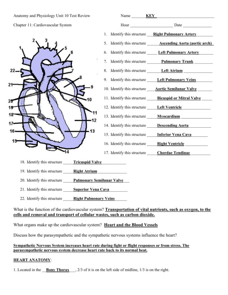
Anatomy and Physiology Unit 10 Test Review Name _______KEY_____________________________ Chapter 11: Cardiovascular System Hour _____________________ Date ______________ 1. Identify this structure ___Right Pulmonary Artery________ 5. Identify this structure ______Ascending Aorta (aortic arch)_ 6. Identify this structure _____ Left Pulmonary Artery_______ 7. Identify this structure _______Pulmonary Trunk _________ 8. Identify this structure _______Left Atrium ______________ 9. Identify this structure _____Left Pulmonary Veins________ 10. Identify this structure ____Aortic Semilunar Valve _______ 11. Identify this structure _____Bicuspid or Mitral Valve _____ 12. Identify this structure _____Left Ventricle______________ 13. Identify this structure _____Myocardium _______________ 14. Identify this structure _____Descending Aorta___________ 15. Identify this structure _____Inferior Vena Cava________ 16. Identify this structure _____Right Ventricle ___________ 17. Identify this structure _____Chordae Tendinae__________ 18. Identify this structure _____Tricuspid Valve____________ 19. Identify this structure _____Right Atrium ______________ 20. Identify this structure _____Pulmonary Semilunar Valve___ 21. Identify this structure _____Superior Vena Cava_________ 22. Identify this structure _____Right Pulmonary Veins______ What is the function of the cardiovascular system? Transportation of vital nutrients, such as oxygen, to the cells and removal and transport of cellular wastes, such as carbon dioxide. What organs make up the cardiovascular system? Heart and the Blood Vessels Discuss how the parasympathetic and the sympathetic nervous systems influence the heart? Sympathetic Nervous System increases heart rate during fight or flight responses or from stress. The parasympathetic nervous system decrease heart rate back to its normal beat. HEART ANATOMY: 1. Located in the __Bony Thorax___, 2/3 of it is on the left side of midline, 1/3 is on the right. 2. Size and shape is similar to a __Human Fist__ 3. Weighs approx. 11 oz. and pumps __Blood___ 4. Where is the heart located? Medial to the lungs and anterior to the spine inside of the bony thorax. 5. What are the three main layers of the heart? Describe them. Epicardium – also called the visceral pericardium, outermost layer of the heart Myocardium – cardiac muscle layer of the heart, thickest layer Endocardium – innermost layer of endothelium 4 CHAMBERS: Right Atrium - receiving chamber (from _Vena Cavae, both superior and inferior_) Left Atrium - receiving chamber (from __left and right pulmonary veins__) Right Ventricle - discharging chamber (to _pulmonary trunk and pulmonary arteries__) Left Ventricle - discharging chamber (to __aorta_) _Septum_ = the wall between chambers _Myocardium_ = walls of twisted and whorled cardiac muscle, the layer that actually contracts _Endocardium__ = smooth lining of heart chambers (inflammation of this is called endocarditis) _Pericardium_ = covering sac containing lubricating fluid 2 layers of Heart’s Protective covering: 1. _Visceral__ pericardium = inner layer (also called epicardium) 2. _Parietal__ pericardium = outer layer HEART VALVES: 1. ____Tricuspid____ Valve = between right atrium and ventricle 2. ____Bicuspid________ (Mitral Valve) = between left atrium and ventricle 3. ____Aortic Semilunar_______ valve = at beginning of aorta 4. ____Pulmonary Semilunar______ valve = at beginning of pulmonary artery Heart Sounds: "lub" = closing of __AV or atrioventricular__valves (ventricles contract) "dub" = closing of __Semilunar_ valves (heart relaxes) VESSELS CONNECTED TO HEART: __Superior Vena Cava___ - brings blood to right atrium from above __Inferior Vena Cava_____ - brings blood to right atrium from below __Pulmonary_ Artery - takes blood from right ventricle to Lungs __Pulmonary veins_ - brings blood from lungs to left atrium __Aorta_- takes blood from left ventricle to body __Myocardial__ INFARCTION = Death or damage to the tissue of the myocardium (usually because of a blocked artery) __Angina__ PECTORIS = Chest pain caused by insufficient oxygen to the heart. (usually occurs during exertion or exercise that increases demand for oxygen) CONDUCTION SYSTEM OF HEART ____Sinoatrial Node___ (SA node): Instability of the nerve membrane generates a nerve impulse by itself. Can be regulated by sympathetic and parasympathetic nervous system. Impulse spreads out over atria causing them to contract. ___Atrioventricular__ Node (AV node): Transmits impulse from atria to the ventricles along special tract of fibers called the __Atrioventricular or branch bundles______. These bundles branch out into special conduction fibers called ____Purkinje Fibers____. If the AV node is destroyed or damaged the atria and ventricles will contract independently. BLOOD VESSELS ARTERIES (also includes arterioles) Primary function: distribute blood away from heart 3 layers: 1. Tunica __Externa__ - outer connective tissue 2. Tunica __Media___ - middle layer; contains vasodilators and vasoconstrictors 3. Tunica __Intima___ - inner layer, single layer of endothelial cells. VEINS Primary function: veins return blood to _Heart_ The muscle layer is much _Thinner_ than arteries. Veins in extremities contain __Valves_. List the factors which assist in returning blood to the heart Contraction of skeletal Muscle, Inhalation (breathing) creating differences in pressure and suction, and valvular shutting to prevent backflow. CAPILLARIES Primary function: _diffusion and exchange__ between blood and cells What are the two types of capillaries and what does each do? Vascular Shunts – create a passageway between arterioles and venules, true capillaries are where nutrient/waste exchange occurs Capillaries are made up of a single layer of endothelial cells. How does that affect their function? One single layer of cells allows for easy diffusion and exchange between nutrients and wastes possible. What would happen if blood pressures were high in the capillaries? They would easily burst open and be destroyed CIRCULATION _Systemic__ - Blood flow from the heart to all parts of the body and then back again. _Pulmonary__ - Blood flow from the heart to lungs and back _Hepatic Portal__ - Blood flows from spleen, stomach, pancreas, gall bladder, and intestines into the liver by way of the portal vein. Don’t worry about this! BLOOD PRESSURE Define the term blood pressure, name the type of blood vessels where blood pressure is significant, and name the normal (average) value in a resting adult. Blood pressure is the pressure that blood exerts against the inner walls of the blood vessels. Usually refers to the pressure of the largest systemic arteries. Normal BP is 120 mmHg/80 mmHg. What is a pulse? Pressure wave resulting from expansion and recoil of an artery that occurs from each left ventricle contraction. Systolic pressure = _Normal is 120 mmHg_ Diastolic pressure = _Normal is 80 mmHg_ What are some of the factors that influence blood pressure? (don’t forget renal and neural factors!) Diet and exercise will influence BP. Sympathetic NS causes vasoconstriction which increases BP. Kidneys alter blood pressure by altering blood volume. Kidneys also release rennin that will cause vasoconstriction. Temperature will cause vessels to shrink and increase BP. Salty food increases BP. Chemicals like nicotine and epinephrine cause vasoconstriction, raising BP, and chemicals like alcohol and histamine cause vasodilation lowering BP. Review Questions Continued Which chamber has the thickest wall (myocardium)? Why? Ventricles because they have to pump the blood away from the heart where the blood has the greatest distance to go. Do pulmonary veins contain oxygenated blood? Yes Do pulmonary arteries contain oxygenated blood? NO Where do coronary veins drain to? Coronary sinus and back into the right atrium Describe the conduction system of the heart. Impulse keeps heart pumping in a consistent manner. Impulse first is generated by SA or sinoatrial node (pacemaker) and sends it across to the AV or atrioventricular node which then sends it to the AV or atrioventricular bundle (bundle of His), then to the bundle branches, then to the purkinje fibers which cause myocardial contraction. What is the central blood-containing space of a blood vessel called? Lumen How does the structure of veins differ from arteries? Veins have larger lumens and thinner tunica media, and arteries have smaller lumens and very thick tunica externa and tunica media. What is the aorta? Major artery that receives blood from left ventricle and delivers it to the body. Define vasodilation and vasoconstriction. Vasodilation is blood vessel dilation or widening, and vasoconstriction is when blood vessels constrict due to factors like cold temperatures, etc. What causes vasoconstriction? Cold temperatures, Sympathetic nervous causes like when we lie down and get up fast, when blood volume drops vessels will constrict to compensate, when we exercise vigorously vessels, except for in skeletal muscle, will constrict. What is congestive heart failure? What are the two types? Briefly describe each and include major symptoms. When pumping efficiency of heart is depressed so that circulation is inadequate to meet body’s needs, the balance between cardiac output and venous return is off. Blood will collect in certain chambers and cause the heart to swell in size. Right side leads to distal/peripheral edema (swelling in legs, hands, feet). Left side leads to pulmonary edema (fluid collection in lungs). Tell if the following are characteristic of arteries, capillaries or veins: Presence of smooth muscle allows them to constrict and dilate. veins and arteries Lumens are largest. Veins Have the thickest tunica media. Veins and Arteries Are able to accommodate a large volume of blood. Veins Exposed to the highest pressures of any vessels. Arteries The link between arteries and veins in the pathway of blood. Capillaries Experience the least pressure. Veins The smallest vessels. Capillaries Vessels that transport blood away from the heart. Arteries The tunica externa is the heaviest wall layer. Arteries Presence of elastin allows them to stretch and recoil. Veins and Arteries Walls consist of just a thin tunica intima. Capillaries Role: the exchange of materials between the blood and the interstitial fluid. Capillaries Define the terms tachycardia and bradycardia. Tachycardia is above normal heart rate (very fast), and bradycardia is below normal heart rate (slower). What hormones are involved in regulation of blood pressure and blood flow? Renin from kidneys, thyroxine from thyroid and epinephrine from adrenal cortex. What is hypertension? High blood pressure What is hypotension? Low blood pressure What is atherosclerosis? When a fatty plaque builds up on the walls of arteries decreasing lumen size and how much blood can flow at a given time. Can lead to blockage and stroke. This disease affects the coronary arteries and aorta What is stroke volume? amount of blood pumped out of each ventricle with each heartbeat. Discuss the factors that affect cardiac output. Cardiac output (CO) is stroke volume (SV) X heart rate (HR). We can influence our cardiac output by changing either of these factors. We can exercise, change our diets, get injured and bleed which would cause our heart rate to jump, and exercising would also create more force and move blood to the heart faster therefore increasing stroke volume. Discuss the factors the regulate heart rate. Bleeding of any kind would lower blood pressure and cause the heart to pump faster. Stress, such as fight or flight would cause the release of epinephrine which would cause increased heart rate. The metabolic hormone thyroxine also increases heart rate. Exercise causes the heart to pump faster and low oxygen and blood glucose would also cause the heart to pump faster. Other factors include gender, diet, temperature, presence of disease like congestive heart failure, and some ions and chemicals. What is the term referring to all of the events associated with one heartbeat called? __Cardiac Cycle_____ Define the terms systole and diastole. Systole is ventricular contraction and diastole is ventricular relaxation. Name the common term for the sinoatrial (SA) node _____Pacemaker____________ What is the function of serous fluid around the heart? To reduce friction between heart beats and prevent heart layers from sticking together. Label the artery, capillary and vein. Also label the layers of each.


