Sonic hedgehog Mediates the PolarizingActivityof the ZPA
advertisement
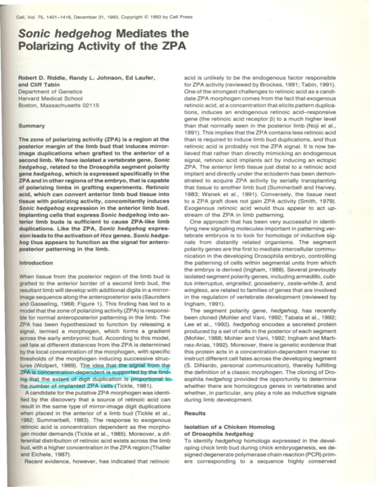
.....Cell, Vol. 75, 1401-1416, December 31, 1993, Copyright @ 1993 by Cell Press Sonic hedgehog Mediates the PolarizingActivityof the ZPA Robert D. Riddle, Randy L. Johnson, and Cliff Tabin Departmentof Genetics HarvardMedical School Boston,Massachusetts02115 Ed Laufer, Summary The zone of polarizing activity (ZPA) is a region at the posterior margin of the limb bud that induces mirrorimage duplications when grafted to the anterior of a second limb. We have isolated a vertebrate gene, Sonic hedgehog, related to the Drosophila segment polarity gene hedgehog, which is expressed specifically in the ZPAand in other regions ofthe embryo, that is capable of polarizing limbs in grafting experiments. Retinoic acid, which can convert anterior limb bud tissue into tissue with polarizing activity, concomitantly induces Sonic hedgehog expression in the anterior limb bud. Implanting cells that express Sonic hedgehog into anterior limb buds is sufficient to cause ZPA-like limb duplications. Like the ZPA, Sonic hedgehog expression leads to the activation of Hox genes. Sonic hedgehog thus appears to function as the signal for anteroposterior patterning in the limb. Introduction When tissue from the posterior region of the limb bud is grafted to the anterior border of a second limb bud, the resultant limb will develop with additional digits in a mirrorimage sequence along the anteroposterior axis (Saunders and Gasseling, 1968; Figure 1). This finding has led to a modelthat the zone of polarizing activity (ZPA) is responsible for normal anteroposterior patterning in the limb. The ZPA has been hypothesized to function by releasing a signal, termed a morphogen, which forms a gradient across the early embryonic bud. According to this model, cell fate at different distances from the ZPA is determined by the local concentration of the morphogen, with specific thresholds of the morphogen inducing successive structures (Wolpert, 1969). T~e idea that the signal from ttw ZPA is concentration-dependent is supported by the find.. ing that toe extent of digit duplication is proportional to.. the number of implanted ZPA cells. (Tickle, 1981). A candidate for the putative ZPA morphogen was identified by the discovery that a source of retinoic acid can result in the same type of mirror-image digit duplications when placed in the anterior of a limb bud (Tickle et aI., 1982; Summerbell, 1983). The response to exogenous retinoic acid is concentration dependent as the morphogen model demands (Tickle et aI., 1985). Moreover, a differential distribution of retinoic acid exists across the limb bud,with a higher concentration in the ZPA region (Thaller and Eichele, 1987). Recent evidence, however, has indicated that retinoic acid is unlikely to be the endogenous factor responsible for ZPA activity (reviewed by Brockes, 1991; Tabin, 1991). One of the strongest challenges to retinoic acid as a candidate ZPA morphogen comes from the fact that exogenous retinoic acid, at a concentration that elicits pattern duplications, induces an endogenous retinoic acid-responsive gene (the retinoic acid receptor (3)to a much higher level than that normally seen in the posterior limb (Noji et aI., 1991). This implies that the ZPA contains less retinoic acid than is required to induce limb bud duplications, and thus retinoic acid is probably not the ZPA signal. It is now believed that rather than directly mimicking an endogenous signal, retinoic acid implants act by inducing an ectopic ZPA. The anterior limb tissue just distal to a retinoic acid implant and directly under the ectoderm has been demonstrated to acquire ZPA activity by serially transplanting that tissue to another limb bud (Summerbell and Harvey, 1983; Wanek et aI., 1991). Conversely, the tissue next to a ZPA graft does not gain ZPA activity (Smith,1979). Exogenous retinoic acid would thus appear to act upstream of the ZPA in limb patterning. One approach that has been very successful in identifying new signaling molecules important in patterning vertebrate embryos is to look for homologs of inductive signals from distantly related organisms. The segment polarity genes are the first to mediate intercellular communication in the developing Drosophila embryo, controlling the patterning of cells within segmental units from which the embryo is derived (Ingham, 1988). Several previously isolated segment polarity genes, including armadillo, cubitus interruptus, engrailed, gooseberry, zeste-white-3, and wingless, are related to families of genes that are involved in the regulation of vertebrate development (reviewed by Ingham, 1991). The segment polarity gene, hedgehog, has recently been cloned (Mohler and Vani, 1992; Tabata et aI., 1992; Lee et aI., 1992). hedgehog encodes a secreted protein produced by a set of cells in the posterior of each segment (Mohler, 1988; Mohler and Vani, 1992; Ingham and Martinez-Arias, 1992). Moreover, there is genetic evidence that this protein acts in a concentration-dependent manner to instruct different cell fates across the developing segment (S. DiNardo, personal communication), thereby fulfilling the definition of a classic morphogen. The cloning of Drosophila hedgehog provided the opportunity to determine whether there are homologous genes in vertebrates and whether, in particular, any playa role as inductive signals during limb development. Results Isolation of a Chicken Homolog of Drosophila hedgehog To identify hedgehog homologs expressed in the developing chick limb bud during chick embryogenesis, we designed degenerate polymerase chain reaction (PCR) primers corresponding to a sequence highly conserved ~ ~ Cell 1402 rs~~ Posterior . J N III [)A)~\~ III IV Figure 1. Limb Patterning and the ZPA A model for anterioposterior patterning, based on Saunders and Gasseling (1968). The left portion of the top panel diagrams a schematized stage 20 limb bud. The somites are illustrated as blocks along the left margin of the limb bud; the right portion of the same panel illustrates the mature wing. The hatched region on the posterior limb is the ZPA. The overlying distal ectoderm is the AER. Normally, the developed wing contains three digits: II, III, and IV. The bottom panel shows the result of transplanting a ZPA from one limb bud to the anterior margin of another. The mature limb now contains six digits (IV, III, II, II, III,and IV)in a mirror-image duplication of the normal pattern. The large arrows in both panels represent the signal produced by the ZPA that acts to specify digit identity. .. .DN" 'v,.. - - - - - - - - - - - -- - - - - - -- - - - - -- - -- -- - - - - - d - - GO","" "",.c-,= H.,n ceHDHC",""'" KH"" """"""."""'"'"' :, ; ~ ; ; Q; ; ; Q; ~ ; ~ ; ~~ ; Q~ ~ ~: ;[]J; : ~: ;[]J: ~; : z ~ ; ; ~~ ~::i~,z;',1;;;::::'~"x ~: ;;; :mill~~~ ~[;j~i ~~[]J~ITIl~ ~[ID~m: :~o~; ~[ID~rn:[;j~rn; :J;J]~[]J:!r:TI:[j]~~~rn~I~; ~rn~rn;;[] ~":,~:i;;:~'"'"' ~~~ ~:1: =,~:i;;:~"" i:~;rn;rn~If]:I; ~::I;m~ ~1B~m;Iill];[ID;:m g;;io":,z;'="oo i~I:;:;:: ~1~rn::~~I;:;;:~:: ~:~::;; ~:~IM~:rn:: =,~:;;;:~'"'"' i:~[]:Ii; ~:I~~:~~ ~~:: :m;;: ~rn~~IDI:~~ :~;::;IDI ;;~ :1; ~~~~I~~~~~~~; ;IT];: ~i~;~: ~::i~,~:;;;::::',,;""' ~: ~~~;~: ~~~[;]~~ ~":,~:i;;:~ between Drosophila hedgehog (Lee et al., 1992; Mohler and Vani, 1992) and mouse Desert hedgehog (Dhh, a mouse homolog of hedgehog not expressed in embryonic limb buds; isolated in a parallel study by Echelard et aI., 1993 [this issue of Celm. Using genomic DNA as a template, a PCR fragment of the expected size was amplified, cloned, and used as a probe to screen an unamplified cDNA library prepared from stage 22 limb bud RNA (Hamburger and Hamilton, 1951). A 1.6 kb cDNA clone containing a single long open reading frame was isolated. Conceptual translation of this open reading frame predicts a protein of 425 amino acids that is highly related to Drosophila hedgehog (Figure 2). The gene encoded by this cDNA was named Sonic hedgehog (after the Sega computer game cartoon character). Over the entire open reading frame, the Drosophila and chicken proteins are 48% identical, a value that rises to 78% when conservative amino acid substitutions are included. The predicted Drosophila protein extends 62 amino acids beyond that of Sonic hedgehog at its amino terminus. This N-terminal extension corresponds to a region just prior to the putative signal peptide of the fly protein and has been postulated to be removed during processing of the secreted form of Drosophila hedgehog (Lee et aI., 1992). Although chicken Sonic hedgehog does not contain sequences corresponding to the N-terminus of Drosophila hedgehog, it is likely to be secreted. The sequence of residues 1-26 of the Sonic hedgehog protein is highly hydrophobic (Kyte and Doolittle, 1982) and matches well with consensus sequences for eukaryotic signal peptides (Landry and Gierasch, 1993). There is also a predicted cleavage site after the first 26 amino acids of the Sonic hedgehog protein (von Heijne, 1986). Southern blot analysis of genomic DNA using Sonic hedgehog as a probe yielded three unique bands, suggesting that there are at least two other hedgehog homologs in the chicken genome (data not shown). Supporting this, multiple hedgehog-related genes have been isolated from the mouse (Echelard et aI., 1993) and zebrafish (Krauss et aI., 1993 [this issue of Celm. Based on sequence and expression patterns, one of the homologs in both the mouse and zebrafish is orthologous to chicken Sonic hedgehog. All of the vertebrate proteins are highly related at the amino acid level and all have similar predicted structures. ;:: : ~~:[]J;;[;]~m; ;~~:?' ~?~~~; :rn~;~~~;[]~: ~:: ~,,:,~:;;"~""" ;:; ;; :~i~; ~::; g:1B;l:I~ ;~: ;~: ~:: ~;~~ :I~:t ;I:;[] ;::.iio":,~:i;;::::'oornoo e ;::m;:i:I:~;I:;i;: ~il:: ~rn~m~~[]~[]~ ~;IB;;;[:J~~;=,~~'"'"' m ; ;[]Ji: ~; ~:~ i ~~~ ~~~[[ill: m ,eG""" mD ~~[TI: ~~~~~; ~~~;C:J=,~:~oornoo ,",C"," ~"c-",oornoo """"'"'~,,=~ Figure 2. Predicted Amino Acid Sequence of the Chick Sonic hedgehog and Its Similarity to Drosophila hedgehog An alignment comparing the amino acid sequences of chick Sonic hedgehog with its Drosophila homolog (Lee et aI., 1992; Mohler and Vani, 1992). Sonic hedgehog residues 1-26 correspond to the proposed signal peptide. In the chick Sonic hedgehog cDNA pHH-2, stop codons precede the first methionine (data not shown). Identical residues are enclosed by boxes, and gaps have been introduced to optimize similarity. The nucleotide sequence of the Sonic hedgehog cDNA has been submitted to GenBank. Sonic hedgehog Expression Colocalizes with ZPA Activity Sonic hedgehog was isolated on the basis of its expression in stage 22 embryonic limbs. To determine whether the Sonic hedgehog message was restricted to a subset of limb bud cells, whole-mount in situ hybridization was performed using a riboprobe corresponding to the entire Sonic hedgehog cDNA clone. By stage 21, Sonic hedgehog expression is apparent in posterior regions of both the forelimb (Figure 3A) and the hindlimb (data not shown). Sections reveal that expression of Sonic hedgehog in limb buds is limited to the mesenchyme (Figure 3B). The tissue of the limb bud that displays Sonic hedgehog expression Sonic 1403 hedgehog Mediates ZPA Activity nQST . 13~ a Figure 3. Sonic hedgehog Is Expressed in the Posterior Mesenchyme of Limb Buds In Figures 3-6, Sonic hedgehog mRNA was detected by whole-mount in situ hybridization. Control hybridizations using a Sonic hedgehog sense probe gave no specific signal. (A) Close-up view of a stage 20-21 left wing bud (WB). Sonic hedgehog message is found in the posterior/proximal region of the bud (arrow). (B) Slightly oblique section through the posterior limb bud of a stage 21 embryo. Sonic hedgehog mRNA is detected in the mesenchyme of the limb bud. No expression is observed in the ectoderm, including the AER. At this stage, staining can also be observed in the notochord (NC) and the floor plate region of the neural tube (NT). Abbreviations: ANT, anterior; and POST, posterior. '-- .. Cell 1404 t , A B A Stage 17 , . A B Stage 21 Stage 19 r= I5 B ~9 . I I L A A B Stage 23 B Stage 25 ~=J t t A B Stage 27 anterior i' i posterior A B Stage 29 .Sonic hedgehog 1405 Mediates ZPA Activity corresponds to the ZPA, a region of the posterior mesenchyme that is capable of causing anteroposterior mirrorimage duplications when transplanted into a second limb bud. ZPA activity has been carefully mapped both spatially and temporally within the limb bud (Honig and Summerbell, 1985). In these experiments, small blocks of limb bud tissue from various locations and stages of chick embryogenesis were grafted to the anterior of host limb buds, and the strength of ZPA activity was quantified according to the degrees of digit duplication (Figure 4A). Polarizing activity is first weakly detectable along the flank prior to limb bud outgrowth. This activity reaches its maximal strength at stage 19 in the proximal posterior margin of the limb bud. By stage 23, polarizing activity extends along the full length of the posterior border of the limb bud and then shifts distally so that by stage 25 it is no longer detectable at the base of the limb. ZPA activity then fades distally until it is last detected at stage 29. This detailed map of endogenous polarizing activity provided the opportunity to determine the extent of the correlation between the spatial pattern of ZP A activity and Sonic hedgehog expression over a range of developmental stages. Whole-mount in situ hybridization was used to assay the spatial and temporal pattern of Sonic hedgehog expression in the limb bud (Figure 4B). Sonic hedgehog expression is not detected until stage 17, during the initiation of limb bud formation, at which time it is weakly observed in a punctate pattern. From that point onwards, the Sonic hedgehog expression pattern exactly matches the location of the ZPA as determined by Honig and Summerbell (1985), both in position and in intensity of expression (Figure 4). Several other embryonic tissues are also able to cause ZPA-like pattern alterations when engrafted into limb buds. These tissues include Hensen's node (Saunders and Gasseling, 1983; Hornbruch and Wolpert, 1986; Stocker and Carlson, 1990), the notochord (Wagner et aI., 1990), and the floor plate of the neural tube (Wagner et aI., 1990). Sonic hedgehog is strongly expressed in each of these tissues (Figure 5). Induction of Sonic hedgehogExpressionby Retinoic Acid A source of retinoic acid placed at the anterior margin of the limb bud can induce ectopic ZPA tissue that is capable of causing mirror-image duplications (Summerbell and Harvey, 1983; Wanek et al., 1991). The commitment to form ZPA tissue is "not an immediate response to retinoic acid but rather takes approximately 14 hr to develop (Wanek et aI., 1991). When it does develop, the polarizing B' , <." Figure 5. Sonichedgehog IsExpressed in Hensen's Node, Notochord, and Floor Plate of the Neural Tube Embryos in (A) and (B) are dorsal views anterior to the top. (A) Stage 4+ embryo. Staining is observed at the anterior end of the primitivestreak correspondingto Hensen's node (HN).Sonic hedgehog expression is also observed in midline tissues anterior to the node. (B) Stage 8+ embryo. Sonic hedgehog expression is observed along the midline of the embryo from just anterior to the node to the rostral extent of the head process. The node itself no longer expresses Sonic hedgehog at this stage. (C) Transverse section of a Stage 8+ embryo at a level just anterior to the somites. Prominent Sonic hedgehog expression is evident in the notochord (NC) and the floor plate (FP). activity is not found surrounding the implanted retinoic acid source; activity is found only distal to the source, in the mesenchyme along the margin of the limb bud. If Sonic hedgehog expression is truly indicative of ZPA tissue, then it should be induced in the ectopic ZPA formed in response to retinoic acid. To test this, we implanted retinoic acid-soaked beads in the anterior of limb buds and assayed for expression of Sonic hedgehog after various lengths of time using whole-mount in situ hybridization. As the limb bud grows, the bead remains embedded proximally. Ectopic Sonic hedgehog expression is detected in the mesenchyme 24 hr after bead implantation (Figure 6A). This expression is found a short distance from the distal edge of the bead. By 36 hr, Sonic hedgehog is strongly expressed distal to the bead in a stripe just under Figure 4. Sonic hedgehog Expression and the ZPA during Limb Bud Outgrowth The regions of the limb that contain polarizing activity were mapped by Honig and Summerbell (1985). (A) Here are shown reproductions of the relative polarizing strength of limb tissue at specific developmental stages as originally drawn by Honig andSummerbell(1985).Theunboxedregionswerefoundnot to havesignificantpolarizingactivity. " (B) Here are shown representative whole-mount in situ analyses of Sonic hedgehog mRNA at the same developmental time points. The arrowhead in the stage 17 photograph points to the location of faint Sonic hedgehog staining. Cell 1406 24 Hours 48 Hours 36 Hours --- ~ "., ........ 'It ~ A ~ .,.., B c Figure 6. Retinoic Acid and Sonic hedgehog Expression Stage 20 limb buds were implanted with beads soaked in 1 mglml retinoic acid. The beads were implanted in the anterior margin under the AER. (A, S, and C) Here are shown Sonic hedgehog expression at 24, 36, and 48 hr post implantation. The black arrowheads indicate ectopic Sonic hedgehog expression along the anterior margin; the white arrowheads indicate the retinoic acid bead implant. Sonic hedgehog expression was visualized by whole-mount in situ analysis. Limbs implanted with control dimethyl sulfoxide-soaked beads showed no ectopic Sonic hedgehog expression (data not shown). the anterior ectoderm in a mirror-image pattern relative to the endogenous Sonic hedgehog expression in the posterior of the limb bud (Figure 68). At 48 hr, the retinoic acidindoced Sonic hedgehog message fades in concert with the endogenous message (Figure 6C). Effects of Ectopic Expression of Sonic hedgehog on Limb Patterning The normal expression pattern of Sonic hedgehog, as well as that induced by retinoic acid, is consistent with Sonic hedgehog being a signal produced by the ZPA. To determine whether Sonic hedgehog expression is sufficient for ZPA activity, we ectopically expressed the gene within the limb bud. 'n most of the experiments, we utilized a variant of a replication-competent retroviral vector called RCAS (Hughes et aI., 1987), both as a vehicle to introduce the Sonic hedgehog cDNA into chick cells and to drive its expression. To control the region infected with the Sonic hedgehog-RCAS virus, we took advantage of the fact that there are subtypes of avian retroviruses that have host ranges restricted to particular strains of chickens (Weiss, 1984; Fekete and Cepko, 1993a). Thus, a vector with a type E envelope protein (RCAS-E, Fekete and Cepko, 1993b) is unable to infect the cells of the standard specific pathogen-free outbred chick embryos routinely used in our lab. However, RCAS-E is able to infect cells from chick embryos of line 15b. In the majority of experiments, we infected primary chick embryo fibroblasts (CEFs) prepared from line 15b embryos in vitro. Infected cells were pelleted and implanted into a slit made in the anterior of virally resistant host limb buds (Figure 7). Due to the restricted host range of the vector, infection was limited to engrafted cells and did not spread into host limb bud tissues (Figure 7). To determine the fate of cells implanted according to our protocols and to control for any effect of our implant procedure, we implanted CEFs infected with an RCAS-E vector expressing human placental alkaline phosphatase. Alkaline phosphatase expression can be easily monitored histochemically, and the location of infected cells can thus be conveniently followed at any stage. Within 24 hr, implanted cells are dispersed proximally and distally along the anterior margin of the limb bud (Figure 8). Subsequently, alkaline phosphatase-positive cells are seen to disperse throughout the anterior portion of the limb and into the flank of the embryo (data not shown). Control limb buds engrafted with alkaline phosphatase-expressing cells or uninfected cells give rise to limbs with structures indistinguishable from unoperated wild-type limbs (Table 1; Figure 9A). Such limbs have the characteristic anteriorto-posterior digit pattern 2-3-4. ZPA grafts give rise to a variety of patterns of digits depending on the placement of the graft within the bud (Tickle et aI., 1975) and the amount of tissue engrafted (Tickle, 1981). In some instances, the result can be;as weak as the duplication of a single digit 2. Howeve~ in optimal cases, the ZPA graft evokes the production of a full mirror-image duplication of digits 4-3-2-2-3-4 ~r 4-3-2-3-4 (Figure 9~ A scoring system has been devised that rates the effectiveness of polarizing activity on the basis of the duplication of the most posterior digit; any graft that leads to the development of a duplication of digit 4 has been defined as reflecting 100% polarizing activity (Honig and Summerbell, 1985). Grafts of 15b fibroblasts expressing Sonic hedgehCifuesuited in a range of ZPA-like phenotypes. In some instances, the resultant limbs deviate from the wild type solely by the presence of a mirror-image duplication of digit " Sonic hedgehog 1407 Mediates ZPA Activity Transfect sonic/RCAS(E) and allow viral spread Infectable strain of donor cells 15bCEF + Pellet + Implant + Anterior Resistant strain of host embryo Posterior Stage 19-23 embryo Figure 7. Assay for Polarizing Activity Sonic hedgehog-RCAS-E1 or Sonic hedgehog-RCAS-E2 were transfected into line 15b CEFs and then incubated until the cells were completely infected (Morgan et aI., 1992). These cells were lightly trypsinized, pelleted, and implanted into the anterior margin of stage 19-23 embryos resistant to RCAS-E infection. Thus, the virus was unable to infect surrounding tissue, and the region expressing levels of Sonic hedgehog was confined Experimental Procedures). to the engrafted high cells (see 2 (Table 1; Figure 9C). The most common digit phenotype resulting from grafting Sonic hedgehog-RCAS-infected CEF cells is a mirror-image duplication of digits 4 and 3 with digit 2 missing: 4-3-3-4. In many such cases, the two central digits appear fused (Table 1; Figure 9E). In a number of the cases, the grafts induced full mirror-image duplications of the digits equivalent to optimal ZPA grafts 4-3-2-2-3-4 (Table 1; Figure 9D). Besides digit duplications,ectopic expression of Sonic hedgehog also gave rise to occasional duplications of proximal elements, including the radius or ulna, humerus, and coracoid (Table 1; Figures 9F and 9G). Many of these are clearly mirror-image duplications (for example, the humerus in Figure 9G). Thus, while these proximal phenotypes are not features of ZPA grafts, they are consistent with an anterior-toposterior respecification of cell fate. In some instances, most commonly when the radius or ulna was duplicated, more complex digit patterns were observed. Typically, an additional digit 3 was formed distal to a duplicated radius (Figure 9F). The mirror-image duplications caused by ZPA grafts are not limited to skeletal elements. For example, feather buds are normally present only along the posterior edge of the limb (Figure 10A). Limbs exhibiting mirror-image duplications as a result of ectopic Sonic hedgehog expression have feather buds on both their anterior and posterior edges, similar to those observed in ZPA grafts (Figures 10B and 10C). While ZPA grafts have a powerful ability to alter limb pattern when placed at the anterior margin of a limb bud, they have no effect when placed at the posterior margin (Saunders and Gasseling, 1968). Presumably, the lack of posterior effect results from polarizing activity already being present in that region of the bud. Consistent with this, grafts of Sonic hedgehog-expressing cells placed in the posterior of limb buds never result in changes in the number of digits (Table 1; see Figure 9H). Some such grafts did produce distortions in the shape of limb elements; most commonly, a slight posterior curvature in the distal tips of digits 3 and 4 was seen when compared with wild-type wings (see Figure 9H). Effect of Ectopic Sonic hedgehog Expression on Hoxd Gene Activity The correct expression of Hoxd genes is part of the process by which specific skeletal limb elements are determined (Morgan et aI., 1992). These genes are normally expressed in a nested pattern emanating from the posterior margin of the limb bud (Dolle et aI., 1989; IzpisuaBelmonte et aI., 1991). A transplant of aZPA into the anterior of a chick limb bud ectopically activates sequential transcription of Hoxd genes in a pattern that mirrors the normal sequence of Hoxd gene expression (Noh no et aI., 1991; Izpisua-Belmonte et aI., 1991). Since ectopic Sonic hedgehog expression leads to the same pattern duplications as a ZPA graft, we reasoned that Sonic hedgehog would. also lead to sequential activation of Hoxd genes. To test this hypothesis, anterior buds were injected with Sonic hedgehog-RCAS-A2, a virus capable of directly infecting host strains of chicken embryos. This approach does not strictly limit the region expressing Sonic hedgehog (being that it is only moderately controlled by the timing, location, and titer of the viral injection), and thus it might be expected to give a more variable result. However, experiments testing the kinetics of viral spread in infected limb buds indicate that infected cells remain localized near the anterior margin of the bud for at least 48 hr (data not shown). Hoxd gene expression was monitored at various times post infection by whole-mount in situ hybridization. As expected, these genes are activated in a mirror-image pattern relative to their expression in the posterior of controllimbs. The temporal sequence of their activation also parallels that seen in anterior limb buds in ZP A transplants. For example, after 24 hr, Hoxd-11 is strongly activated, while Hoxd-13 is barely detectable (data not shown). However, by 36 hr, Hoxd-13 is expressed in a mirror-image \ -- -- ~ Cell 1408 24 Hours 0 Hours " .. A B Figure 8. Location of Implanted Cells during Development Cells expressing RCASBP/AP(E) were implanted into a viral-resistant limb as described in Figure 4. (A) This panel shows the implant (stained with Nile Blue) immediately after implantation. (B) This panel shows the same cells 24 hr after implantation (visualized by alkaline phosphatase staining, Fekete and Cepko, 1993a). symmetrical pattern in the broadened distal region of infected limb buds (Figure 11). Discussion The Predicted Properties of the Protein Encoded by Sonic hedgehog Are Consistent with It Being an Intercellular Signal We have cloned a cDNA related to the Drosophilagene hedgehog.There is strong genetic evidence that hedge- hog functions as an intercellular signal during Drosophila embryogenesis (Ingham, 1991). Consistent with such a role, the Drosophila hedgehog protein was predicted to contain an effective signal sequence, and this peptide was demonstrated to direct secretion in vitro into microsomes (Lee et aI., 1992). Moreover, hedgehog has been shown to be secreted in vivo (Taylor, 1993). Computer analysis of the predicted Sonic hedgehog protein suggests that it too is a secreted protein, consistent with it also serving as an intercellular signal during vertebrate embryogenesis. Table 1. Digit and Proximal Limb Bone Duplications Induced by Sonic hedgehog Grafts Percentage Most Posterior Digit Duplicated (n) Percentage Proximal Element Duplicated (n) Implant (n) II III IV WT Radius/Ulnac Humerus Coracoid WTd Anterior Sonic hedgehog" (54) Alkaline phosphatase (10) Posterior Sonic hedgehog (7) Anterior stage 22" (6) Anterior stage 23" (4) 11 (6) 0 0 0 0 20 (11) 0 0 0 44(24) 0 0 100 (6) 50(2) 24(13) 100 (10) 100 (7) 0 (0) 0 (0) 28 (15) 0 0 11 (6) 0 0 9 (5) 0 0 63(34) 100 (10) 100 (7) 50(2) Grafts of line 15b CEFs infected with the Sonic hedgehog-RCAS-E1, Sonic hedgehog-RCAS-E2, or RCASBP/AP E) viruses were implanted into the anterior or posterior margin of stage 19-23 limb buds. Sonic hedgehog-RCAS-E1-infected and Sonic hedgehog-RCAS-E2-infected cells gave equivalent results and are tabulated together. Embryos were harvested at day 10, seven days after grafting, stained with alcian green, cleared in methyl salicylate, and scored. (Left side) The numerical identity of the most posterior digit duplicated in each limb scored. The percentage of limbs with each particular value is shown, with the absolute number indicated in parentheses. (Right side) The number of limbs with each of the indicated long bones duplicated is indicated. To be scored as a duplication, >50% of the length of the bone had to be duplicated. " Three grafts were placed at the distal tip of the limb bud and are scored in the anterior graft category. These embryos are also scored in the anterior sonic hedgehog row of the table. c Radial and ulnar duplications consist of a combination of limbs with duplicated radii or ulnae, as well as a single ulna or radius, and limbs in which the radius was apparently transformed into an ulna, as judged by morphological criteria. " d Doesnot equal100% minusthe sumof the previousthreecolumnsbecausesomelimbshad morethanone proximalelementduplicated. Sonic hedgehog Mediates ZPA Activity 1409 III III A. Control B. ZPA Graft III ,... III C. Son ic Graft D. 'sonic Graft E. Sonic Graft ~ III F. Sonic Graft G. Sonic Graft H. Posterior Sonic Graft Figure 9. Morphology of Grafted Limbs Grafts of ZPA tissue or line 15b CEFs infected with the Sonic hedgehog-RCAS-E2 virus were implanted into the anterior or posterior margin of stage 19-23 limb buds (see Figure 4). Embryos were harvested at day 10, seven days after grafting, stained with alcian green, and cleared in methyl salicylate. The identities of digits (II, III, or IV) and long bones (H [humerus], R [radius], or U [ulna]) are indicated. (A) Unimplanted control limb, digit pattern 2-3-4; (8) anterior ZPA graft, digit pattern 4-3-2-3-4; (C) anterior Sonic hedgehog graft, digit pattern 2-2-3-4; (D) anterior Sonic hedgehog graft, digit pattern 4-3-2-2-3-4; (E) anterior Sonic hedgehog graft, digit pattern 4-3-3-4 (fused digit 3); (F) anterior Sonic hedgehog graft, digit pattern 3-3-3-4; duplicated radius; (G) anterior Sonic hedgehog graft, digit pattern 4-4-3-3-4 (additional digit 4 is hidden from view); duplicated humerus; and (H) posterior Sonic hedgehog graft, digit pattern 2-3-4. Further support for this functional homology is provided by the finding that the zebrafish homolog of Sonic hedgehog is capable of acting equivalently to Drosophila hedgehog when ectopically expressed in the developing Drosophila embryo (Krauss et aI., 1993). Sonic hedgehog Is Coexpressed with ZPA Polarizing Activity in the Limb Bud Analysis of the expression pattern of Sonic hedgehog in the embryonic limb reveals a striking correlation with the region mapped as the ZPA. While surgical manipulations have previously defined these spatial and temporal boundaries, the region is morphologically indistinguishable from the rest of the undifferentiated limb bud, and a molecular marker for the ZPA has been lacking. The discovery of Sonic hedgehog provides a powerful molecular marker for identifying ZPA cells in various mutant backgrounds and experimental situations. The strong correlation between tissue defined as the ZPA and the expression of Sonic hedgehog begins at the earliest stages of limb bud outgrowth. Yet, prior to that, the posterior region of the presumptive limb bud along the flank also has polarizing activity (Hornbruch and Wolpert, 1991). At that time, the mesenchymal cells do not express Sonic hedgehog. However, since these cells are fated to express Sonic hedgehog later, they are likely to activate Sonic hedgehog expression after transplantion to the anterior limb bud margin. ~ Cell 1410 Control ZPA Graft Sonic Graft It ;~' ~i,r !;! '~' ,:j , e [ ~.:i :t, ;, J, , [ "' 1; '' 11 't , , , '" '¥ "" . ' '"~ f ',1; '" I ' '' ' "" ' " '.':' . '"~ ..' .; , .,, " ., ' tLJ".".",." ,, ' ., B A , '~ . ",~, , , .",.,.,. ,,' ,,". .',' ' "!! ;;' ~i/ " ' .' j.. '!!, . , , .,..", ,, C Figure 10, Effect of Ectopic Sonic hedgehog on Feather Bud Formation Grafts of ZPA tissue or line 15b CEFs infected with the Sonic hedgehog-RCAS-E2 virus were implanted into the anterior margin of stage 19-23 limb buds (see Figure 4), Embryos were harvested at day 10, seven days after grafting, stained with alcian green, and cleared in methyl salicylate, Photographs show the region of the radius and ulna. Anterior is to the left and posterior to the right. Note the location of the feather buds that are solely on the posterior edge of the control limb (A), but on both anterior and posterior of the ZPA-grafted (B) and Sonic hedgehog-grafted (C) limbs. I I III A B Figure 11. Expression of Hoxd-13 after Ectopic Sonic hedgehog Expression I I ~ The anterior margins of stage 20 limb buds were infected with the Sonic hedgehog-RCAS-A2 virus. Thirty-six hours after infection, the embryos were harvested, fixed, and assayed for Hoxd-13 expression by whole-mount in situ analysis. (A) Control limb bud and (B) infected limb bud. ~ "''Sonic hedgehog 1411 Mediates ZPA Activity Other embryonic regions that also possess ZPA activity also express Sonic hedgehog. These regions include Hensen's node (Saunders and Gasseling, 1983; Hornbruch and Wolpert, 1986; Stocker and Carlson, 1990), the notochord (Wagner et aI., 1990), and the floor plate of the neural tube (Wagner et aI., 1990). All of these tissues are known to be powerful signaling centers in their own right, each involved in patterning embryonic structures along the midline (Jessell and Melton, 1992). Thus, Sonic hedgehog is likely to playa role in the inductive interactions regulated by those centers. Moreover, the fact that they all express Sonic hedgehog provides an explanation for the common effect observed when they are each grafted into the anterior of a limb bud. Sonic hedgehog Expression Is Sufficient for ZPA Activity The intriguing colocalization of the ZPA with Sonic hedgehog expression in the limb bud suggested that Sonic hedgehog might be part of the mechanism through which the ZPA exerts its influence. To determine whether Sonic hedgehog is sufficient to polarize the limb bud and induce digit duplications, we ectopically expressed Sonic hedgehog in the limb bud. By implanting CEFs expressing Sonic hedgehog in the anterior of limb buds, we obtained mirrorimage duplications similar to those resulting from ZPA transplants. Most phenotypically altered limbs include a duplicated digit 4. Mirror-image duplication of digit 4 has been used as the criterion for attributing full ZPA activity to the donor tissue (Honig and Summerbell, 1985). Several implanted limbs developed a digit pattern, 4-3-2-2-3-4, resembling those obtained from optimal ZPA grafts. The mostfrequent resultant digit pattern was 4-3-3-4. This pattern has previously been interpreted as a response to extremely high ZPA activity because dose response to retinoic acid treatment produces (in response to increasing concentrations of retinoic acid) the following: 2-3-4, 2-2-3-4, 3-2-2-34,4-3-2-2-3-4, and 4-3-3-4 (Tickle et aI., 1985). In the present case, the hyper-ZPA response may be due to the large number of Sonic hedgehog-expressing cells we were able to implant. Alternatively, the extreme degree of pattern modification may be due to the fact that, unlike the endogenous ZPA, the cells implanted in these experiments do not turn off Sonic hedgehog expression late in limb bud development. The phenotypes obtained in Sonic hedgehog grafts also differ from the results of ZPA grafts in that they occasionally produce duplication of proximal elements such as the humerus and coracoid. This is also likely to be a consequence of the persistence of !1onic hedgehog expression when the implanted cells disperse proximally in the limb bud. In ZPA grafts, the polarizing activity (and presumably Sonic hedgehog expression) are only maintained distally, adjacent to the apical ectodermal ridge (AER). The existence of mirror-image-duplicated proximal elements in Sonic hedgehog grafts provides a strong indication that proximal elements are specified along the anteroposterior axis by the same mechanism as are digits. The results of implanting Sonic hedgehog-expressing CEF cells strongly suggest that Sonic hedgehog expression is sufficient to induce the pattern alterations. An alternative explanation is that the CEF cells fortuitously express other required factor(s) normally produced by the ZPA that, in concert with Sonic hedgehog, affect limb pattern. However, this possibility seems unlikely since polarizing activity can also be produced by unrelated cas cells e1<pressingSonic hedgehog (data not shown), as well as by anterior limb bud cells directly infected with Sonic hfdgehog virus. Sonic hedgehog Acts Upstream of Hox Genes in Regulating Anteroposterior Limb Pattern Both ZPA grafts and retinoic acid induce Hoxd gene expression as part of the polarizing process (Nohno et aI., 1991; Izpisua-Belmonte et aI., 1991). Similarly, anterior misexpression of Sonic hedgehog leads to ectopic activation of Hoxd genes. The identification of Sonic hedgehog as an upstream signal in Hoxd gene induction is important both'for understanding the regulation of Hox genes during embryogenesis as well for understanding the mechanisms of action of the ZPA. More work is needed to address the exact sequence of events by which the nested Hoxd gene expression pattern is established in response to Sonic hedgehog activity. The Hoxd genes may be downstream targets of Sonic hedgehog in the appendages of lower vertebrates as well. The homolog of Sonic hedgehog is expressed along the posterior margin of the fin buds in zebrafish in a pattern similar to that found in chicken limb buds (Krauss et aI., 1993). This expression in fish indicates that the signaling system used to pattern the anteroposterior axis of the limb is not novel to tetrapods but rather is very ancient, having been conserved at least since the evolutionary divergence of tetrapods from the line that led to the teleost fish. This is consistent with the suggestion that the evolutionary emergence of the tetrapod limbs made use of a preexisting system for specifying positional differences in the fin based on the expression pattern of the Hox genes (Tabin, 1992; Tabin and Laufer, 1993). Ectopic Retinoic Acid Acts via Sonic hedgehog in the Limb The mirror-image duplications of the limb induced by retinoic acid appear to be.mediated through the induction of Sonic hedgehog. When a retinoic acid bead is implanted in the anterior of a limb bud, it induces ZPA activity in tissue distal to the bead along the edge of the ectoderm, but not in any of the other tissue surrounding the bead (Wanek et aI., 1991). Maintenance of this activity does not require continuous exposure to retinoic acid. Retinoic acid bead implants activate Sonic hedgehog expression in exactly the same region as the induced ZPA activity and in a distribution that mirrors that of the endogenous Sonic hedgehog expression domain in the posterior of the limb bud. While anterior tissue becomes determined to form an ectopic ZPA in response to retinoic acid after as little as 14 hr, it takes 24 hr before phenotypic consequences of this commitment are observed in the adjacent limb bud tissue (for example, the activation of Hox genes). The ki- -----Cell 1412 ZPA -. Sonic Polarization AER-. Figure Proliferation Growth Factors 12. Model for How ZPA Activity is Mediated Sonic hedgehog mesenchyme is proposed to act directly and to indirectly AER. The AER produces affect growth by Sonic hedgehog as a signal mesodermal factors (which to polarize growth through the the are likely to include members of the FGF family, see Discussion) that stimulate the proliferation of the mesenchyme. AER factors also act in a reciprocal fashion to induce the maintenance of the ZPA and thereby support expression of Sonic hedgehog. The result of the combined indirect actions of Sonic hedgehog is the coordinated continued direct and formation of limb pattern. netics of induced Sonic hedgehog expression parallel the induction of ZPA activity; Sonic hedgehog is detectable by 24 hr and is strongly activated by 36 hr. Interestingly, Sonic hedgehog induction in the mesenchyme appears to be dependent on an activity of the AER. Whether retinoic acid is acting directly on the mesenchyme, the AER, or both is not clear. Since ectopic expression of Sonic hedgehog in this region of the limb bud is sufficient to induce mirror-image duplications, it seems very likely that this is the mechanism through which exogenous retinoic acid is acting. Consistent with this idea, the limb bud is competent to respond to Sonic hedgehog at least until stages 22 and 23 (Table 1), yet retinoic acid is not able to induce pattern alterations after stage 21 (Summerbell, 1983). While exogenous retinoic acid can induce Sonic hedgehog expression and ZPA activity in the anterior of the limb bud, its endogenous role, if it has any, in regulating Sonic hedgehog expression is unknown. Sonic hedgehog May Be Involved in Communication between the Limb Mesenchyme and the AER The phenotypic consequences of a ZPA graft actually reflecttwodistinct activities. First, aZPA transplant polarizes the limb bud such that regions in proximity to the graft take on a posterior character. Second, a ZPA transplant results in expanded growth along the distal tip of the limb bud, ultimately producing additional digits. The number of digits in a limb and the anteroposterior identity of each digit are determined separately (reviewed by Tabin, 1992; Laufer, 1993). The ability of the ZPA to influence both of these traits reflects the fact that these two processes are coordinated during normal limb development. One mechanism for achieving this would be for both ZPA activities to be mediated by a single factor. Consistent with this idea, ectopic expression of Sonic hedgehog both broadens the limb bud, (see Figure 11b) leading to the formation of additional digits, and strongly polarizes it, resulting in mirrorimage digit duplications. Thus, Sonic hedgehog appears to be the factor that unifies these activities of the ZPA. While limb polarization induced by Sonic hedgehog could be a direct action on the mesenchyme, Sonic hedge- hog probably induces the formation of additional digits indirectly by acting through the ectoderm. This indirect action is implied by two observations. First, the posterior mesenchyme is required to maintain the AER. Second, the AER is known to produce factors required for limb outgrowth (reviewed by Laufer, 1993). The best candidates for these mitogenic factors are members of the fibroblast growth factor (FGF) family. FGFs are produced by the AER, and exogenous FGFs can stimulate outgrowth and proximodistal patterning of the limb. However, FGFs themselves do not alter digit identity or limb polarity (Niswander and Martin, 1992; Suzuki et aI., 1992; Niswander and Martin, 1993; Riley et aI., 1993; Niswander, et aI., 1993; B. alwin, personal communication). Thus, Sonic hedgehog may be involved in AER maintenance and thereby may regulate production of growth factors required for mesenchymal proliferation. In a reciprocal interaction, the AER is known to be required for maintenance of ZPA activity (Vogel and Tickle, 1993). FGF-4, which is expressed in the posterior AER, can replace the AER in terms of maintaining ZPA activity (Niswander and Martin, 1992; Suzuki et aI., 1992; Vogel and Tickle, 1993). Since Sonic hedgehog is produced by the ZPA, we would expect its expression to be dependent on AER factors. This appears to be the case, since both endogenous Sonic hedgehog expression at the posterior margin of the limb bud and ectopic expression in response to retinoic acid are restricted to cells in close proximity to the AER. Furthermore, Sonic hedgehog expression in the mesenchyme appears to be temporally correlated with that described for FGF-4 in the posterior AER (Niswander and Martin, 1992). FGF-4 may thus be the AER factor that is required for Sonic h.edgehog expression. It will be interesting to learn whether the expression of Sonic hedgehog and FGF-4 are truly codependent. While other growth factors are known to be expressed in the developing limb, Sonic hedgehog and FGFs appear to have primary functions in anteroposterior patterning. A model depicting their potential interactions is shown in Figure 12. Is Sonic hedgehog a Morphogen? The model for limb patterning that has historically held the most favor is based on a diffusible long-range signal. Thus, the discovery that Sonic hedgehog encodes a signal protein produced by the ZPA raises the possibility that it is the long-hypothesized ZPA morphogen (Wolpert, 1969). While the transcription pattern of Sonic hedgehog does not appear to be graded within its domain of expression, the protein it produces could form a concentration gradient by diffusion, at least over short distances. Recently, a fate map of the limb bud has indicated that early in limb development, the total width of the digit-producing field extends a total of only 300 J.1Mfrom the ZPA (H. Haack and P. Gruss, personal communication). Thus, if Sonic hedgehog does encode a diffusible morphogen, initially the distance across which it has to diffuse is not prohibitive. As the limb bud grows, the digit-producing fields expand considerably. If Sonic hedgehog acts as a diffusible morphogen at later stages, it must do so over longer distances. Alternatively, if it acts at short range, the proportion of cells exposed to ,...... Sonic hedgehog 1413 Mediates ZPA Activity II II hedgehog encodes a secreted factor that is produced by the ZPA and that is sufficient for mediating the effects of the ZPA. Sonic hedgehog is therefore extremely likely to encode the key signal responsible for controlling the anteroposterior axis. Itundoubtedly acts in a complex regulatory network, which can now be investigated at a molecular level. Experimental Procedures A Figure 13. Possible to Pattern Mechanisms Unless otherwise noted, all standard cloning techniques were performed according to Ausubel et at (1989), and all enzymes were obtained from Boehringer Mannheim Biochemicals. c B by Which Sonic hedgehog May Act the Mesenchyme Sonic hedgehog may function gen (A) or it may function (B). This instructive in limb patterning by initiating a series signal could directly as a diffusible of cell-cell morpho- interactions affect limb mesenchyme or it could act through an AER intermediate (C). In each panel, the hatched regions along the posterior margin (the bottom of the limb bud) are ZPA cells expressing Sonic hedgehog. (A) Here the intensity of the stippling in the limb bud is meant to suggest a graded distribution of the Sonic hedgehog protein. (B and C) Here the arrows are meant by Sonic hedgehog. to suggest a potential signal cascade, initiated PCR Cloning of Sonic hedgehog Genomic Fragments Degenerate oligonucleotides corresponding to a portion EcoRI, Clal, and Xbal sites, respectively, on their 5' ends to facilitate subcloning. The nucleotide sequence of these oligos is as follows: vHH50, 5'-GGAATTCCCAG(CA)GITG(CT)AA(AG)GA(AG)(CA)(AG)I- (GCT)TIAA-3'; vHH30, 5'-TCA TCGA TGGACCCA(GA)TC(GA)AAICCIGC(TC)TC-3'; and vHH31, 5'-GCTCT AGAGCTCIACIGCIA(GA)IC(GT)IGinosine. Nested PCR was performed by first amplifying chicken genomic DNA using the vHH50 and vHH30 primer pair and then further amplifying that product using the vHH50 and vHH3i CIA-3'. I represents primer Sonic hedgehog will decrease as the bud grows. This could allow Sonic hedgehog to act differentially based on the time in contact with a given cell population rather than on actual concentration. In ZPA grafts, the number of digits duplicated is propol"J' tional to the number of implanted cells, suggesting that' the activity of Sonic hedgehog is indeed concentrationt dependent (Tickle, 1981).~lf this is the case, then im- . planting additional Sonic hedgehog-expressing cells iAto the posterior limb..budshould result in a higher concenk'ation of Sonic hedgehog protein at the posterior margin and an anterior shift in the resultant gradient. We observed no effect on digit pattern as a result of posterior implants. One explanation for the lack of phenotypes, if Sonic hedgehog is indeed acting as a concentration-dependent morphogen, is that the limb bud may be able to regulate its response to the shifted gradient. A precedent for this exists in that Drosophila embryos can regulate their response to an increase in the bicoid gradient to produce a morphologically normal adult (Driever and NOssleinVolhard, 1989). Sonic hedgehog patterns the anteroposterior limb axis. The data presented here are consistent with at least three models for the mechanism of its action. Sonic hedgehog protein may act in a concentration-dependent manner, instructing cells of their position and thereby determining their fate along the anteroposterior limb axis (Figure 13A). Alternatively, Sonic hedgehog may provide a local signal that is only the first step in a series of intercellular interactions that act in a cascade to pattern the limb bud (Figure 138). Finally, the effect of Sonic hedgehog on the mesenchymal pattern c()uld be exclusively indirect, acting through the ectoderm (Figure 13C). There is a wealth of evidence that the ZPA regulates anteroposterior patterning within the limb bud. Sonic- of the Dro- sophila hedgehog protein (amino acid residues 161-237, Lee et at, 1992) were synthesized. vHH50, vHH30, and vHH3i also contained pair. In each case, the reaction conditions were as follows: initial denaturation at 93°C for 2.5 min., followed by 30 cycles of 94°C for 45 s, 50°C for 1 min, noc for 1 min, and a final incubation of noc for 5 min. The 220 bp PCR product was subcloned into pGEM7zf (Promega). Two unique clones, pCHA and pCHB, were identified. Isolation of Chicken Sonic hedgehog cDNA Clones A stage 22 limb bud cDNA library was constructed in )..gt10using Eco RI-Notllinkers (Stratagene). Unamplified phage plaques (1 x 10") were transferred to nylon filters (Colony/Plaque screen, NEN) and screened with a mixture of 32P-labeledinserts from PCR clones pCHA and pCHB. Hybridization was performed at 42°C in 50% formamide, 2x SSC, 10% dextran sulfate, and 1% SDS and were washed at 63°C once in 0.5% bovine serum albumin, 40 mM NaHPO. (pH 7.2), 5% SDS, and 1 mM EDTA, and twice in 40 mM NaHPO. (pH 7.2), 1% SDS, and 1mM EDTA. Positively hybridizing plaques were then visualized on Kodak XAR-5 film. Eight were identified, purified, and their cDNA inserts excised with EcoRland subcloned into pBluescript SK(+) (Stratagene). All eight had approximately 1.6 kb inserts with identical restriction patterns. One, pHH-2, was chosen for sequencing and used in all further manipulations. DNA Sequence Analysis Nucleotide sequences were determined bythe dideoxy chain termination method (Sanger et at, 1977) using Sequenase v2.0 T7 DNA polymerase (U.S. Biochemicals). 5' and 3' nested deletions of pHH-2 were generated by using the nucleases Exolll and S1 (Erase-a-Base, Promega), and individual subclones were sequenced. DNA and amino acid sequences were analyzed using both Genetics Computer Group (Devereux et at, 1984) and DNAstar software (Madison, Wisconsin). Searches for related sequences were done through the BLAST network service (Altschul et at, 1990) provided by the National Center for Biotechnology Information. Preparation Plasmid of Digoxigenin-Labeled Rlboprobes pHH-2 was linearized with Hindlll and transcribed RNA polymerase (for antisense probes) or was linearized with T3 with BamHI and transcribed with T7 RNA pOlymerase (for sense control probes), according to the instructions of the manufacturer for the preparation of nonradioactive digoxigenin transcripts. To detect a Hoxd-13 message, an antisense riboprobe (gift of A. Burke, C. Nelson, and B. Mor- gan) derived from the 3' region of a Hoxd-13 cDNA was used. Following the transcription reaction, RNAase-free water. RNA was precipitated and resuspended in .- ~ ---...... Cell 1414 Plasmlds pHH-2 is a cDNA containing the entire coding region of chicken Sonic hedgehog. RCASBP(A) and RCASBP(E)are replication-competent retroviral vectors that encode viruses with differing host ranges (see below). RCANBP(A) is a variant of RCASBP(A) from which the second splice acceptor has been removed. This results in a virus that cannot express the inserted gene and acts as a control for the effects of viral infection (Hughes et aI., 1987; Fekete and Cepko, 1993a). RCASBPI AP(E) is a version of RCASBP(E) containing a human placental alkaline phosphatase cDNA (Fekete and Cepko, 1993b). SLAX13 (a gift from C. Nelson) is a pBluescript SK(+)-derived plasmid with a second Clal restriction site and the 5' untranslated region of v-src (from the adaptor plasmid CLA12-Nco, Hughes et aI., 1987) cloned 5' of the EcoRt (and Clal) sites in the pBluescript polylinker. Because the first two methionine codons appear equally likely to function as the translational initiator, based on their sequence context (Kozak, 1987),RCASBP plasmids encoding Sonic hedgehog from either the first (M1) or second (M2) methionine (at position 4) were constructed. First, a 1.4 kb Sonic hedgehog fragment containing the coding regions of pHH-2 was shuttled into SLAX-13 using oligonucleotides to modify the 5' end of the cDNA such that either the first or second methionine is in frame with the Ncol site of SLAX-13. The amino acid sequence of Sonic hedgehog is not mutated in these constructs. The M1 and M2 Sonic hedgehog Clal fragments (v-src 5'UTR:Sonic hedgehog) were each then subcloned into RCASBP(A), RCANBP(A), and RCASBP(E), generating Sonic hedgehog-RCAS-A 1, Sonic hedgehog-RCAS-A2, Sonic hedgehog-RCAN-A 1, Sonic hedgehog-RCAN-A2, Sonic hedgehog-RCASE1, and Sonic hedgehog-RCAS-E2. Whole-Mount In Situ Hybridization Whole-mount in situ hybridization was performed using protocols modified from Parr et al. (1993), Sasaki and Hogan (1993), and Rosen and Beddington (1993). Embryos were removed from the egg, and extraembryonic membranes were dissected in calcium-free and magnesium-free phosphate-buffered saline (PBS) at room temperature. Unless otherwise noted, all washes are for 5 min at room temperature. Embryos were fixed overnight at 4°C with 4% paraformaldehyde in PBS, washed twice with PBT (PBS with 0.1% Tween 20) at 4°C, and dehydrated through an ascending methanol series in PBT (25%, 50%,75%, 2x 100% methanol). Embryos were stored at -20°C until further use. Both prelimb bud and limb bud stage embryos were rehydrated through a descending methanol series followed by two washes in PBT. Limb bud stage embryos were bleached in 6% hydrogen peroxide in PBT, washed three times with PBT, permeabilized with proteinase K (Boehringer Mannheim, 2 I1g/ml) for 15 min, washed with 2 mglml glycine in PBT for 10 min, and washed twice with PBT. Prelimb bud stage embryos were permeabilized (without prior incubation with hydrogen peroxide) by three 30 min washes in RIPA buffer (150 mM NaCI, 1% Nonidet P-40, 0.5% deoxycholate, 0.1% SDS, 1 mM EDTA, 50 mM Tris-HCI [pH 8.0)). In all subsequent steps, prelimb bud and limb bud stage embryos were treated equivalently. Embryos were fixed with 4% paraformaldehyde plus 0.2% gluteraldehyde in PBT, washed four times with PBT, washed once with prehybridization buffer (50% formam ide, 5x SSC, 1% SDS, 50 I1glml total yeast RNA, 50 I1glml heparin [pH 4.5)), and incubated with fresh prehybridization buffer for 1 hr at 70°C. The prehybridization buffer was then replaced with hybridization buffer (prehybridization buffer with digoxigenin-Iabeled riboprobe at 1 I1g/ml)and incubated overnight at 70°C. Following hybridization, embryos were washed three times for 30 min each time at 70°C with solution 1 (50°A>formam ide, 5 x SSC, 1% SDS [pH 4.5)), three times for 30 min each time at 70°C with solution 3 (50% formam ide, 2 x SSC [pH 4.5)), and three times at room temperature with tris-buffered saline (TBS, with 2 mM levamisole) containing 1% Tween 20. Nonspecific binding of antibody was prevented by preblocking embryos in TBS plus 1% Tween 20 containing 10% heatinactivated sheep serum for 2.5 hr at room temperature and by preincubating anti-digoxigenin Fab alkaline phosphatase conjugate (Boehringer Mannheim) in TBS plus 1% Tween 20 containing heatinactivated 1°A> sheep serum and approximately 0.3% heat-inactivated chick embryo powder. After an overnight incubation at 4°C with the preadsorbed antibody in TBS plus 1% Tween 20 containing 1% sheep serum, embryos were washed three times for 5 min each time at room temperature with TBS plus 1% Tween 20, five times for 1.5 hr each time at room temperature with TBS plus 1% Tween 20, and overnight with TBS plus 1% Tween 20 at 4°C. The buffer was exchanged by washing three times for 10 min each time with NTMT (100 mM NaCI, 100 mM Tris-HCI, 50 mM MgCI', 1% Tween 20, 2 mM levamisole). The antibody detection reaction was performed by incubating embryos with detection solution (NTMT with 0.25 mglml nitroblue tetrazolium and 0.13 mg/mI5-bromo-4-chloro-3-indolyl-phosphate toluidinium). In general, prelimb bud stage embryos were incubated for 5-15 hr and limb bud stage embryos for 1-5 hr. After the detection reaction was deemed complete, embryos were washed twice with NTMT, once with PBT (pH 5.5), postfixed with 4% paraformaldehyde/0.1 % gluteraldehyde in PBT, and washed several times with PBT. In some cases, embryos were cleared through a series of 30%, 50%, 70%, and 80% glycerol in PBT. Whole embryos were photographed under transmitted light using a Nikon zoom stereo microscope with Kodak Ektar 100 ASA film. Selected embryos were processed for frozen sections by dehydration in 30% sucrose in PBS followed by embedding in gelatin and freezing. Cryostat sections (25 11m)were collected on superfrost plus slides (Fisher), rehydrated in PBS, and mounted with gelvatol. Sections were photographed with Nomarski optics using a Zeiss Axiophot microscope and Kodak Ektar 25 ASA film. Chick Embryos, Cell Lines, and Virus Production All experimental manipulations were performed on standard specific pathogen-free white Leghorn chick embryos from closed flocks provided fertilized by SPAFAS (Norwich, Connecticut). Eggs were incubated at 37.5°C and staged according to Hamburger and Hamilton (1951). All CEFs were provided by C. Cepko. Standard specific pathogen-free embryos and CEFs have previously been shown to be susceptible to RCASBP(A) infection but resistant to RCASBP(E) infection (Fekete and Cepko, 1993b). Line 15b CEFs are susceptible to infection by both RCASBP(A) and RCASBP(E). These viral host ranges were confirmed in control experiments (data not shown). CEF cultures were grown and transfected with retroviral vector DNA as described (Morgan et aI., 1992; Fekete and Cepko, 1993b). All viruses were harvested and concentrated as previously described (Morgan et aI., 1992; Fekete and Cepko, 1993b) and had titers of approximately 10" cfulml. Cell Implants A single 60 mm dish containing line 15b CEFs that had been infected with either RCASBP/AP(E), Sonic hedgehog-RCAS-E1, or Sonic hedgehog-RCAS-E2 were grown to 50%-90% confluence, lightlytrypsinized, and then spun at 1000 rpm for 5 min in a clinical centrifuge. The pellet was resuspended in 1 ml media, transferred to a microcentrifuge tube, and then microcentrifuged for 2 min at 2000 rpm. Following a 30 min incubation at 37° C, the pellet was respun for 2 min at 2000 rpm and then lightly stained in media containing 0.01% nile blue sulfate. Pellet fragments approximately 300 11mx 100 11mx 50 11min size were implanted into the anterior region of stage 19-23 wing buds as described by Riley et al. (1993). At embryonic day 10, the embryos were harvested, fixed in 4% paraformaldehyde in PBS, stained with alcian green, and cleared in methyl salicylate (Tickle et aI., 1985). Viral Infections Concentrated Sonic hedgehog-RCAS-A2 or Sonic hedgehog-RCANA2 was injected under the AER on the anterior margin of stage 2022 wing buds. At 24 or 36 hr post infection, the embryos were harvested, fixed in 4% paraformaldehyde in PBS, and processed for whole-mount in situ analysis as described above. Retinoic Acid Bead Implants Fertilized white Leghorn chicken eggs were incubated to stage 20 and then implanted with AG1-X2 ion exchange beads (Bio-Rad) soaked in 1 mglml retinoic acid (Sigma) as described by Tickle et al. (1985). Briefly, the beads were soaked for 15 min in 1 mglml retinoic acid in dimethyl sulfoxide, washed twice, and implanted under the AER on the anterior margin of the limb bud. After 24 or 36 hr, some of the implanted embryos were harvested and fixed overnight in 4% paraformaldehyde in PBS and were then processed for whole-mount in situ analysis as described above. The remainder of the animals were allowed to develop to embryonic day 10 to confirm that the dose of Sonic hedgehog Mediates ZPA Activity 1415 retinoic acid used was capable of inducing mirror-image duplications. Control animals were implanted with dimethyl sulfoxide-soaked beads; they showed no abnormal phenotype or gene expression. within the Drosophila body segment. Curr. Op. Gen. and Dev. 1,261267. Acknowledgments Izpisua-Belmonte, J. C., Tickle, C., Dolle, P., Wolpert, L., and Duboule, D. (1991). Expression of the homeobox Hox-4 genes and the specification of position in chick wing development. Nature 350, 585-589. The first two authors, work described Ingham, R. D. R. and R. L. J., contributed in this paper. and members We are grateful of their respective equally Morgan, Ann Burke, and Connie Phil Jessell, T. M., and Melton, D. A. (1992). Diffusible factors in vertebrate embryonic induction. Cell 68, 257-270. for generously sharing ideas and data prior to publication. We thank Elizabeth and members of the Tabin lab for comments on the manuscript; Nelson, Bruce to the to Andy McMahon, laboratories Ingham, P. W., and Martinez Arias, A. (1992). Boundaries and fields in early embryos. Cell 68, 221-235. Cepko Wilder Craig Kozak, M. (1987). An analysis of 5'-noncoding sequences from 699 vertebrate messenger RNAs. Nucl. Acids Res. 15,8125-8132. for supplying Krauss, S., Concordet, J.-P., Taylor, A. M., and Ingham, p.w. (1993). A functionally conserved homolog of the Drosophila segment polarity gene hedgehog is expressed in tissues with polarizing activity in Zebrafish embryos. Cell, this issue. reagents; Enrico DiMambro for technical assistance; Julia Khorana for artwork; and Helen Cassels for secretarial assistance. This work was supported by grants from the National Institutes of Health and the Human Frontier Science Program, by Postdoctoral fellowships from the National Institutes of Health to R. L. J. and R. D. R., and by a Postdoctoral fellowship Kyte, J., and Doolittle, R. F. (1982). A simple method for displaying the hydropathic character of a protein. J. Mol. BioI. 157, 133-148. from Merck to E. L. Received November 11, 1993; revised November 23, 1993. Landry, S. J., and Gierasch, L. M. (1993). Recognition of nascent polypeptides for targeting and folding. Trends Biochem. Sci. 16, 159163. References Laufer, E. (1993). Factoring in the limb. Curr. BioI. 3, 306-308. Altschul, S. F., Gish, W., Miller, W., Myers, E. W., and Lipman, D. J. (1990). Basic local alignment search tool. J. Mol. BioI. 215, 403-410. Lee, J. J., von Kessler, D. P., Parks, S., and Beachy, P. A. (1992). Secretion and localized transcription suggest a role in positional signaling for products of the segmentation gene hedgehog. Cell 71, 33-50. Ausubel, F. M., Brent, R., Kingston, R. E., Moore, D. D., Seidman, J. G., Smith, J. A., and Struhl, K. (1989). Current Protocols in Molecular Biology. (New York: Greene Publishing Associates and Wiley Intersci- Mohler, J. (1988). Requirements for hedgehog, a segment polarity gene, in patterning larval and adult cuticle of Drosophila. Genetics 120, 1061-1072. ence). Brockes,J. P. (1991).We may not have a morphogen. Nature 350..~ 15. Mohler, J., and Vani, K. (1992). Molecular organization and embryonic expression of the hedgehog gene involved in cell-cell communication in segmental patterns in Drosophila. Development 115, 957-971. Devereux, J., Haeberli, P., and Smithies, O. (1984). A comprehensive set of sequence analysis programs for the VAX. Nucl. Acids Res. 12, 387-394. Morgan, B. A., Izpisua-Belmonte, J.-C., Duboule, D., and Tabin, C. J. (1992). Targeted misexpression of Hox-4.6 in the avian limb bud causes apparent homeotic transformations. Nature 358, 236-239. Driever, W., and Niisslein-Volhard, The bicoid protein is a in early Drosophila em- Niswander, L., and Martin, G. R. (1992). FGF-4 expression during gastrulation, myogenesis, limb and tooth development in the mouse. Development 114, 755-768. Dolle, P., Izpisua-Belmonte, J. C., Falkenstein, H., Renucci, A., and Duboule, D. (1989). Coordinate expression of the murine Hox-5 com- Niswander, L., and Martin, G. R. (1993).FGF-4and BMP-2have opposite effects on limb growth. Nature 361, 68-71. plex homeobox-containing ture 342, 767-772. Niswander, L., Tickle, C., Vogel, A., Booth, I., and Martin, G. R. (1993). FGF-4 replaces the apical ectodermal ridge and directs outgrowth and positive regulator of hunchback bryo. Nature 337, 138-143. C. (1989). transcription genes during limb pattern formation. Na- Echelard, Y., Epstein, D. J., St.Jacques, B., Shen, L., Mohler, J., McMahon, J. A., and McMahon, A. (1993). Sonic hedgehog, a member of a family of putative signaling molecules, tion of CNS polarity. Cell, this issue. Fekete, D., and Cepko, C. (1993a). is implicated Replication-competent retroviral 2604-2613. D., and Cepko, C. (1993b). Retroviral infection Nohno, T., Noji, S., Koyama, E., Ohyama, K., Myokai, F., Kuroiwa, A., Saito, T., and Tanaguchi, S. (1991). Involvement of the Chox-4 chicken homeobox genes in determination of anteroposterior axial polarity during limb development. Cell 64, 1197-1205. in the regula- vectors encoding alkaline phosphatase reveal spatial restriction of viral gene expression/transduction in the chick embryo. Mol. Cell. BioI. 13, Fekete, patterning of the limb. Cell 75, 579-587. coupled with tissue transplantation limits gene transfer in the chicken embryo. Proc. Natl. Acad. Sci. USA 90, 2350-2354. - Noji, S., Nohno, T., Koyama, E., Muto, K., Ohyama, K., Aoki, Y., Tamura, K., Ohsugi, K., Ide, H., Taniguchi, S., and Saito, T. (1991). Retinoic acid induces polarizing activity but is unlikely to be a morpho- gen in the chick limb bud. Nature Parr, B. A., Shea, 350, 80-86. M. J., Vassileva, G., and McMahon, Mouse Wnt genes exhibit discrete domains of expression A. P. (1993). in the early Hamburger, V. and Hamilton, H. L. (1951). A series of normal stages in the development of the chick embryo. J. Exp. Morph. 88, 49-92. embryonic Honig,L.S., and Summerbell,D.(1985).Maps of strength of positional J. F. (1993). Retroviral expression of FGF-2 (bFGF) affects patterning signallingactivity in the developing chick wing bud. J. Embryol. Exp. Morph. 87, 163-174. in chick limb buds. Development Hornbruch,A., and Wolpert, L. (1986). Positional signalling by ization in the mouse embryo: gene expression in three dimensions. Hensen'snode whim grafted to the chick limb bud. J. Embryol. Exp. Morph. 94, 257-265. Trends Genet. 9, 162-167. Hornbruch,A., and Wolpert, L.(1991).The spatial and temporal distributionof polarizing activity in the flank of the pre-limb bud stages in the chick embryo. Development 111, 721-731. Hughes, S. H., Greenhouse, J. J., Petropoulos, C. J., and Sutrave, P.(1987).Adaptor plasmids simplitythe insertion offoreign DNA into helper-independent retroviral vectors. J. Virol. 61, 3004-3012. Ingham,P. W. (1988). The molecular genetics of embryonic pattern formation in Drosophila. Nature 335, 25-33. Ingham, P. W. (1991). Segment polarity genes and cell patterning CNS and limb buds. Development Riley, B. B., Savage, Rosen, M. P., Simandl, B., and Beddington, 119, 247-261. B. K., Olwin, B. B., and Fallon, 118, 95-104. R. S. P. (1993). Whole-mountin situ hybrid- Sanger, F., Nicklen, S., and Coulson, A. R. (1977). DNA sequencing with chain termination inhibitors. Proc. Natl. Acad. Sci. USA 74, 54635467. Sasaki, H., and Hogan, B. L. M. (1993). Differentialexpression of multiplefork head-related genes during gastrulation and axial pattern formation in the mouse embryo. Development 118,47-59. Saunders, J. W.,and Gasseling, M.(1968).Ectodermal-mesenchymal interaction in the origin of limb symmetry. In Epithelial-Mesenchymal Interaction, R. Fleischmayer and R. E. Billingham, eds. (Baltimore: Williams and Wilkins), pp 78-97. Cell 1416 Saunders, J. W., and Gasseling, M. (1983). New insights into the problem of pattern regulation in the limb bud of the chick embryo. In Limb Development and Regulation, Part A. J. F. Fallon and A. I. Caplan, eds. (New York: Alan R Liss), pp 67-76. Smith, J. C. (1979). Evidence for a positional memory in the development of the chick wing bud. J. Embryol. Exp. Morphol. 52, 105-113. Stocker, K. M., and Carlson, B. M. (1990). Hensen's node, but not other biological signallers can induce supernumerary digits in the developing chick limb bud. Roux's Arch. Dev. BioI. 198,371-381. Summerbell, D. (1983). The effects of local application of retinoic acid to the anterior margin of the developing chick limb. J. Embryol. Exp. Morph. 78,269-289. Summerbell, D., and Harvey, F. (1983). Vitamin A and the control of pattern in developing limbs. In Limb Development and Regeneration, J. F. Fallon and A. I. Caplan, eds. (New York: Alan R. Liss), pp 109118. Suzuki, H. R, Sakamoto, H., Yoshida, T., Sugimura, T., Terada, M., and Solursh, M. (1992). Localization of Hst1 transcripts to the apical ectodermal ridge in the mouse embryo. Dev. BioI. 150,219-222. Tabata, T., Eaton, S., and Kornberg, T. B. (1992). The Drosophila hedgehog gene is expressed specifically in posterior compartment cells and is a target of engrailed regulation. Genes Dev. 6, 2635-2645. Tabin, C. J. (1991). Retinoids, homeoboxes, and growth factors: toward molecular models for limb development. Cell 66, 199-217. Tabin, C. J. (1992). Why we have (only)five fingers per hand: Hox genes and the evolution of paired limbs. Development 116, 289-296. Tabin, C. J., and Laufer, E. (1993). Hoxgenes and serial homology. Nature 361, 692~93. Taylor, A. M., Nakano, Y., Mohler, J., and Ingham, P. W. (1993). Contrasting distributions of patched and hedgehog proteins in the Drosophila embryo. Mech. Dev. 42, 89-96. Thaller, C., and Eichele, G. (1987).Identificationand spatial distribution of retinoids in the developing chick limb bud. Nature 327, 625628. Tickle, C. (1981). The number of polarizing region cells required to specify additional 298. digits in the developing chick wing. Nature 289, 295- Tickle, C., Alberts, B. M., Wolpert, L., and Lee, J. (1982). Local application of retinoic acid to the limb bud mimics the action of the polarizing region. Nature 296, 564-565. Tickle, C., Lee, J., and Eickele, G. (1985). A quantitative analysis of the effect of all-trans-retinoic acid on the pattern of chick wing development. Dev. Bioi. 109, 82-95. Tickle, C., Summerbell, ling and specification 254, 199-202. D., and Wolpert, of digits in chick L. (1975). Positional limb morphogenesis. signal- Nature Vogel, A., and Tickle, C. (1993). FGF-4 maintains polarizing activity of posterior limb bud cells in vivo and in vitro. Development 119, 199206. von Heijne, G. (1986). A new method for predicting signal sequence sites. Nucl. Acids Res. 11, 1986. Wagner, M., Thaller, C., Jessell, T., and Eichele, G. (1990). Polarizing activity and retinoid synthesis Nature in the floor plate of the neural tube. 345, 819-822. Wanek, N., Gardiner, D. M., Muneoka, K., and Bryant, S. V. (1991). Conversion by retinoic acid of anterior cells into ZPA cells in the chick wing bud. Nature 350, 81-83. Weiss, R (1984). Experimental ruses. In RNA Tumor Viruses, Varmus, Spring and J. Coffin, biology and assay of RNA tumor viVolume One. R Weiss, N. Teich, H. eds. (Cold Spring Harbor Laboratory Harbor, New York: Cold Press), pp 209-260. Wolpert, L. (1969). Positional information and the spatial pattern of cellular differentiation. J. Theor. Bioi. 25, 1-47.
