EPITHELIUM: TYPES, LOCATION, FUNCTIONS (II). Learning
advertisement
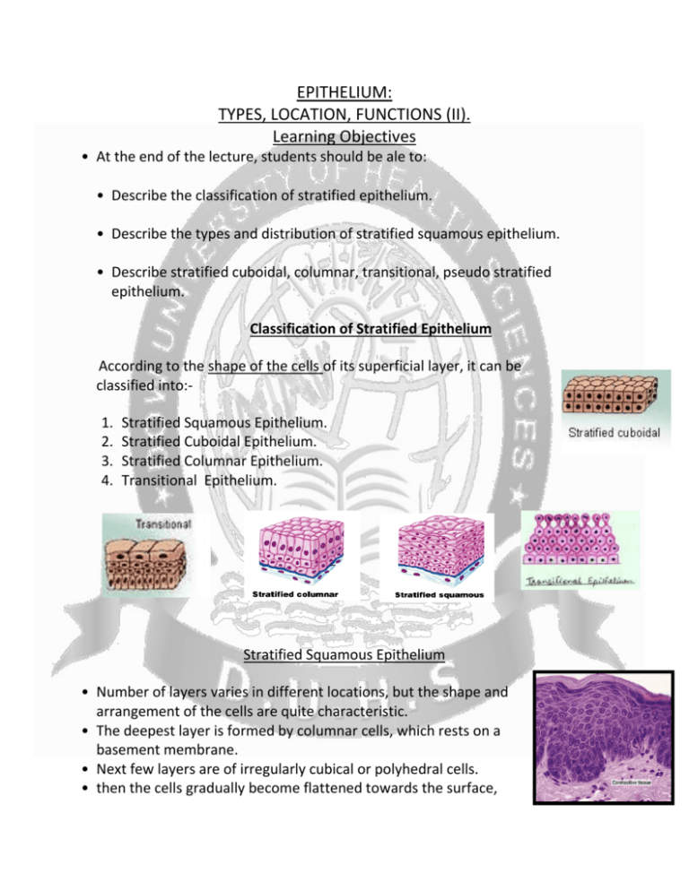
EPITHELIUM: TYPES, LOCATION, FUNCTIONS (II). Learning Objectives • At the end of the lecture, students should be ale to: • Describe the classification of stratified epithelium. • Describe the types and distribution of stratified squamous epithelium. • Describe stratified cuboidal, columnar, transitional, pseudo stratified epithelium. Classification of Stratified Epithelium According to the shape of the cells of its superficial layer, it can be classified into:1. 2. 3. 4. Stratified Squamous Epithelium. Stratified Cuboidal Epithelium. Stratified Columnar Epithelium. Transitional Epithelium. Stratified Squamous Epithelium • Number of layers varies in different locations, but the shape and arrangement of the cells are quite characteristic. • The deepest layer is formed by columnar cells, which rests on a basement membrane. • Next few layers are of irregularly cubical or polyhedral cells. • then the cells gradually become flattened towards the surface, thus the most superficial layer consists of squamous (flat) cells. • Mitosis is frequently observed in the basal layer of the epithelium. Types & Distribution Stratified Squamous Epithelium (A) Keratinized or cornified:1. Entire free surface of the body. 2. Orifices of cavities on the body. Stratified Squamous Epithelium (B) Non-keratinized or non-cornified :It lines the mucous membrane of: • Oral cavity. • Pharynx. • Esophagus. • Vagina. • Some parts of male & female urethra. Stratified Cuboidal Epithelium • Stratified cuboidal epithelium is a rare type of epithelial tissue. • It is composed of cuboidally shaped cells arranged in multiple layer Distribution • Epithelium surrounding the antra (cavity) of ovarian follicle. • Epithelium lining the large ducts of sweat & salivary glands. • Epithelium of seminiferous tubules of Testis. Stratified Columnar Epithelium • • It is rare in occurrence. Consists of columnar cells which rest on several layers of roughly cuboidal cells. Distribution: • • • Typical example – conjunctival (ocular) epithelium. Found chiefly at the junction of the stratified squamous epithelium with the pseudo-stratified epithelium, eg. Junction of nasopharynx with oropharynx. In some parts of penile urethra. Transitional Epithelium • It has a remarkable ability to increase or decrease the general surface area according to the degree of distention of the organ. • Distribution:• Calyces & pelvis of Kidney. • Ureter. • Urinary bladder. • Prostatic urethra. Photomicrograph showing the main consists of an inner layer of transitional epithelium, a highly vascularized connective tissue, a smooth muscle layer, and an outer layer of connective tissue. Pseudostratified Epithelium • It is actually a layer of single cells, but some cells are broader near the base and others are near the apex. • Nuclei lie in the broader part of each cell, thus the nuclei lie at different level, giving a false appearance of stratification. • All cells are attached to the basal lamina but some cells do not reach the free apical surface. Distribution :• Mainly in Respiratory System. • Male Reproductive System. REFERENCES BASIC HISTOLOGY BY JUNQUEIRA PAGE NO 74-80 THANK YOU ALL.


