Franck-Condon Principle: I2 Vibronic Absorption Lab
advertisement
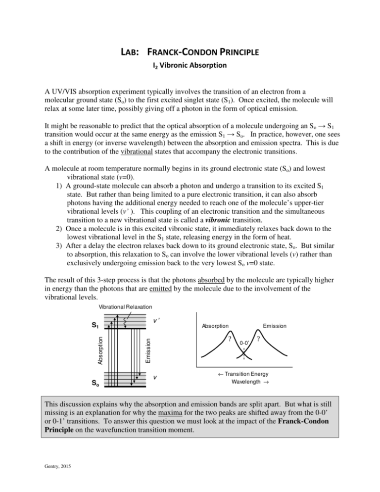
LAB: FRANCK-CONDON PRINCIPLE I2 Vibronic Absorption A UV/VIS absorption experiment typically involves the transition of an electron from a molecular ground state (So) to the first excited singlet state (S1). Once excited, the molecule will relax at some later time, possibly giving off a photon in the form of optical emission. It might be reasonable to predict that the optical absorption of a molecule undergoing an So → S1 transition would occur at the same energy as the emission S1 → So. In practice, however, one sees a shift in energy (or inverse wavelength) between the absorption and emission spectra. This is due to the contribution of the vibrational states that accompany the electronic transitions. A molecule at room temperature normally begins in its ground electronic state (So) and lowest vibrational state (v=0). 1) A ground-state molecule can absorb a photon and undergo a transition to its excited S1 state. But rather than being limited to a pure electronic transition, it can also absorb photons having the additional energy needed to reach one of the molecule’s upper-tier vibrational levels (v’ ). This coupling of an electronic transition and the simultaneous transition to a new vibrational state is called a vibronic transition. 2) Once a molecule is in this excited vibronic state, it immediately relaxes back down to the lowest vibrational level in the S1 state, releasing energy in the form of heat. 3) After a delay the electron relaxes back down to its ground electronic state, So. But similar to absorption, this relaxation to So can involve the lower vibrational levels (v) rather than exclusively undergoing emission back to the very lowest So v=0 state. The result of this 3-step process is that the photons absorbed by the molecule are typically higher in energy than the photons that are emitted by the molecule due to the involvement of the vibrational levels. Vibrational Relaxation v’ So Absorption ? Emission Absorption S1 v Emission 0-0’ ? ← Transition Energy Wavelength → This discussion explains why the absorption and emission bands are split apart. But what is still missing is an explanation for why the maxima for the two peaks are shifted away from the 0-0’ or 0-1’ transitions. To answer this question we must look at the impact of the Franck-Condon Principle on the wavefunction transition moment. Gentry, 2015 OPTICAL SELECTION RULES FOR VIBRATIONAL TRANSITIONS Pure Vibrational Transition: In a typical FTIR experiment, the instrument monitors the transition between different vibrational states in a single potential energy well. Under these conditions the intensity of an optical transition from one vibrational state to another is controlled by the transition moment. (1) where ψ i and ψ f are the wavefunctions for the initial and final states respectively, and µ is the molecular dipole moment. So long as the system stays within the same potential energy well the only time that the integral in (1) is non-zero is if the vibrational transition obeys the selection rule ∆v = ±1 where v is the vibrational quantum number. For all other transitions, e.g. v = 0→2, the transition moment goes to zero and the transition will not be visible in an absorption experiment. Single Potential Well Potential Energy, V(r) µif = ∫ψ *f ⋅ µ ⋅ψ i dτ 3 v 2 1 ∆v =+1 0 re Internuclear Distance, r Two Potential Wells Excited Electronic State, E ’ v’ Energy Vibronic Transition: This ∆v = ±1 selection rule breaks down, however, if the vibrational transition is coupled to an electronic transition. The process of moving an electron from one molecular orbital to another causes a change in the electron distribution around the nuclei. This change in electron distribution, in turn causes a change in the bond strength, and thus a change in the bond distance, the bond dissociation energy, and the vibrational frequency. Ground Electronic State, E v re re’ Internuclear Distance FRANCK-CONDON PRINCIPLE FOR VIBRONIC TRANSITIONS This shift in equilibrium bond length and the timing for when it occurs are very important when it comes to how vibrational wavefunctions interact with one another. Franck-Condon Principle The Franck-Condon Principle states that an optical vibronic transition is essentially instantaneous compared to the relatively slow response of the nuclei shifting to new equilibrium positions. 2) a much slower step that occurs at a later time when the nuclei shift to their new equilibrium positions. Franck-Condon Factors 1) Absorption 1) very fast optical absorption to the excited electronic state while the nuclei remain in their ground-state positions; and Energy This means that a vibronic transition occurs in two discrete steps: 2) Shift in Nuclei re re’ Internuclear Distance Page 2 of 10 Franck-Condon Factor The sideways translation in the potential energy curves that occurs between two different vibronic states leads to optical absorbance values that are very different in strength than those seen with pure vibrational transitions. The Franck-Condon Principle says that the optical absorption (or emission) for a vibronic transition will be proportional to the square of the overlap integral of the two vibrational wavefunctions. This squared integral is called the Franck-Condon Factor (FCF). The total optical strength is then given by the FCF times a constant electronic interaction term. 2 FCF = ∫ψ *f ⋅ψ i dτ vib (2) The overlap integral is strongly affected by the offset between the equilibrium bond lengths for the ground-state and excited-state potential energy wells. This can be seen in the figure below which looks at the overlap between v=1 and v’=4 states for wavefunctions that are either centered at the same re position or with one function offset to the side from the other. In the first case the overlaps cancel and FCF = 0. In the second case the offset in positions means that the overlaps no longer cancel and there is a sizeable FCF interaction. Two wavefunctions centered at same r position One wavefunction offset (centered at different r position v1 v1 v4 v4 offset v1* v4 ∫ (v1*v4) = 0 overlaps cancel out v1* v4 overlaps do not cancel ∫ (v1*v4) > 0 The result of the offset in vibronic wavefunctions is that : - There will be a large number of v→v’ transitions that all have sizeable absorbance values. - The maximum absorbance will occur for the transition that has the largest overlap between the ground-state v=0 wavefunction and the offset excited-state v’ wavefunction. This can occur at a relatively large v’ value. Franck-Condon Factors Page 3 of 10 Approximation Technique to Locate the Maximum Franck-Condon Overlap An approximate way of finding the excited quantum state having the maximum overlap with the ground state v=0 state is shown below. This quantum level should correspond to where the edge of the excited-state Morse potential curve has the same internuclear separation distance as the center of the ground-state well. That is to say, one can draw a vertical line from the center of the bottom ground-state potential well and look for where it intersects the upper excited-state potential curve. The excited vibrational quantum level that matches the energy of the intersection point should correspond to the observed maximum in an absorption spectrum, v’max. Absorption Experiment FranckCondon intersection point Excited Electronic State vmax’ Energy v’max 0’ v’ 0 0-vmax’ Ground Electronic State optical transition strong overlap 0’ v little overlap 0 0-0’ 0 ← Transition Energy re re’ Internuclear Distance Wavelength → MORSE POTENTIAL A Franck-Condon analysis requires one to be able to calculate the vibrational energies for the different electronic states. The potential energy surface for molecular vibrations can be described using the Morse potential. ( 1 − e −α ( r − re ) ) 2 (3) De is the dissociation energy. It describes the difference in energy from the very bottom of the potential energy well up to the dissociation plateau. re is the equilibrium bond length, i.e. the internuclear distance corresponding to the bottom of the potential energy well. α is a factor that controls the width of the well and is dependent on the vibrational frequency and the reduced mass of the molecule. Franck-Condon Factors Potential Energy, V(r) V ( r ) = De Morse potential 3 2 1 0 v De re Internuclear Distance, r Page 4 of 10 Given that a molecular vibration is controlled by the Morse potential, the energies for the various vibrational quantum levels can then be determined. 1 ɶ vib = ɶve v + 1 − ɶve χ ɶ v + E 2 2 2 v = 0,1,2,… (4) When the energy equation is written this way with the use of the ~ headers it is based on spectroscopic wavenumbers (cm-1) rather than pure SI-unit energy. The transition energy, ∆Eɶ v −v ' , from a ground-state vibrational state ( v) to an excited electronic/vibrational state ( v’ ) depends on the vibrational energies of the initial and final states (Evib and E’vib) as well as on the electronic transition energy, Te’. Te is measured from the bottom of the ground electronic potential well to the bottom of the excited-state electronic potential well. ∆Eɶ v −v ' = Tɶe ' + Eɶ 'vib − Eɶ vib (5) The inter-relationships between diatomic molecular constants and the constants used in Eqn’s 3 and 4 can be shown to give the following equations: Eɶ vib , ɶve , Dɶ e , and Tɶe µ = ma ⋅ mb ma + mb α = ɶve π ɶ = χ Franck-Condon Factors ɶve ɶe 4D 2µ c ɶe hD given in units of cm-1 ma and mb are mass of atoms a and b in kg α calculated in units of meters-1 c is the speed of light, in cm/s h is Planck’s constant, in J·s ɶ = anharmonicity constant, with χ dimensionless units in this format Page 5 of 10 Experimental In this experiment you will experimentally measure the vibronic absorption spectrum for Iodine, I2(g). You will then compare your experimental result to calculated predictions based on the Franck-Condon Principle. If you already measured the spectrum of I2 earlier in the semester you can skip over the experimental section. To measure the spectrum, put a few crystals of I2 in a cuvette and then seal it with a cap. You will then need to warm the iodine so that it sublimes to vapor. This can be done with a heat gun. It helps to warm both the cuvette and the sample holder in the spectrometer otherwise the large thermal mass of the sample holder will cool the cuvette before you have a chance to record the spectrum. Use the Unicam UV/Vis spectrometer to record the absorbance spectrum. Do a quick spectral run to observe where the primary absorption peak is located. This can be done by setting the instrument to scan speed = “Intelliscan”, data interval = normal, and bandwidth = 1nm. Try a wavelength range of 350 – 700 nm first. Once you have a spectrum, record a second, higher-quality spectrum over the active absorption region for your sample. You will want to record the entire span of the absorption band. For this scan, set the instrument so that the data interval is set to the highest resolution and the bandwidth to 0.2 nm. Export the data as an ASCII batch file when done. • Include a copy of the spectrum in your report as well as report the wavelength location of the maximum absorbance. Do this even if you included the spectrum in your earlier lab report. Calculations We will analyze the data using two different techniques. We will first use Excel to calculate the ground-state and excited-state Morse potentials for I2. We will use these curves along with the Franck-Condon Approximation technique to estimate the energy, wavelength, and quantum number for the location of maximum absorbance. Secondly, we will use a Java application1 to explore the individual Franck-Condon Factors. These calculations require that you know the vibronic constants for Iodine. Literature Values2 for I2(g) Term Symbol 1 Ground State, X Excited State, B 3 Σ + g Π 0 +u Dɶ e (cm-1) (Å)* Tɶe (cm-1) 18713. vɶe (cm-1) 214.502 2.6663 0 5168.7 125.697 3.0247 15769.01 re * Note that the equation for alpha (α) for use in Eqn (3) will give a result in m-1. This will need to be converted to Angstroms-1 (Å-1) if the units are going to cancel with re in the equation for α. Franck-Condon Factors Page 6 of 10 A) Excel Analysis • Use the equation for the Morse potential (Eqn. 3) to calculate the potential energies V(r) for both the ground-state and excited state vibrations. Do this over a range of r = 2.2 to 6.0Å. Use increments of 0.01 Å for your data table, but insert an additional row in the middle so as to include the ground state equilibrium distance r = re = 2.6663Å point in your calculations. • Add the excited-state electronic energy, Te’ to the excited-state vibrational curve to get the total energy for the excited vibronic system. E’tot = E’vib+Te’ • Plot the ground-state and excited-state energy curves on the same graph showing energy on the y axis and internuclear separation distance (r) on the x axis. Limit your y-axis scale to a maximum of 30,000 cm-1 so that you can more easily see the differences in the potential wells for the two states. Your graph should resemble the figure that shows the two Morse potentials on page 2. To double check your calculations, for r = 4.0 Å, Ground X state: Evib ≈ 14113cm-1, Excited B State: E’vib+Te’ ≈ 19149cm-1 Use Franck-Condon Approximation Technique to Locate Maximum Wavefunction Overlap • On your graph of energy vs. internuclear distance, draw a straight vertical line on your graph starting from the center re position of the lower potential energy curve and proceeding upwards until it intersects the upper energy curve. Include this graph and line in your report. • Use your table of calculated values to actually determine the values of E’vib+Te’ and E’vib at that intersection point of r = re,ground state . • Use Eqn. 4 to determine the quantum number corresponding to that excited vibrational state. [The easiest way to do this may be to try various values of quantum number v’ until the quantum energy E’vib(v) approximately matches your observed intersection E’vib value. Alternatively you could use the Solver add-in for Excel.] • Using Eqn. 5, what is the energy of transition ( ∆Eɶ v −v ' , in cm-1) for that point? [Do not forget to account for the zero-point energy of the ground-state vibration and the electronic transition energy.] • What is the wavelength of light corresponding to that 0-v’max transition? • By contrast, what is the energy and the wavelength for the 0-0’ transition? • What is the shift in wavelength (in nm) from the 0-0’ origin to the 0-v’max peak? • Report these results in a table in your report and compare the transition wavelengths to the observed locations and absorbances seen in your experimental spectra. Franck-Condon Factors Page 7 of 10 B) Java Analysis S.W. North and his colleagues at Texas A&M University have prepared a handy Java program that plots and analyzes Franck-Condon factors. 1 • Go to our class website and do a right-click “Save Target as…” to download the “Java.FCIntensity.jar” file onto your computer. If you are using one of the departmental computers you may need to have me do this for you (with my administrator access). • Start the program by double clicking on the icon. • You should see two panels appear. The main panel on the left is where you enter the molecular constants. In order to model your absorption results you will want to put the ground-state values into the boxes marked “Initial State”. The excited-state constants are entered in the boxes for “Final State”. You will also need to tell the program what the starting vibrational constant is. This should be v=0. (Why?) The final state should be a range of v’ = 0 to 60. Lastly you will need to insure that the masses for the two nuclei in the diatomic model are entered correctly. • Click on “Calculate”. You should now see a plot of the two energy curves at the top of the panel and the relative intensities for all of the different 0-v’ transitions in the center of the panel. Estimate the wavelength corresponding to the peak maximum? How do your experimental peak location and peak width compare to this calculated spectrum? • The “Wavefunction” panel on the right allows you to take a closer look at specific transitions based on the molecular constants that you entered on the other panel. In the center of the Wavefunction panel you can enter a starting quantum number (v=0) and an excited-state final quantum number (v’ ). When you click on the “Plot” button at the bottom, the program will show you the two wavefunctions (offset in space according to re and re’ ). The graph at the bottom shows the result of multiplying one wavefunction times the other wavefunction. At the very bottom of the panel you will see boxes that report the wavelength and the integrated Franck-Condon Factor for that transition. • Using the Wavefunction panel, repeatedly try various values of v’ until you find the maximum value for the Franck-Condon Factor. As you change the value of v’ note how the overlap changes between the two wavefunctions. Once you have found the v’ value that gives the largest FCF, report the wavelength, and quantum number for the transition. The wavelength should match the location of the peak maximum on the left panel. • The main panel on the left has a File menu tab that allows you to print the two panels. Your lab report should include a print of the calculated spectrum on the left and the wavefunction transition on the right showing the maximum overlap. • When you are done with the analysis of the maximum overlap, use the Wave Function panel to determine the FCF and wavelength for the 0-0’ transition. Note the difference in how the peaks do or do not overlap as compared to the maximum value. Franck-Condon Factors Page 8 of 10 Questions Comment on the comparison of your experimental data to the two calculated results (Excel and Java). How closely do the locations of the peak maxima agree? Discuss the advantages and shortcomings of the different approaches. How far in wavelength (nm) did the peak maxima shift from the original 0-0’ transition? What would your absorption spectrum have looked like if the system limited itself to the ∆v = ±1. What did you observe with the Java program when you compared the wavefunctions and their overlaps for the 0-0’ versus 0-v’max transitions? Observation of Hot Bands in Experimental Spectra If you take a closer look at your experimental spectrum in the range of 530-590nm, you will see the emergence of a second set of peaks, and eventually a third set, appearing at longer wavelengths. Normally molecules start in their lowest vibrational level because there is not enough thermal energy available to populate higher vibrations. In the case of Iodine, however, the vibrational energy gap is small enough that now there is enough thermal energy to generate some small amount of additional population in the next two higher vibrational levels. Vibronic transitions starting from these higher vibrational levels are called “hot bands” . 0-v’ Iodine ( I2 ) v’ Energy Absorbance S1 1-v’ 0-v’ 1-v’ hot band 2-v’ v So 530 540 550 560 570 Wavelength (nm) 580 590 - Plot your high-resolution UV/Vis data in the range of 530-590nm. - Use the Boltzmann distribution (beginning of first semester Thermodynamics) to calculate the population probability ratio of the v=1 versus v=0 vibrational levels in the ground electronic state in I2. Report your results in terms of the Boltzmann ratio, P1 / P0 = e − ( E1 − E0 )/ kT . Comment on the amount of population seen in the higher vibrational levels for the I2 molecule and how this affects your ability to observe hot bands. - Given that you have shown that I2 has a sizeable population distribution in the v=1, return to the Java program and on the main panel on the left change the starting quantum number from v=0 to v=1. [You may get an error message that will require you to limit your range of excited-state quantum numbers. You might recall that there is an upper limit v’ level, beyond which the bond breaks rather than vibrates.] Franck-Condon Factors Page 9 of 10 - Similarly change the starting wavelength on the Wavefunction panel on the right to find the maximum wavelengths and quantum numbers for the two hot bands. - Lab-report question: Is the observed long-wavelength shoulder in your experimental spectrum generally consistent with the predicted locations and populations of the hot bands? References 1 Gucchese, G., Lucchese, R.R., North, S.W.; J. Chem. Ed. 2010, 87, 345. 2 Bother, J.R., Tainter, C.J.; J Phys Chem Lab 2006, 10, 55. Franck-Condon Factors Page 10 of 10
