Vol 447j17 May 2007jdoi:10.1038/nature05775
REVIEWS
Origins of major human infectious diseases
Nathan D. Wolfe1, Claire Panosian Dunavan2 & Jared Diamond3
Many of the major human infectious diseases, including some now confined to humans and absent from animals, are ‘new’
ones that arose only after the origins of agriculture. Where did they come from? Why are they overwhelmingly of Old World
origins? Here we show that answers to these questions are different for tropical and temperate diseases; for instance, in the
relative importance of domestic animals and wild primates as sources. We identify five intermediate stages through which a
pathogen exclusively infecting animals may become transformed into a pathogen exclusively infecting humans. We propose
an initiative to resolve disputed origins of major diseases, and a global early warning system to monitor pathogens infecting
individuals exposed to wild animals.
uman hunter/gatherer populations currently suffer, and
presumably have suffered for millions of years, from infectious diseases similar or identical to diseases of other wild
primate populations. However, the most important infectious diseases of modern food-producing human populations also
include diseases that could have emerged only within the past 11,000
years, following the rise of agriculture1,2. We infer this because, as
discussed below, these diseases can only be sustained in large dense
human populations that did not exist anywhere in the world before
agriculture. What were the sources of our major infectious diseases,
including these ‘new’ ones? Why do so many animal pathogens,
including virulent viruses like Ebola and Marburg, periodically
infect human hosts but then fail to establish themselves in human
populations?
A tentative earlier formulation1 noted that major infectious diseases of temperate zones seem to have arisen overwhelmingly in the
Old World (Africa, Asia and Europe), often from diseases of Old
World domestic animals. Hence one goal of this article is to reappraise that conclusion in the light of studies of the past decade.
Another goal is to extend the analysis to origins of tropical diseases3.
We shall show that they also arose mainly in the Old World, but for
different reasons, and mostly not from diseases of domestic animals.
These results provide a framework for addressing unanswered
questions about the evolution of human infectious diseases—questions not only of practical importance to physicians, and to all the rest
of us as potential victims, but also of intellectual interest to historians
and evolutionary biologists. Historians increasingly recognize that
infectious diseases have had major effects on the course of history;
for example, on the European conquest of Native Americans and
Pacific Islanders, the inability of Europeans to conquer the Old
World tropics for many centuries, the failure of Napoleon’s invasion
of Russia, and the failure of the French attempt to complete construction of a Panama Canal4–6. Evolutionary biologists realize that
infectious diseases, as a leading cause of human morbidity and mortality, have exerted important selective forces on our genomes2,7.
We begin by defining five stages in the evolutionary transformation of an animal pathogen into a specialized pathogen of humans,
and by considering why so many pathogens fail to make the transition from one stage to the next. We then assemble a database of 15
temperate and 10 tropical diseases of high evolutionary and/or historical impact, and we compare their characteristics and origins. Our
concluding section lays out some unresolved questions and suggests
two expanded research priorities. We restrict our discussion to
H
unicellular microbial pathogens. We exclude macroparasites (in
the sense of ref. 7), as well as normally benign commensals that cause
serious illness only in weakened hosts. The extensive Supplementary
Information provides details and references on our 25 diseases,
robustness tests of our conclusions, factors affecting transitions
between disease stages, and modern practices altering the risk of
emergence of new diseases.
Evolutionary stages
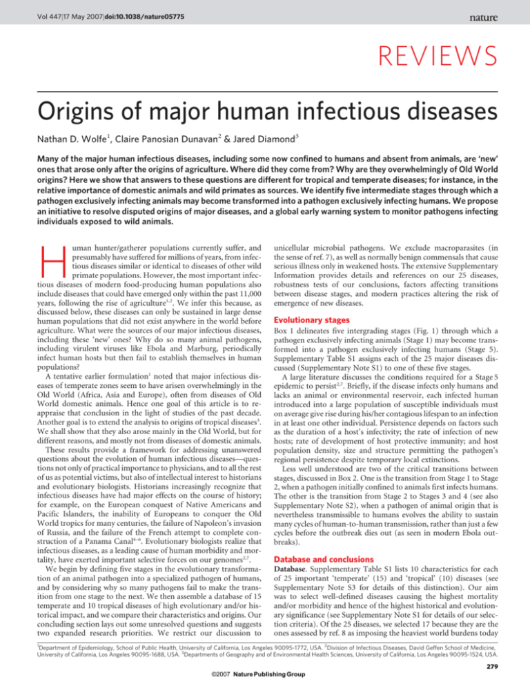
Box 1 delineates five intergrading stages (Fig. 1) through which a
pathogen exclusively infecting animals (Stage 1) may become transformed into a pathogen exclusively infecting humans (Stage 5).
Supplementary Table S1 assigns each of the 25 major diseases discussed (Supplementary Note S1) to one of these five stages.
A large literature discusses the conditions required for a Stage 5
epidemic to persist2,7. Briefly, if the disease infects only humans and
lacks an animal or environmental reservoir, each infected human
introduced into a large population of susceptible individuals must
on average give rise during his/her contagious lifespan to an infection
in at least one other individual. Persistence depends on factors such
as the duration of a host’s infectivity; the rate of infection of new
hosts; rate of development of host protective immunity; and host
population density, size and structure permitting the pathogen’s
regional persistence despite temporary local extinctions.
Less well understood are two of the critical transitions between
stages, discussed in Box 2. One is the transition from Stage 1 to Stage
2, when a pathogen initially confined to animals first infects humans.
The other is the transition from Stage 2 to Stages 3 and 4 (see also
Supplementary Note S2), when a pathogen of animal origin that is
nevertheless transmissible to humans evolves the ability to sustain
many cycles of human-to-human transmission, rather than just a few
cycles before the outbreak dies out (as seen in modern Ebola outbreaks).
Database and conclusions
Database. Supplementary Table S1 lists 10 characteristics for each
of 25 important ‘temperate’ (15) and ‘tropical’ (10) diseases (see
Supplementary Note S3 for details of this distinction). Our aim
was to select well-defined diseases causing the highest mortality
and/or morbidity and hence of the highest historical and evolutionary significance (see Supplementary Note S1 for details of our selection criteria). Of the 25 diseases, we selected 17 because they are the
ones assessed by ref. 8 as imposing the heaviest world burdens today
1
Department of Epidemiology, School of Public Health, University of California, Los Angeles 90095-1772, USA. 2Division of Infectious Diseases, David Geffen School of Medicine,
University of California, Los Angeles 90095-1688, USA. 3Departments of Geography and of Environmental Health Sciences, University of California, Los Angeles 90095-1524, USA.
279
©2007 Nature Publishing Group
REVIEWS
NATUREjVol 447j17 May 2007
Box 1 j Five stages leading to endemic human diseases
We delineate five stages in the transformation of an animal pathogen
into a specialized pathogen of humans (Fig. 1). There is no inevitable
progression of microbes from Stage 1 to Stage 5: at each stage many
microbes remain stuck, and the agents of nearly half of the 25
important diseases we selected for analysis (Supplementary Table S1)
have not reached Stage 5.
. Stage 1. A microbe that is present in animals but that has not
been detected in humans under natural conditions (that is,
excluding modern technologies that can inadvertently transfer
microbes, such as blood transfusion, organ transplants, or
hypodermic needles). Examples: most malarial plasmodia, which
tend to be specific to one host species or to a closely related
group of host species.
. Stage 2. A pathogen of animals that, under natural conditions,
has been transmitted from animals to humans (‘primary
infection’) but has not been transmitted between humans
(‘secondary infection’). Examples: anthrax and tularemia bacilli,
and Nipah, rabies and West Nile viruses.
. Stage 3. Animal pathogens that can undergo only a few cycles of
secondary transmission between humans, so that occasional
human outbreaks triggered by a primary infection soon die out.
Examples: Ebola, Marburg and monkeypox viruses.
. Stage 4. A disease that exists in animals, and that has a natural
(sylvatic) cycle of infecting humans by primary transmission
from the animal host, but that also undergoes long sequences of
secondary transmission between humans without the
involvement of animal hosts. We arbitrarily divide Stage 4 into
three substages distinguished by the relative importance of
primary and secondary transmission:
Stage 4a. Sylvatic cycle much more important than direct
human-to-human spread. Examples: Chagas’ disease and (more
frequent secondary transmission approaching Stage 4b) yellow
fever.
Stage 4b. Both sylvatic and direct transmission are important.
Example: dengue fever in forested areas of West Africa and
Southeast Asia.
Stage 4c. The greatest spread is between humans. Examples:
influenza A, cholera, typhus and West African sleeping sickness.
. Stage 5. A pathogen exclusive to humans. Examples: the agents
causing falciparum malaria, measles, mumps, rubella, smallpox
and syphilis. In principle, these pathogens could have become
confined to humans in either of two ways: an ancestral pathogen
already present in the common ancestor of chimpanzees and
humans could have co-speciated long ago, when the chimpanzee
and human lineages diverged around five million years ago; or
else an animal pathogen could have colonized humans more
recently and evolved into a specialized human pathogen. Cospeciation accounts well for the distribution of simian foamy
viruses of non-human primates, which are lacking and
presumably lost in humans: each virus is restricted to one
primate species, but related viruses occur in related primate
species19. While both interpretations are still debated for
falciparum malaria, the latter interpretation of recent origins is
widely preferred for most other human Stage 5 diseases of
Supplementary Table S1.
(they have the highest disability-adjusted life years (DALY) scores).
Of the 17 diseases, 8 are temperate (hepatitis B, influenza A, measles,
pertussis, rotavirus A, syphilis, tetanus and tuberculosis), and 9 are
tropical (acquired immune deficiency syndrome (AIDS), Chagas’
disease, cholera, dengue haemorrhagic fever, East and West African
sleeping sicknesses, falciparum and vivax malarias, and visceral leishmaniasis). We selected eight others (temperate diphtheria, mumps,
plague, rubella, smallpox, typhoid and typhus, plus tropical yellow
fever) because they imposed heavy burdens in the past, although
modern medicine and public health have either eradicated them
(smallpox) or reduced their burden. Except for AIDS, dengue fever,
and cholera, which have spread and attained global impact in modern
times, most of these 25 diseases have been important for more than
two centuries.
Are our conclusions robust to variations in these selection criteria?
For about a dozen diseases with the highest modern or historical
burdens (for example, AIDS, malaria, plague, smallpox), there can
be little doubt that they must be included, but one could debate some
of the next choices. Hence we drew up three alternative sets of diseases sharing a first list of 16 indisputable major diseases but differing
in the next choices, and we performed all 10 analyses described below
on all three sets. It turned out that, with one minor exception, the
three sets yielded qualitatively the same conclusions for all 10 analyses,
although differing in their levels of statistical significance (see Supplementary Note S4). Thus, our conclusions do seem to be robust.
Temperate/tropical differences. Comparisons of these temperate
and tropical diseases yield the following conclusions:
. A higher proportion of the diseases is transmitted by insect
vectors in the tropics (8/10) than in the temperate zones (2/
15) (P , 0.005, x2-test, degrees of freedom, d.f. 5 1). This difference may be partly related to the seasonal cessations or
declines of temperate insect activity.
. A higher proportion (P 5 0.009) of the diseases conveys longlasting immunity (11/15) in the temperate zones than in the
tropics (2/10).
. Animal reservoirs are more frequent (P , 0.005) in the tropics
(8/10) than in the temperate zones (3/15). The difference is in
the reverse direction (P 5 0.1, NS, not significant) for environmental reservoirs (1/10 versus 6/15), but those environmental reservoirs that do exist are generally not of major
significance except for soil bearing tetanus spores.
. Most of the temperate diseases (12/15) are acute rather than
slow, chronic, or latent: the patient either dies or recovers
within one to several weeks. Fewer (P 5 0.01) of the tropical
diseases are acute: 3/10 last for one or two weeks, 3/10 last for
weeks to months or years, and 4/10 last for many months to
decades.
. A somewhat higher proportion of the diseases (P 5 0.08, NS)
belongs to Stage 5 (strictly confined to humans) in the temperate zones (10/15 or 11/15) than in the tropics (3/10). The
paucity of Stage 2 and Stage 3 diseases (a total of only 5 such
diseases) on our list of 25 major human diseases is noteworthy,
because some Stage 2 and Stage 3 pathogens (such as anthrax
and Ebola) are notoriously virulent, and because theoretical
reasons are often advanced (but also denied) as to why Stage 5
microbes with long histories of adaptation to humans should
tend to evolve low morbidity and mortality and not cause
major diseases. We discuss explanations for this outcome in
Supplementary Note S5.
Most (10/15) of the temperate diseases, but none of the tropical
diseases (P , 0.005), are so-called ‘crowd epidemic diseases’ (asterisked in Supplementary Table S1), defined as ones occurring locally
as a brief epidemic and capable of persisting regionally only in large
human populations. This difference is an immediate consequence of
the differences enumerated in the preceding five paragraphs. If a
disease is acute, efficiently transmitted, and quickly leaves its victim
either dead or else recovering and immune to re-infection, the epidemic soon exhausts the local pool of susceptible potential victims. If
in addition the disease is confined to humans and lacks significant
animal and environmental reservoirs, depletion of the local pool of
potential victims in a small, sparse human population results in local
termination of the epidemic. If, however, the human population is
large and dense, the disease can persist by spreading to infect people
in adjacent areas, and then returning to the original area in a later
year, when births and growth have regenerated a new crop of previously unexposed non-immune potential victims. Empirical epidemiological studies of disease persistence or disappearance in isolated
human populations of various sizes have yielded estimates of the
population required to sustain a crowd disease: at least several hundred thousand people in the cases of measles, rubella and pertussis2,7.
But human populations of that size did not exist anywhere in the
280
©2007 Nature Publishing Group
REVIEWS
NATUREjVol 447j17 May 2007
Transmission
to humans
Stage
Stage 5:
exclusive
human agent
Only from
humans
Stage 4:
long outbreak
From animals
or (many cycles)
humans
Stage 3:
limited
outbreak
From animals
or (few cycles)
humans
Stage 2:
primary
infection
Only from animals
Stage 1:
agent only
in animals
None
Rabies
Ebola
Dengue
HIV-1 M
Figure 1 | Illustration of the five stages through which pathogens of
animals evolve to cause diseases confined to humans. (See Box 1 for
details.) The four agents depicted have reached different stages in the
process, ranging from rabies (still acquired only from animals) to HIV-1
(now acquired only from humans).
world until the steep rise in human numbers that began around
11,000 years ago with the development of agriculture1,9. Hence the
crowd epidemic diseases of the temperate zones must have evolved
since then.
Of course, this does not mean that human hunter/gatherer communities lacked infectious diseases. Instead, like the sparse populations of our primate relatives, they suffered from infectious diseases
with characteristics permitting them to persist in small populations,
unlike crowd epidemic diseases. Those characteristics include: occurrence in animal reservoirs as well as in humans (such as yellow fever);
incomplete and/or non-lasting immunity, enabling recovered
patients to remain in the pool of potential victims (such as malaria);
and a slow or chronic course, enabling individual patients to continue to infect new victims over years, rather than for just a week or
two (such as Chagas’ disease).
Pathogen origins. (See details for each disease in Supplementary
Note S10). Current information suggests that 8 of the 15 temperate
diseases probably or possibly reached humans from domestic animals (diphtheria, influenza A, measles, mumps, pertussis, rotavirus,
smallpox, tuberculosis); three more probably reached us from apes
(hepatitis B) or rodents (plague, typhus); and the other four (rubella,
syphilis, tetanus, typhoid) came from still-unknown sources (see
Supplementary Note S6). Thus, the rise of agriculture starting
11,000 years ago played multiple roles in the evolution of animal
pathogens into human pathogens1,4,10. Those roles included both
generation of the large human populations necessary for the evolution and persistence of human crowd diseases, and generation of
large populations of domestic animals, with which farmers came into
much closer and more frequent contact than hunter/gatherers had
with wild animals. Moreover, as illustrated by influenza A, these
domestic animal herds served as efficient conduits for pathogen
transfers from wild animals to humans, and in the process may have
evolved specialized crowd diseases of their own.
It is interesting that fewer tropical than temperate pathogens originated from domestic animals: not more than three of the ten tropical diseases of Supplementary Table S1, and possibly none (see
Supplementary Note S7). Why do temperate and tropical human
diseases differ so markedly in their animal origins? Many (4/10)
tropical diseases (AIDS, dengue fever, vivax malaria, yellow fever)
but only 1/15 temperate diseases (hepatitis B) have wild non-human
primate origins (P 5 0.04). This is because although non-human
primates are the animals most closely related to humans and hence
pose the weakest species barriers to pathogen transfer, the vast majority of primate species is tropical rather than temperate. Conversely,
few tropical but many temperate diseases arose from domestic animals, and this is because domestic animals live mainly in the temperate zones, and their concentration there was formerly even more
lop-sided (see Supplementary Note S8).
A final noteworthy point about animal-derived human pathogens
is that virtually all arose from pathogens of other warm-blooded
vertebrates, primarily mammals plus in two cases (influenza A and
ultimately falciparum malaria) birds. This comes as no surprise, considering the species barrier to pathogen transfer posed by phylogenetic distance (Box 2). An expression of this barrier is that primates
constitute only 0.5% of all vertebrate species but have contributed
about 20% of our major human diseases. Expressed in another way,
the number of major human diseases contributed, divided by the
number of animal species in the taxonomic group contributing those
diseases, is approximately 0.2 for apes, 0.017 for non-human primates other than apes, 0.003 for mammals other than primates,
0.00006 for vertebrates other than mammals, and either 0 or else
0.000003 (if cholera really came from aquatic invertebrates) for animals other than vertebrates (see Supplementary Note S9).
Geographic origins. To an overwhelming degree, the 25 major
human pathogens analysed here originated in the Old World. That
proved to be of great historical importance, because it facilitated
the European conquest of the New World (the Americas). Far more
Native Americans resisting European colonists died of newly introduced Old World diseases than of sword and bullet wounds. Those
invisible agents of New World conquest were Old World microbes to
which Europeans had both some acquired immunity based on individual exposure and some genetic resistance based on population
281
©2007 Nature Publishing Group
REVIEWS
NATUREjVol 447j17 May 2007
Box 2 j Transitions between stages
Transition from Stage 1 to Stage 2. Most animal pathogens are not
transmitted to humans, that is, they do not even pass from Stage 1 to Stage
2. This problem of cross-species infection has been discussed
previously20–23. Briefly, the probability-per-unit-time (p) of infection of an
individual of a new (that is, new recipient) host species increases with the
abundance of the existing (that is, existing donor) host, with the fraction of
the existing host population infected, with the frequency of ‘encounters’
(opportunities for transmission, including indirect ‘encounters’ via
vectors) between an individual of the existing host and of the new host,
and with the probability of transmission per encounter. p decreases with
increasing phylogenetic distance between the existing host and new host.
p also varies among microbes (for example, trypanosomes and
flaviviruses infect a wide taxonomic range of hosts, while plasmodia and
simian foamy viruses infect only a narrow range), and this variation is
related to a microbe’s characteristics, such as its ability to generate
genetic variability, or its ability to overcome host molecular barriers of
potential new hosts (such as humoral and cellular defenses or lack of cell
membrane receptors essential for microbe entry into host cells).
These considerations illuminate different reasons why a given
animal host species may or may not become a source of many
infections in humans. For instance, despite chimpanzees’ very low
abundance and infrequent encounters with humans, they have
donated to us numerous zoonoses (diseases that still mainly afflict
animals) and one or two established human diseases (AIDS and
possibly hepatitis B) because of their close phylogenetic relationship to
humans. Despite their large phylogenetic distance from humans, many
of our zoonoses and probably two of our established diseases (plague
and typhus) have been acquired from rodents, because of their high
abundance and frequent encounters with humans in dwellings.
Similarly, about half of our established temperate diseases have been
acquired from domestic livestock, because of high local abundance and
very frequent contact. Conversely, elephants and bats are not known to
have donated directly to us any established diseases and rarely donate
zoonoses, because they are heavily penalized on two or three counts:
large phylogenetic distance, infrequent encounters with humans, and
(in the case of elephants) low abundance. One might object that Nipah,
severe acute respiratory syndrome (SARS) and rabies viruses do infect
humans from bats, but these apparent exceptions actually support our
conclusion. While bats may indeed be the primary reservoir for Nipah
and SARS, human infections by these viruses are acquired mainly from
intermediate animal hosts that frequently encounter humans
(respectively, domestic pigs, and wild animals sold for food). The rare
cases of rabies transmission directly to humans from bats arise
because rabies changes a bat’s behaviour so that it does encounter and
bite humans, which a healthy bat (other than a vampire bat) would
never do.
Transition from Stage 2 to Stage 3 or 4. Although some Stage 2 and 3
pathogens, such as the anthrax and Marburg agents, are virulent and
feared, they claim few victims at present. Yet if they made the transition
to Stage 4 or 5, their global impact would be devastating. Why do animal
pathogens that have survived the initial jump across species lines into a
human host (Stages 1 to 2) usually reach a dead end there, and not
evolve past Stages 3 and 4 into major diseases confined to humans
(Stage 5)? Barriers between Stages 2 and 3 (consider the rabies virus)
include differences between human and animal behaviour affecting
transmission (for example, animals often bite humans but humans
rarely bite other humans); a pathogen’s need to evolve adaptations to
the new human host and possibly also to a new vector; and obstacles to
a pathogen’s spread between human tissues (for example, BSE is
restricted to the central nervous system and lymphoid tissue). Barriers
between Stages 3 and 4 (consider Ebola virus) include those related to
human population size and to transmission efficiency between humans.
The emergence of novel pathogens is now being facilitated by modern
developments exposing more potential human victims and/or making
transmission between humans more efficient than before24–27. These
developments include blood transfusion (hepatitis C), the commercial
bushmeat trade (retroviruses), industrial food production (bovine
spongiform encephalitis, BSE), international travel (cholera),
intravenous drug use (HIV), vaccine production (simian virus 40,
SV40), and susceptible pools of elderly, antibiotic-treated,
immunosuppressed patients (see Supplementary Note S2 for details).
exposure over time, but to which previously unexposed Native
American populations had no immunity or resistance1,4–6. In contrast, no comparably devastating diseases awaited Europeans in the
New World, which proved to be a relatively healthy environment for
Europeans until yellow fever and malaria of Old World origins
arrived11.
Why was pathogen exchange between Old and New Worlds so
unequal? Of the 25 major human diseases analysed, Chagas’ disease
is the only one that clearly originated in the New World. For two
others, syphilis and tuberculosis, the debate is unresolved: it remains
uncertain in which hemisphere syphilis originated, and whether
tuberculosis originated independently in both hemispheres or was
brought to the Americas by Europeans. Nothing is known about the
geographic origins of rotavirus, rubella, tetanus and typhus. For all of
the other 18 major pathogens, Old World origins are certain or
probable.
Our preceding discussion of the animal origins of human pathogens may help explain this asymmetry. More temperate diseases arose
in the Old World than New World because far more animals that
could furnish ancestral pathogens were domesticated in the Old
World. Of the world’s 14 major species of domestic mammalian
livestock, 13, including the five most abundant species with which
we come into closest contact (cow, sheep, goat, pig and horse), originated in the Old World1. The sole livestock species domesticated in
the New World was the llama, but it is not known to have infected us
with any pathogens1,2—perhaps because its traditional geographic
range was confined to the Andes, it was not milked or ridden or
hitched to ploughs, and it was not cuddled or kept indoors (as are
some calves, lambs and piglets). Among the reasons why far more
tropical diseases (nine versus one) arose in the Old World than the
New World are that the genetic distance between humans and New
World monkeys is almost double that between humans and Old
World monkeys, and is many times that between humans and Old
World apes; and that much more evolutionary time was available for
transfers from animals to humans in the Old World (about 5 million
years) than in the New World (about 14,000 years).
Outlook and future research directions
Many research directions on infectious disease origins merit more
effort. We conclude by calling attention to two such directions: clarifying the origins of existing major diseases, and surveillance for
early detection of new potentially major diseases.
Origins of established diseases. This review illustrates big gaps in
our understanding of the origins of even the established major infectious diseases. Almost all the studies that we have reviewed were
based on specimens collected opportunistically from domestic animals and a few easily sampled wild animal species, rather than on
systematic surveys for particular classes of agents over the spectrum
of domestic and wild animals. A case in point is our ignorance even
about smallpox virus, the virus that has had perhaps the greatest
impact on human history in the past 4,000 years. Despite some
knowledge of poxviruses infecting our domestic mammals, we know
little about poxvirus diversity among African rodents, from which
those poxviruses of domestic mammals are thought to have evolved.
We do not even know whether ‘camelpox’, the closest known relative
of smallpox virus, is truly confined to camels as its name implies
or is instead a rodent virus with a broad host range. There could be
still-unknown poxviruses more similar to smallpox virus in yet
unstudied animal reservoirs, and those unknown poxviruses could
be important not only as disease threats but also as reagents for drug
and vaccine development.
Equally basic questions arise for other major pathogens. While
falciparum malaria, an infection imposing one of the heaviest global
burdens today, seems to have originated from a bird parasite whose
descendants include both the Plasmodium falciparum infecting
humans and the P. reichenowii infecting chimpanzees, malaria
researchers still debate whether the bird parasite was introduced to
282
©2007 Nature Publishing Group
REVIEWS
NATUREjVol 447j17 May 2007
both humans and chimpanzees12 a few thousand years ago in association with human agriculture, or instead more than five million
years ago before the split of humans and chimpanzees from each
other13. Although resolving this debate will not help us eradicate
malaria, it is fascinating in its own right and could contribute to
our broader understanding of disease emergence. In the case of
rubella, a human crowd disease that must have emerged only in
the past 11,000 years and for which some close relative may thus still
exist among animals, no even remotely related virus is known; one or
more may be lurking undiscovered somewhere. Does the recent identification of porcine rubulavirus and the Mapuera virus in bats as the
closest known relatives of mumps virus mean that pigs infected
humans, or that human mumps infected pigs, or that bats independently infected both humans and pigs? Is human tuberculosis descended from a ruminant mycobacterium that recently infected
humans from domestic animals (a formerly prevalent view), or from
an ancient human mycobacterium that has come to infect domestic
and wild ruminants (a currently popular view)?
To fill these and other yawning gaps in our understanding of
disease origins, we propose an ‘origins initiative’ aimed at identifying
the origins of a dozen of the most important human infectious diseases: for example, AIDS, cholera, dengue fever, falciparum malaria,
hepatitis B, influenza A, measles, plague, rotavirus, smallpox, tuberculosis and typhoid. Although more is already known about the
origins of some of these agents (AIDS, influenza A and measles) than
about others (rotavirus, smallpox and tuberculosis), more comprehensive screening is still likely to yield significant new information
about even the most studied agents, as illustrated by the recent
demonstration that gorillas rather than chimpanzees were probably
the donor species for the O-group of human immunodeficiency virus
(HIV)-114. The proposed effort would involve systematic sampling
and phylogeographic analysis of related pathogens in diverse animal
species: not just pigs and other species chosen for their ready availability, but a wider range of wild and domestic species whose direct
contact (for example, as bushmeat) or indirect contact (for example,
vector-mediated) with humans could plausibly have led to human
infections. In addition to the historical and evolutionary significance
of knowledge gained through such an origins initiative, it could yield
other benefits such as: identifying the closest relatives of human
pathogens; a better understanding of how diseases have emerged;
new laboratory models for studying public health threats; and perhaps clues that could aid in predictions of future disease threats.
A global early warning system. Most major human infectious diseases have animal origins, and we continue to be bombarded by novel
animal pathogens. Yet there is no ongoing systematic global effort to
monitor for pathogens emerging from animals to humans. Such an
effort could help us to describe the diversity of microbial agents to
which our species is exposed; to characterize animal pathogens that
might threaten us in the future; and perhaps to detect and control a
local human emergence before it has a chance to spread globally.
In our view, monitoring should focus on people with high levels of
exposure to wild animals, such as hunters, butchers of wild game,
wildlife veterinarians, workers in the wildlife trade, and zoo workers.
Such people regularly become infected with animal viruses, and their
infections can be monitored over time and traced to other people
in contact with them. One of us (N.D.W.) has been working in
Cameroon to monitor microbes in people who hunt wild game, in
other people in their community, and in their animal prey15. The
study is now expanding to other continents and to monitor domestic
animals (such as dogs) that live in close proximity to humans but
are exposed to wild animals through hunting and scavenging.
Monitoring of people, animals, and animal die-offs16 will serve as
an early warning system for disease emergence, while also providing
a unique archive of pathogens infecting humans and the animals to
which we are exposed. Specimens from such highly exposed human
populations could be screened specifically for agents known to be
present in the animals they hunt (for example, retroviruses among
hunters of non-human primates), as well as generically using broad
screening tools such as viral microarrays17 and random amplification
polymerase chain reaction (PCR)18. Such monitoring efforts also
provide potentially invaluable repositories, which would be available
for study after future outbreaks in order to reconstruct an outbreak’s
origin, and as a source of relevant reagents.
1.
2.
3.
4.
5.
6.
7.
8.
9.
10.
11.
12.
13.
14.
15.
16.
17.
18.
19.
20.
21.
22.
23.
24.
25.
26.
27.
Diamond, J. Guns, Germs, and Steel: the Fates of Human Societies (Norton, New
York, 1997).
Dobson, A. P. & Carper, E. R. Infectious diseases and human population history.
Bioscience 46, 115–126 (1996).
Diamond, J. & Panosian, C. in When Disease Makes History: Epidemics and Great
Historical Turning Points (ed. Hämäläinen, P.) 17–44 (Helsinki Univ. Press, 2006).
McNeill, W. H. Plagues and Peoples (Anchor, Garden City, 1976).
Crosby, A. W. Ecological Imperialism: the Biological Expansion of Europe 900–1900
(Cambridge Univ. Press, Cambridge, UK, 1986).
Ramenofsky, A. Vectors of Death: the Archaeology of European Contact (New
Mexico Press, Albuquerque, 1987).
Anderson, R. M. & May, R. M. Infectious Diseases of Humans: Dynamics and Control
(Oxford Univ. Press, Oxford, UK, 1991).
Lopez, A. D., Mathers, C. D., Ezzati, N., Jamison, D. T. & Murray, C. J. L. (eds)
Global Burden of Disease and Risk Factors (Oxford Univ. Press, New York, 2006).
Bellwood, P. First Farmers: the Origins of Agriculture Societies (Blackwell, Oxford,
2005).
Diamond, J. Evolution, consequences, and future of plant and animal
domestication. Nature 418, 34–41 (2002).
McNeill, J. R. in When Disease Makes History: Epidemics and Great Historical Turning
Points (ed. Hämäläinen, P.) 81–111 (Helsinki Univ. Press, Helsinki, 2006).
Waters, A. P., Higgins, D. G. & McCutchan, T. F. Plasmodium falciparum appears to
have arisen as a result of lateral transfer between avian and human hosts. Proc.
Natl Acad. Sci. USA 88, 3140–3144 (1991).
Ayala, F. J., Escalante, A. A. & Rich, S. M. Evolution of Plasmodium and the recent
origin of the world populations of Plasmodium falciparum. Parassitologia 41, 55–68
(1999).
Van Heuverswyn, F. et al. Human immunodeficiency viruses: SIV infection in wild
gorillas. Nature 444, 164 (2006).
Wolfe, N. D. et al. Naturally acquired simian retrovirus infections in central African
hunters. Lancet 363, 932–937 (2004).
Kuiken, T. et al. Pathogen surveillance in animals. Science 309, 1680–1681 (2005).
Wang, D. et al. Viral discovery and sequence recovery using DNA microarrays.
PLoS Biol. 1, E2 (2003).
Jones, M. S. et al. New DNA viruses identified in patients with acute viral infection
syndrome. J. Virol. 79, 8230–8236 (2005).
Switzer, W. M. et al. Ancient co-speciation of simian foamy viruses and primates.
Nature 434, 376–380 (2005).
Taylor, L. H., Latham, S. M. & Woolhouse, M. E. Risk factors for human disease
emergence. Phil. Trans. R. Soc. Lond. B 356, 983–989 (2001).
Moya, A., Holmes, E. C. & Gonzalez-Candelas, F. The population genetics and
evolutionary epidemiology of RNA viruses. Nature Rev. Microbiol. 2, 279–288
(2004).
Antia, R., Regoes, R. H., Koella, J. C. & Bergstrom, C. T. The role of evolution in the
emergence of infectious diseases. Nature 426, 658–661 (2003).
May, R. M., Gupta, S. & McLean, A. R. Infectious disease dynamics: what
characterizes a successful invader? Phil. Trans. R. Soc. Lond. B 356, 901–910
(2001).
Morens, D. M., Folkers, G. K. & Fauci, A. S. The challenge of emerging and reemerging infectious diseases. Nature 430, 242–249 (2004).
Morse, S. S. Factors in the emergence of infectious diseases. Emerg. Infect. Dis. 1,
7–15 (1995).
Wilson, M. E. Travel and the emergence of infectious diseases. Emerg. Infect. Dis. 1,
39–46 (1995).
Weiss, R. A. & McMichael, A. J. Social and environmental risk factors in the
emergence of infectious diseases. Nature Med. 10, S70–S76 (2004).
Supplementary Information is linked to the online version of the paper at
www.nature.com/nature.
Acknowledgements We thank L. Krain for assistance with Supplementary Note
S10; M. Antolin, D. Burke, L. Fleisher, E. Holmes, L. Real, A. Rimoin, R. Weiss and
M. Woolhouse for comments; and many other colleagues for providing
information. This work was supported by an NIH Director’s Pioneer Award and
Fogarty International Center IRSDA Award (to N.D.W.), a W. W. Smith Foundation
award (to N.D.W.), and National Geographic Society awards (to J.D. and N.D.W.).
Author Information Reprints and permissions information is available at
www.nature.com/reprints. The authors declare no competing financial interests.
Correspondence should be addressed to N.W. (nwolfe@ucla.edu) or J.D.
(jdiamond@geog.ucla.edu).
283
©2007 Nature Publishing Group
Origins of HIV and the Evolution of Resistance to
AIDS
Jonathan L. Heeney, et al.
Science 313, 462 (2006);
DOI: 10.1126/science.1123016
The following resources related to this article are available online at
www.sciencemag.org (this information is current as of April 4, 2008 ):
A list of selected additional articles on the Science Web sites related to this article can be
found at:
http://www.sciencemag.org/cgi/content/full/313/5786/462#related-content
This article cites 47 articles, 22 of which can be accessed for free:
http://www.sciencemag.org/cgi/content/full/313/5786/462#otherarticles
This article has been cited by 14 article(s) on the ISI Web of Science.
This article has been cited by 2 articles hosted by HighWire Press; see:
http://www.sciencemag.org/cgi/content/full/313/5786/462#otherarticles
This article appears in the following subject collections:
Virology
http://www.sciencemag.org/cgi/collection/virology
Information about obtaining reprints of this article or about obtaining permission to reproduce
this article in whole or in part can be found at:
http://www.sciencemag.org/about/permissions.dtl
Science (print ISSN 0036-8075; online ISSN 1095-9203) is published weekly, except the last week in December, by the
American Association for the Advancement of Science, 1200 New York Avenue NW, Washington, DC 20005. Copyright
2006 by the American Association for the Advancement of Science; all rights reserved. The title Science is a
registered trademark of AAAS.
Downloaded from www.sciencemag.org on April 4, 2008
Updated information and services, including high-resolution figures, can be found in the online
version of this article at:
http://www.sciencemag.org/cgi/content/full/313/5786/462
REVIEW
Jonathan L. Heeney,1* Angus G. Dalgleish,2 Robin A. Weiss3
The cross-species transmission of lentiviruses from African primates to humans has selected viral
adaptations which have subsequently facilitated human-to-human transmission. HIV adapts not
only by positive selection through mutation but also by recombination of segments of its genome
in individuals who become multiply infected. Naturally infected nonhuman primates are relatively
resistant to AIDS-like disease despite high plasma viral loads and sustained viral evolution. Further
understanding of host resistance factors and the mechanisms of disease in natural primate hosts
may provide insight into unexplored therapeutic avenues for the prevention of AIDS.
uman immunodeficiency viruses HIV-1
and HIV-2, the causes of AIDS, were introduced to humans during the 20th century and as such are relatively new pathogens. In
Africa, many species of indigenous nonhuman
primates are naturally infected with related
lentiviruses, yet curiously, AIDS is not observed
in these hosts. Molecular phylogeny studies
reveal that HIV-1 evolved from a strain of
simian immunodeficiency virus, SIVcpz, within
a particular subspecies of the chimpanzee (Pan
troglodytes troglodytes) on at least three separate occasions (1). HIV-2 originated in SIVsm
of sooty mangabeys (Cercocebus atys), and its
even more numerous cross-species transmission
events have yielded HIV-2 groups A to H (2, 3).
The relatively few successful transfers, in
contrast to the estimated 935 different species
of African nonhuman primates that harbor
lentivirus infections, indicate that humans must
have been physically exposed to SIV from
other primate species, such as African green
monkeys. However, these SIV strains have not
been able to establish themselves sufficiently to
adapt and be readily transmitted between
humans. Thus, it is important to understand
the specific properties required for successful
cross-species transmission and subsequent adaptation necessary for efficient spread within
the new host population. Notably, among the
three SIVcpz ancestors of HIV-1 that have
successfully crossed to humans, only one has
given rise to the global AIDS pandemic: HIV-1
group M with subtypes A to K. Here, we
survey genetically determined barriers to
primate lentivirus transmission and disease
H
1
Department of Virology, Biomedical Primate Research
Centre, Rijswijk 2280 GH, Netherlands. 2St. George’s
Hospital Medical School, Division of Oncology, Department
of Cellular and Molecular Medicine, Cranmer Terrace, London
SW17 0RE, UK. 3Wohl Virion Centre, Division of Infection
and Immunity, University College, London W1T 4JF, UK.
*To whom correspondence should be addressed. E-mail:
heeney@bprc.nl
462
and how this has influenced the evolution of
disease and disease resistance in humans.
Origins and Missing Links
A new study of SIVcpz not only confirms that
HIV-1 arose from a particular subspecies of
chimpanzee, P. t. troglodytes, but also suggests
that HIV-1 groups M and N arose from
geographically distinct chimpanzee populations
in Cameroon. Keele et al. (1) combined painstaking field work collecting feces and urine
from wild chimpanzee troupes with equally
meticulous phylogenetic studies of individual
animals and the SIV genotypes that some of
them carry. These data have enabled a more
precise origin of HIV-1 M and N to be determined. The origin of group O remains to be
identified, but given the location of human
cases, cross-species transmission may have
occurred in neighboring Gabon.
Although HIV-1 has clearly come from
SIVcpz, only some of the extant chimpanzee
populations harbor SIVcpz. SIVcpz itself appears to be a recombinant virus derived from
lentiviruses of the red capped mangabey (SIVrcm)
and one or more of the greater spot-nosed monkey
(SIVgsn) lineage or a closely related species (4).
Independent data reveal that chimpanzees can
readily become infected with a second, distantly related lentivirus (5), suggesting that
recombination of monkey lentiviruses occurred
within infected chimpanzees, giving rise to a
common ancestor of today’s variants of SIVcpz,
which were subsequently transmitted to humans
(Fig. 1A).
It is tempting to speculate that the chimeric
origin of SIVcpz occurred in chimpanzees before subspeciation of P. t. troglodytes and P. t.
schweinfurthii. However, this proposed scenario
raises several questions: Why is SIVcpz not
more widely distributed in all four of the
proposed chimpanzee subspecies? Why is it so
focal in the two subspecies in which it is currently found? These issues raise further questions regarding the chimpanzee’s anthropology,
28 JULY 2006
VOL 313
SCIENCE
Diversity
Although the interspersal of SIVcpz and SIVsm
in the molecular phylogeny of HIV-1 and
HIV-2, respectively, reveals successful crossspecies transmission events, there are a surprisingly limited number of documented cases, and
direct evidence of a simian-to-human transmission is still missing. This suggests that, in contrast to a fulminant zoonotic (a pathogen
regularly transmitted from animals to humans),
a complex series of events (for instance, adaptations and acquisition of viral regulatory genes
such as vpu, vif, nef, and tat and structural
genes gag and env) was required for these SIVs
to infect a human and to sustain infection at
levels sufficient to become transmissible within
the local human population. Closer examination
of HIV-1 and HIV-2 groups and subgroups
reveals differences in variants and genetic
groups and rates of transmission in different
populations even after infection is well established. This complex picture is beginning to
merge with our understanding of the dynamics
of evolving lentiviral variants that infect the
natural nonhuman primate hosts. For instance,
within the eight HIV-2 groups, A and B are
endemic, whereas the others represent single
infected persons clustering closely to SIVsm
strains (2, 6). These observations reinforce the
notion that important adaptations have been
necessary for the virus to acquire the ability to
be efficiently transmitted.
Since its emergence, HIV-1 group M has
diverged into numerous clades or subtypes (A to
K) as well as circulating recombinant forms
(CRFs) (7). There appears to have been an early
‘‘starburst’’ of HIV-1 variants leading to the
different subtypes. CRFs have segments of the
genome derived from more than one subtype,
and two of these—CRF01_AE in Southeast
Asia and CRF02_AG in West Africa—have
relatively recently emerged as fast-spreading
epidemic strains. Currently, subtype C and
subtype A þ CRF02_AG account for approximately 75% of the 14,000 estimated new
infections that occur daily worldwide.
Regarding HIV in the Americas, subtype B
was the first to appear in the United States and
the Caribbean, heralding the epidemic when
AIDS was first recognized in 1981. Subtype B
remains the most prevalent (980%) throughout
the Americas, followed by undetermined CRFs
(9%), F (8%), and C (1.5%) (7). There is a
particularly high degree of genetic diversity of
HIV-1 in Cuba, unparalleled in the Americas
and similar to Central Africa (8), perhaps be-
www.sciencemag.org
Downloaded from www.sciencemag.org on April 4, 2008
Origins of HIV and the Evolution of
Resistance to AIDS
its natural history, the modes of transmission of
SIVcpz among chimpanzees, and the reasons
that it is not a severe pathogen (5). These questions lead to other hypotheses that speculate
about the intermediate hosts that might have
given rise to SIVcpz and ultimately to HIV-1
(Fig. 1, B and C).
REVIEW
www.sciencemag.org
SCIENCE
VOL 313
28 JULY 2006
Downloaded from www.sciencemag.org on April 4, 2008
individuals as well as between
species affect the susceptibility or
resistance of disease progression,
revealing a clinical spectrum of
rapid, intermediate, or slow progression or, more rarely, nonprogression to AIDS within
infected populations. A range of
distinct genetic host factors,
linked to the relative susceptibility or resistance to AIDS, influence disease progression. In
addition to those genes that affect
innate and adaptive immune responses, recently identified genes
block or restrict retroviral infections in primates (including the
human primate). These discoveries provide a new basis for
detailed study of the evolutionary
selection and species specificity
of lentiviral pathogens.
Among the most important
antiviral innate and adaptive immune responses of the host postFig. 1. Possible cross-species transmission events giving rise to SIVcpz as a recombinant of different monkey-derived infection are those regulated by
SIVs. Three different scenarios are considered. (A) P. t. troglodytes as the intermediate host. Recombination of two specific molecules of the maor more monkey-derived SIVs [likely SIVs from red capped mangabeys (rcm), and the greater spot-nosed (gsn) or jor histocompatibility complex
related SIVs, and possibly a third lineage]. Recombination requires coinfection of an individual with one or more (MHC) (13). It is conceivable
SIVs. Chimpanzees have not been found to be infected by these viruses. (B) Unidentified intermediate host. The that in the absence of a vaccine or
SIVcpz recombinant develops and is maintained in a primate host that has yet to be identified, giving rise to the antiviral drugs, the human populaancestor of the SIVcpz/HIV-1 lineage. P. t. troglodytes functions as a reservoir for human infection. (C) An tion will evolve and ultimately
intermediate host that has yet to be identified, which is the current reservoir of introductions of SIVcpz into adapt to HIV infection, in much
current communities of P. t. troglodytes and P. t. schweinfurthii, as a potential source of limited foci of diverse the same way that HIV is evolving
SIVcpz variants.
and adapting to selective pressures
within its host. Indeed, examples
cause Cuban troops served there for the United humans tend to inflict minor parenteral injuries of similar host-viral adaptation and coevolution
are evident in lentivirus infections of domestic
Nations. Less than 50% of Cuban infections are on each other less frequently then simians.
Whether genetic properties of the virus animals. Nevertheless, greater insight into CD4
subtype B, and sequences of all subtypes are
represented either as subtypes or in CRFs. The determine the rapid spread of HIV-1 subtypes tropic lentiviruses and acquired resistance to
incidence of subtype C appears to be increasing such as C and CRF02_AG is not clear, AIDS has come from African nonhuman prirapidly in Brazil, just as it has in Africa and in although relative to other subtypes, subtype C mates, which are not only reservoirs giving rise to
appears to be present at higher load in the the current human lentivirus epidemic but also
East Asia.
vaginas of infected women (11). It is not yet possible reservoirs of past and future retroviral
Host-Pathogen Evolution
apparent whether certain subtypes are more plagues.
Upon adaptation of the virus to a new host, virulent than others for progression to AIDS,
Darwinian selection would not only apply to although some indications of differences do Host Resistance Factors Influencing HIV
Infection and Progression to AIDS
the virus and host, but also to the modes of exist (12).
SIVs do not appear to cause AIDS in their In humans, a spectrum of disease progression
transmission between individuals in the new
species, as well as to efficient replication within natural African hosts (Table 1). Similar to hu- has emerged. Within the infected population,
the infected individual (9). The modes of trans- mans, however, several species of Asian ma- there are individuals with increased susceptibilmission of SIV likely differ from species to caques (Macaca spp.) develop AIDS when ity as well as increased resistance to infection,
species. For example, parenteral transmission infected with a common nonpathogenic lentivi- who display rapid or slow progression to AIDS,
from bites and wounds as a consequence of rus of African sooty mangabeys (SIVsm became respectively. Analyses of several large AIDS
aggression may be the main route of transmis- SIVmac). This observation demonstrates the cohorts have revealed polymorphic variants in
sion in many nonhuman primates (5), whereas pathogenic potential of such viruses after cross- loci that affect virus entry and critical processes
the major current mode of HIV transmission species transmission from an asymptomatic for the intracellular replication of lentivirions as
among humans is sexual. Nevertheless, par- infected species to a relatively unexposed naı̈ve well as subsequent early innate and especially
enteral transmission may well have played a host species. Furthermore, SIV infection of ma- highly specific adaptive host responses (14). To
more important role early in the emergence of caques has provided a powerful experimental date, there is a growing list of more than 10
the African epidemic (10), and it remains a risk model system in which specific host as well as genes and more than 14 alleles that have a
today when nonsterile injecting equipment is viral factors can be controlled and independently positive or negative effect on infection and
disease progression (Table 2).
used. Thus, efficient HIV transmission across studied (13).
Polymorphic loci that limit HIV infection
During the AIDS pandemic, it has become
mucosal surfaces may be a strongly selected
secondary adaptation by the virus, given that clear that host genetic differences between include the well-described CCR5D32 variants
463
(15, 16). The chemokine ligands for these receptors also influence disease progression: One
example is Regulated on Activation Normal
T Cell Expressed and Secreted (RANTES)
(encoded by CCL5), with which elevated circulating levels have been associated with resistance to infections and disease. Moreover, it is
the combination of polymorphisms controlling
levels of expression of ligands and their specific
receptors that exerts the most profound effect
on HIV susceptibility and progression to AIDS;
for example, gene dosage of CCL3L1 acts together with CCR5 promoter variants in human populations (17).
After retrovirus entry into target cells, intracellular ‘‘restriction factors’’ provide an additional barrier to viral replication. To date,
three distinct antiviral defense mechanisms
effective against lentiviruses have been identified: TRIM5a, a tripartite motif (TRIM) family
protein (18); apolipoprotein B editing catalytic
polypeptide (APOBEC3G), a member of the
family of cytidine deaminases (19); and Lv-2
(20). TRIM5a restricts post-entry activities of
the retroviral capsids in a dose-dependent manner (18, 21), and the human form of this protein
has apparently undergone multiple episodes of
positive selection that predate the estimated
origin of primate lentiviruses (22). The speciesspecific restriction of retroviruses is due to a
specific SPRY domain in this host factor,
which appears to have been selected by previous ancestral retroviral epidemics and their
descendant endogenous retroviral vestiges.
TRIM5a proteins from human and nonhuman
primates are able to restrict further species of
lentiviruses and gamma-retroviruses, revealing
a host-specific effect on recently emerged
lentiviruses.
The cytidine deaminase enzymes APOBEC3G
and APOBEC3F also represent post-entry restriction factors that act at a later stage of reverse
transcription than TRIM5a and are packaged
into nascent virions. The APOBEC family in
primates consists of nine cytosine deaminases
(cystosine and uracil) and two others that possess
in vivo editing functions (19, 23). In the absence
of the lentivirus accessory gene ‘‘virion infectivity factor’’ (vif ), APOBEC3G becomes
incorporated into nascent virions and inhibits
HIV activity by causing hypermutations that are
incompatible with further replication. At the
same time, this represents a potentially risky
strategy for the host, given that in some circumstances it might provide an opportunity for
viral diversification (24). As with the primate
TRIM5a family, APOBEC3G activity shows
species-specific adaptations (25) emphasizing
that coevolution of lentiviruses was a prerequisite for adaptation to a new host after
cross-species transmission (26). Thus, although
APOBEC3G clearly possessed an ancient role
in defense against RNA viruses, a function that
predates estimates of the emergence of today’s
primate lentiviruses, APOBEC3G appears to re-
464
main under strong positive selection by exposure to current RNA viral infections (27).
Evolving Host Resistance in the Face of
New Lentiviral Pathogens
Failing the establishment of productive infection
by the earliest innate defenses, natural killer
(NK) cells of the immune system sense and destroy virus-infected cells and modulate the subsequent adaptive immune response. At the same
time, the potentially harmful cytotoxic response
of NK cells means that they are under tight
regulation (28), which is centrally controlled by
a raft of activating and inhibitory NK receptors
and molecules encoded by genes of the MHC.
Viruses have a long coevolutionary history with
molecules of the immune system and a classical
strategy for evading the cytotoxic T cell response of the adaptive immune system is by
altering antigen presentation by MHC class I-A,
I-B, or I-C molecules (29). In turn, the NK response has evolved to sense and detect viral infection by activities such as the down-regulation
of class I MHC proteins.
Human lymphoid cells protect themselves
from NK lysis by expression of the human MHC
proteins human lymphocyte antigen (HLA)–C
and HLA-E as well as by HLA-A and HLA-B.
HIV-1, however, carries accessory genes, including nef, that act to differentially decrease
the cell surface expression of HLA-A and
HLA-B but not HLA-C or HLA-E (30). Such
selective down-regulation may not only facilitate escape from cytotoxic T lymphocytes (CTLs)
that detect antigens presented in the context of
these MHC proteins but also escape from NK
surveillance that might be activated by their loss
of expression. However, within human MHC
diversity, there may be an answer to the
deception of NK cells by HIV. Certain alleles
of HLA (HLA-Bw4) have been found to act as
ligands for the NK inhibitory receptor (KIR)
KIR3DSI and correlations with slower rates of
progression to AIDS in individuals with the
HLA-Bw4 ligand have been made with the
corresponding expression of KIR3DSI expression on NK cells (31). The strength of this
association between increased NK cell killing
and HIV progression will have to bear the test
of time as well as the test of the epidemic.
In the event that rapidly evolving pathogens
such as HIV are able to evade innate defenses,
adaptive defenses such as CTLs provide mechanisms for the recognition and lysis of new
virus-infected targets within the host. This recognition depends on the highly polymorphic
MHC class I molecules to bind and present
viral peptides. However, a long-term CTL response will only be successful if the virus does
not escape it through mutation. Additionally, it
is advantageous to maintain MHC variability
for controlling HIV replication and slowing disease progression (32), given that a greater number of viral peptides will be recognized if the
infected individual is heterozygous for HLA
antigens.
More importantly, there are qualitative differences in the ability of individual class I molecules to recognize and present viral peptides from
highly conserved regions of the virus. These
differences are observed in the spectrum of rapid,
intermediate, and slow progressors in the HIVinfected human population (Table 2). Independent
cohort studies have demonstrated the effects of
specific HLA class I alleles on the rate of
progression to AIDS with acceleration conferred
by a subset of HLA-B*35 (HLA-B*3502, HLAB*3503, and HLA-B*3504) specificities (33, 34).
Most notably, HLA-B*27 and HLA-B*57 have
been associated with long-term survival. Both of
these class I molecules restrict CTL responses to
HIV by presenting peptides selected from highly
conserved regions of Gag. Mutations that allow
escape from these CTL-specific responses arise
Table 1. Natural lentivirus infections without immunopathology in African nonhuman primates.
Naturally resistant species and features of resistance
Examples
Chimpanzees (P. troglodytes), SIVcpz (HIV-1 in humans)
Sooty mangabeys (C. atys), SIVsm (HIV-2 in humans)
African green monkeys (AGMs) (Chlorocebus sp.), SIVagm
Common features of asymptomatic lifelong infection
Persistent plasma viremia
Maintenance of peripheral CD4 T cell levels
Sustained lymph node morphology
High mutation rate in vivo
Marginal increase in apoptosis returning to normal range
Transient low-level T cell activation and proliferation, returning to normal range
Less rigorous T cell responses than those in disease-susceptible species
Observed in one of these species, awaiting confirmation in others
High replication of virus in gastrointestinal tract, transient loss of CD4 T cells
CTL responses to conserved viral epitopes
Maintenance of dendritic cell function
Early induction of transforming growth factor–b1 and FoxP3 expression in AGMs with renewal of CD4
and increase in IL-10
28 JULY 2006
VOL 313
SCIENCE
www.sciencemag.org
Downloaded from www.sciencemag.org on April 4, 2008
REVIEW
REVIEW
host resistance genes and virus infection. This
would perhaps be similar to the asymptomatic
lentivirus infections currently observed in naturally infected African nonhuman primates.
Disease Resistance in African
Nonhuman Primates
Studies of SIVs in their natural hosts have been
difficult and limited because of ethical issues and
the endangered status of some species. For the
most part, SIV natural history studies have been
restricted to chimpanzees, sooty mangabeys, and
African green monkeys. The chimpanzee is the
closest living relative of humans, and two of its
subspecies—P. t. schweinfurthii in East Africa
and P. t. troglodytes in Western Central Africa—
have certain wild communities with infected
individuals (1). Although we should be cautious
with generalizations, differences in transmission
patterns may exist between the naturally infected monkey and ape populations (5). The prevalence of naturally occurring SIVsm in sooty
mangabeys and SIVagm in African green
monkeys appears to be relatively high, between
30 and 60%, increasing with age. However,
SIVcpz infection across remaining free-ranging
chimpanzee populations appears to be relatively
low and regionally focal, restricted to certain
troupes or communities in which it may reach
levels greater than 20% (1, 37, 38).
Table 2. Human genes identified that influence HIV infection and disease.
Gene products
TRIM5a
ABOBEC3G
Allele(s)
Barriers to retroviral infection
SPRY species
specific
Polymorphisms
Effect
Infection resistance,
capsid specific
Infection resistance,
hypermutation
Influence on HIV-1 infection
Coreceptor/ligand
CCR5
CCL2, CCL-7, CCL11
(MCP1, MCP3, eotaxin), H7
Cytokine
IL-10
Coreceptor/ligand
CCR5
CCR2
CCL5 (RANTES)
CCL3L1 (MIP1a)
DC-SIGN
Cytokine
IL-10
IFN-g
Innate
KIR3DS1 (with HLA-Bw4)
Adaptive
HLA-A, HLA-B, HLA-C
HLA-B*5802, HLA-B*18
HLA-B*35-Px
HLA-B*27
HLA-B*57, HLA-B*5801
D32 homozygous
5¶A dominant
Influence on development of AIDS
, Infection
j Infection
, Infection
D32 heterozygous
164 dominant
ln1.1c dominant
Copy number
Promoter variant
,
,
j
,
,
5¶A dominant
179T dominant
j Disease progression
j Disease progression
3DS1 epistatic
, Disease progression
Homozygous
Codominant
Codominant
Codominant
Codominant
j
j
j
,
,
www.sciencemag.org
SCIENCE
Disease progression
Disease progression
Disease progression
Disease progression
Parenteral infection
Disease
Disease
Disease
Disease
Disease
progression
progression
progression
progression
progression
VOL 313
Few naturally infected chimpanzees have
been available for study (1), and much of the
knowledge of the immune responses to lentivirus in this species has come from animals infected with HIV-1 strains in the late 1980s and
1990s (39). In contrast to pathogenic HIV-1
infection of humans or SIVmac infection of
rhesus macaques, the hallmarks of lentivirus
infection in chimpanzees include the absence of
overt CD4 T cell loss, a lack of generalized
immune activation, and the preservation of
secondary lymphoid structure, specifically with
respect to MHC class II antigen presenting cells
(APCs) in infected lymph nodes (39, 40). In
addition, there is little increase in apoptosis or
anergy and no marked loss of interleukin (IL)-2–
producing CD4þ T cells after infection (Table 1)
(41, 42). These findings further underscore the
importance of maintaining intact dendritic cell
function and CD4 T cell interaction, which are
symptoms of early immune dysfunction in infected AIDS-susceptible species (40).
Notably, CD8þ CTLs in chimpanzees recognize highly conserved HIV-1 Gag epitopes,
which correspond to almost identical epitopes
presented by HLA-B*57 and HLA-B*27 alleles
of humans with nonprogression or slow progression to AIDS (43). A phylogenetic analysis
of MHC class I alleles in chimpanzees as compared with humans reveals an overall reduction
of HLA-A, HLA-B, and HLA-C lineages in
chimpanzees. Furthermore, comparative analysis of intron 2 sequences strongly supports
marked reduction in the MHC class I repertoire,
especially in the HLA-B locus (44). These data
imply that chimpanzees may have experienced
a selective sweep, possibly caused by a viral
epidemic in the distant past. We could envision
such a selective sweep of the modern day human population in the HIV-1 pandemic (in the
absence of antiretroviral therapy), with a strong
positive selection for HLA-B alleles beneficial
for long-term survival (36).
It is becoming clearer that infected chimpanzees are relatively resistant to developing AIDS,
not because they control virus load better than
humans (45), but because they avoid the
immunopathological events that affect the function of lymphoid tissue in humans and macaques
that progress to AIDS. Thus, certain African
nonhuman primates, such as chimpanzees, serve
as natural lentivirus reservoirs and sustain
lentivirus infection without the immunopathology (40, 42) (Table 1). Mature CD4 T cells
of chimpanzees are susceptible to SIVcpz or
HIV-1 infection and cytopathology, but unlike
macaques and humans, chimpanzees retain the
renewal capacity to replace and sustain sufficient
numbers of immunologically competent CD4 T
cells to maintain immunological integrity (39).
Downloaded from www.sciencemag.org on April 4, 2008
only at great cost to viral fitness, reflected in
lower viral loads (13) and survival benefit.
Evidence is emerging that HIV-1 is continuing to adapt under pressure from HLArestricted immune responses in the human
population. In a study that examined the relationship between HIV reverse transcriptase
sequence polymorphisms and HLA genotypes,
virus load was found to be predicted by the
degree of HLA-associated selection of viral reverse transcriptase sequence (35). In a broader
context, these results indicate that HLA alleles
in the host population play an important role
in shaping patterns of adaptation of viral sequences both within the host and at large.
Recent data have also started to suggest a
potential influence that the HIV-1 epidemic may
have on descendants of the HIV-infected population. In examining the relative contributions of
HLA-A, HLA-B, and HLA-C alleles on restricting effective antiviral CTL, Kiepiela et al. (36)
observed that HLA-B but not HLA-A allele
expression influenced the rate of disease progression in that cohort. Thus, certain HLA-B alleles
that favor long-term survival with HIV infection,
in the absence of treatment, will be positively
selected and will continue to evolve more rapidly
over time. This coevolution of virus and host
would be predicted to continue over generations
until a relative equilibrium is reached between
How Will Humans Evolve in the
Era of Medical Intervention?
New generations of more effective antiviral
drug combinations are being developed, as are
28 JULY 2006
465
strategies to reduce virus load and facilitate
restoration of CD4þ T cell numbers. The opportunity to convert an HIV-1 viremic patient
into an aviremic individual by antiviral chemotherapy is an achievable clinical aim (46).
Concern remains over the resident proviral population in long-lived lymphocytes and in APCs.
Under antiretroviral treatment, aviremic CD4þ
T cell tropic primate lentiviruses may also share
features with the true ‘‘slow’’ replicating lentiviruses of ruminants. The prototypic lentiviruses
of sheep and goats infect and persist in APCs
such as dendritic and monotype/macrophage
lineages without overt plasma viremia (47). Disease development is asymptomatic until late
stages and is extremely protracted. Even in the
absence of viremia and CD4 T cell loss, symptoms associated with chronic inflammation
develop insidiously in diverse tissues resulting
in a range of clinical conditions including encephalitis, pneumonia, and arthritis. It is important to consider that after solving the side effects
of antiviral therapies such as lipodystrophy,
HIV-infected aviremic humans might develop
such classical lentivirus symptoms over a longer
period of time.
Clearly, prophylactic strategies such as
vaccines to prevent infection are the ultimate
public health goals. Failing this, there is abundant evidence of previous retroviral epidemics
embedded within the human genome. These
suggest that there are further undisclosed anti-
466
retroviral defenses, which have coevolved and
will continue to coevolve in human populations
in response to retroviral insurgents.
References and Notes
1. B. F. Keele et al., Science 313, 523 (2006); published
online 25 May 2006 (10.1126/science.1126531).
2. F. Damond et al., AIDS Res. Hum. Retroviruses 20, 666
(2004).
3. M. L. Santiago et al., J. Virol. 79, 12515 (2005).
4. E. Bailes et al., Science 300, 1713 (2003).
5. J. L. Heeney et al., J. Virol. 80, 7208 (2006).
6. F. Gao et al., J. Virol. 68, 7433 (1994).
7. S. Osmanov, C. Pattou, N. Walker, B. Schwardlander,
J. Esparza, J. Acquir. Immune Defic. Syndr. 29, 184
(2002).
8. M. T. Cuevas et al., AIDS 16, 1643 (2002).
9. P. Kellam, R. A. Weiss, Cell 124, 695 (2006).
10. E. Drucker, P. G. Alcabes, P. A. Marx, Lancet 358, 1989
(2001).
11. G. C. John-Stewart et al., J. Infect. Dis. 192, 492
(2005).
12. P. Kaleebu et al., J. Infect. Dis. 185, 1244 (2002).
13. P. J. Goulder, D. I. Watkins, Nat. Rev. Immunol. 4, 630
(2004).
14. S. J. O’Brien, G. W. Nelson, Nat. Genet. 36, 565
(2004).
15. W. A. Paxton et al., Nat. Med. 2, 412 (1996).
16. R. Liu et al., Cell 86, 367 (1996).
17. E. Gonzalez et al., Science 307, 1434 (2005).
18. M. Stremlau et al., Nature 427, 848 (2004).
19. A. M. Sheehy, N. C. Gaddis, J. D. Choi, M. H. Malim,
Nature 418, 646 (2002).
20. C. Schmitz et al., J. Virol. 78, 2006 (2004).
21. G. J. Towers, Hum. Gene Ther. 16, 1125 (2005).
22. S. L. Sawyer, L. I. Wu, M. Emerman, H. S. Malik, Proc.
Natl. Acad. Sci. U.S.A. 102, 2832 (2005).
23. P. Turelli, D. Trono, Science 307, 1061 (2005).
24. V. Simon et al., PLoS Pathog. 1, e6 (2005).
28 JULY 2006
VOL 313
SCIENCE
25. S. L. Sawyer, M. Emerman, H. S. Malik, PLoS Biol. 2, E275
(2004).
26. H. P. Bogerd, B. P. Doehle, H. L. Wiegand, B. R. Cullen,
Proc. Natl. Acad. Sci. U.S.A. 101, 3770 (2004).
27. J. Zhang, D. M. Webb, Hum. Mol. Genet. 13, 1785 (2004).
28. L. L. Lanier, Annu. Rev. Immunol. 23, 225 (2005).
29. B. N. Lilley, H. L. Ploegh, Immunol. Rev. 207, 126
(2005).
30. G. B. Cohen et al., Immunity 10, 661 (1999).
31. M. P. Martin et al., Nat. Genet. 31, 429 (2002).
32. M. Carrington, S. J. O’Brien, Annu. Rev. Med. 54, 535
(2003).
33. M. Carrington et al., Science 283, 1748 (1999).
34. H. Hendel et al., J. Immunol. 162, 6942 (1999).
35. C. B. Moore et al., Science 296, 1439 (2002).
36. P. Kiepiela et al., Nature 432, 769 (2004).
37. E. Nerrienet et al., J. Virol. 79, 1312 (2005).
38. M. L. Santiago et al., J. Virol. 77, 7545 (2003).
39. J. L. Heeney, Immunol. Today 16, 515 (1995).
40. E. Rutjens et al., Front. Biosci. 8, d1134 (2003).
41. M. L. Gougeon et al., J. Immunol. 158, 2964 (1997).
42. K. F. Copeland, J. L. Heeney, Microbiol. Rev. 60, 722
(1996).
43. S. S. Balla-Jhagjhoorsingh et al., J. Immunol. 162, 2308
(1999).
44. N. G. de Groot et al., Proc. Natl. Acad. Sci. U.S.A. 99,
11748 (2002).
45. P. ten Haaft et al., AIDS 15, 2085 (2001).
46. J. E. Gallant et al., N. Engl. J. Med. 354, 251 (2006).
47. S. Ryan, L. Tiley, I. McConnell, B. Blacklaws, J. Virol. 74,
10096 (2000).
48. We thank T. de Koning and H. van Westbroek for
assistance. This work was supported in part by grants
from the NIH–Office of AIDS Research and NIH PO1
A148225-01A2 to J.L.H. R.A.W. is in part supported by
the Medical Research Council. This work was in part
facilitated by the Royal Society of Medicine.
10.1126/science.1123016
www.sciencemag.org
Downloaded from www.sciencemag.org on April 4, 2008
REVIEW
SCIENCESCOPE
AIDS RESEARCH
CREDIT: ADAPTED FROM GILBERT ET AL., PNAS (29 OCTOBER 2007)
Five HIV isolates that had been forgotten in
freezers for 2 decades are revealing new
details about how and when the virus spread
from Africa to Haiti and then exploded on
the world scene. Evolutionary biologist
Michael Worobey of the University of Arizona in Tucson led the new study, which
analyzed HIV saved from f ive Haitian
AIDS patients treated in Miami in 1982 and
1983. “It was the next best thing to being
able to travel back in time,” says
Worobey, who obtained the samples through the U.S. Centers for
Disease Control and Prevention
(CDC) in Atlanta, Georgia.
In a paper published online this week in
the Proceedings of the National Academy of
Sciences, Worobey and co-workers focused
on what’s known as HIV-1 subtype B. “This
was the variant that led to the discovery of
AIDS and so much of the story that reared its
head after 1981,” says Worobey.
Much controversy has swirled around the
origins of the AIDS epidemic. Because some
of the first AIDS cases surfaced in Haitian
immigrants to the United States, CDC—to
the consternation of many—once included
Haitians as a special risk group. Some prominent Haitian researchers have rejected the
idea that the virus spread from Haiti to the
United States, contending that it likely moved
in the other direction.
Molecular analyses of the archival isolates
confirmed earlier reports that subtype B traveled from central Africa to Haiti about 1966,
entering the United States 3 years later. The
researchers’ estimated probability that the
virus instead traveled from the United States
to Haiti—0.00003—is infinitesimal. “The
methods are beautiful, and the analysis is elegant,” says Bette Korber, an immunologist at
Los Alamos National Laboratory in New
Mexico, who published similar results in
Science in 2000 (9 June, p. 1789).
Some are not persuaded. Jean “Bill” Pape,
who heads the largest AIDS research program
in Haiti, says Worobey and co-workers simply
“restate prejudices advanced 2 decades ago.”
Pape notes that the authors offer no details
about the sexual histories of the five Haitian
immigrants, who he contends could have
been infected by Americans. He also questions whether HIV arrived in 1966, pointing
to retrospective studies in Haiti that did not
find an AIDS case until 1978.
Other AIDS researchers counter that the
Worobey paper offers the clearest picture yet
of how the young epidemic matured. “It’s a
very nice piece of evolutionary sleuthing,”
says Beatrice Hahn, a virologist at the University of Alabama, Birmingham, and a coauthor of the Korber study. One provocative
finding, says Hahn, suggests that although
several different isolates of subtype B came
from Haiti to the United States, only one got a
China Wants More Enviros …
BEIJING—At last month’s Communist Party
Congress, China’s leaders enshrined environmental protection in the country’s constitution. Now China’s State Environmental Protection Administration (SEPA) has inked a deal to
train grassroots conservationists. SEPA’s China
Environmental Culture Promotion Association
and Rare, a conservation group in Arlington,
Virginia, will train budding Chinese conservationists in techniques—such as festivals and
puppetry—that can stir public interest and
pride in biodiversity in order to “translate
knowledge into personal, meaningful
change,” says Brett Jenks, president of Rare.
Southwest Forestry University in Kunming City
in China will help launch projects at 10 sites
next year, most likely in some of China’s
roughly 2000 nature reserves.
Downloaded from www.sciencemag.org on April 4, 2008
Reconstructing the Origins of the AIDS
Epidemic From Archived HIV Isolates
–RICHARD STONE
… And Heads to the Moon
Descent of HIV. A new analysis of stored blood
samples from early AIDS patients shows Haiti (green)
as a steppingstone between central Africa and the
rest of the world.
foothold. It had not evolved ways to transmit
more readily, says Worobey, and appears to
have been “lucky” to have spread among
high-risk populations—primarily, gay males
in the United States. It then spread to Canada,
South America, Europe, Asia, and even back
to Africa (see figure).
Anne-Mieke Vandamme, a molecular
epidemiologist at the Rega Institute for
Medical Research in Leuven, Belgium, and
co-author of a 2003 study that arrived at
similar conclusions, says the new work
underscores a fundamental feature of HIV
epidemiology. Most of the early isolates
found in Haitians quickly “died out,” she
notes. “You need an event that boosts the
transmission, and the epidemic takes off.” In
this case, Vandamme says the promiscuity
of gay men appears to have boosted the
prevalence above a threshold that allowed
the virus to thrive.
–JON COHEN
www.sciencemag.org
SCIENCE
VOL 318
Published by AAAS
The first spacecraft launched beyond Earth
orbit by a developing nation is on its way to
the moon. Chang’e 1, named for the Chinese
goddess who flew to the moon, will arrive in
lunar orbit 5 November. The 24 October
launch drew large crowds near the Sichuan
launch center, was broadcast live on national
television, and prompted senior Chinese
officials to declare plans to share culled science data. The 2300-kg satellite will circle
the moon for a year and send back threedimensional images of the lunar surface and
an analysis of moon dust. India and the
United States plan to launch moon orbiters
next year, and Japan announced this week
that it will launch a robotic rover in the
next decade.
–ANDREW LAWLER
Oceans Are Nickel-and-Dimed
HONOLULU—A dozen marine scientists gathered here last week at the behest of the International Seabed Authority to design safeguards against the anticipated damage from
the industrial harvesting of potato-sized nodules rich in nickel and copper sitting on a part
of the Western Pacific sea floor with great biodiversity. “Practically every individual [organism] is a new species,” said Alex D. Rogers of
the Zoological Society of London. The scientists inserted a patchwork of nine 400-km-by400-km protected areas, in between mining
claims in an area nearly the size of Australia.
Harvesting is expected to start within a
decade. If adopted, as expected, the restrictions would be the first such sanctuaries in
international waters. –CHRISTOPHER PALA
2 NOVEMBER 2007
731
Edward C. Holmes*
Department of Biology, Center for Infectious Disease Dynamics, Pennsylvania State University, University Park, PA 16802; and
Fogarty International Center, National Institutes of Health, Bethesda, MD 20892
S
ince the first cases of AIDS were
described in the United States in
1981, the origin of this devastating disease has intrigued scientists and the general public alike. This
fascination is reflected in the numerous theories put forth for the emergence of HIV, the most infamous of
which involves the alleged use of HIVcontaminated oral polio vaccine in
Africa during the late 1950s (1). Thankfully, a steady stream of virological data
and phylogenetic analyses now means
that the oral polio vaccine theory has
rightly been assigned to the back shelves
of science fiction (2). The article by Gilbert et al. in this issue of PNAS (3) similarly uses an elegant combination of
virology and phylogeny to shed light on
another key moment in the history of
HIV: its spread from an origin in Africa
to the Americas.
Haiti: Sink or Source?
From the earliest days of AIDS reporting, it was clear that the Caribbean nation of Haiti was particularly significant
in this epidemic. Indeed, HIV/AIDS
was initially found to be relatively frequent in persons of Haitian origin, and
some Haitian isolates of HIV-1 fell on
relatively deep branches in phylogenetic
trees, suggesting that the virus took an
early foothold in that country. Until
now, however, the connection among
Africa, Haiti, and industrialized nations
like the United States has largely remained the stuff of speculation. By
deploying an impressive armory of phylogenetic techniques, Gilbert et al. (3)
provide the first solid evidence for the
role of Haiti in the emergence and evolution of HIV.
Like many RNA viruses and retroviruses, HIV-1 is genetically very diverse,
falling into a series of phylogenetically
defined clades, or subtypes, that have
differing geographic distributions, as
well as an ever-expanding set of intersubtype recombinants. The focus of this
particular study is HIV-1 subtype B, the
form of virus that was first described in
U.S. populations in the early 1980s and
that still dominates infections in most
industrialized nations to the present day.
The global spread of subtype B is considered a major event in the history of
HIV/AIDS because it marks the point
when the virus first entered the large,
wealthy, and highly mobile populations
of the Western world.
www.pnas.org兾cgi兾doi兾10.1073兾pnas.0709179104
Two factors contribute to the power
of the Gilbert et al. (3) study: (i) the use
of sophisticated methods of sequence
analysis that are able to account for
some of the idiosyncrasies of HIV evolution and (ii) the retrieval of gene
sequence data from ‘‘archival’’ HIV
samples, notably those from patients of
Haitian origin who carried the virus in
the early 1980s. Although far older samples are available from a number of
other viruses (for example, those for
human influenza A virus date back to
1918; ref. 4), these HIV viruses are certainly old with respect to the spread of
HIV outside of Africa and so provide
a unique window into the timescale of
viral evolution. With this happy marriage of new sequence data and state-ofthe-art bioinformatics, Gilbert et al. first
show that those subtype B viruses in
Haiti have their origins in Africa. They
then provide compelling evidence that
Haiti has unwittingly acted as the conduit for the spread of HIV to the
United States and a wide range of other
localities rather than being simply a regional sink.
Of more interest are the attempts of
Gilbert et al. (3) to put these evolutionary events into an historical time frame.
To achieve this result, the authors estimated the time to the most recent common ancestor (TMRCA) of HIV-1
subtype B using a ‘‘relaxed’’ molecular
clock (5), which allows the rate of evolutionary change to vary in a lineagespecific manner, a major factor in the
evolution of HIV. They estimated that
the date for the spread of HIV-1 to
Haiti from its ancestry in Africa is between 1962 and 1970 (with a mean of
1966). Importantly, this timescale corresponds well with a period when many
Haitians returned to their home country
from the Congo, after the latter’s independence from Belgium and subsequent
political crises. Because the Congo region has been shown to play a pivotal
role in the genesis of HIV (6), the correspondence between the travel data
and the inferred epidemiological timescale of Gilbert et al. provides strong
circumstantial evidence that the timescale is broadly correct. This study
therefore highlights the role played by
socioeconomic factors such as human
migration in the history of infectious
disease. In addition, the migration of
HIV from Haiti to the United States
and beyond is dated to the period 1966–
1972, perhaps 30–40 years after the virus first established itself in the human
population in Africa.
A Slow Fuse for AIDS in the Americas?
Perhaps the most fascinating insight
from this exercise in viral archeology is
that HIV was spreading in the United
States for at least 9 years before its first
clinical description. This finding is
bound to spark a lively debate because
it may seem untenable that such a long
period of ‘‘cryptic’’ transmission could
be possible in a nation with such an advanced health care system. Some may
argue that the now-characteristic symptoms of severe immunodeficiency would
have been spotted sooner, even given
the time lag between initial infection
and the onset of AIDS. Although it is
theoretically possible that the high virulence of HIV infection—manifest as
AIDS—did not evolve until later in the
U.S. epidemic, such that HIV virulence
has increased through time, a far more
likely explanation for a long history of
HIV in the United States before the
discovery of ‘‘patient zero’’ is simply
that it went undetected or was misdiagnosed. Indeed, this cryptic period is far
shorter than an equivalent period in
Africa, where the disease may have remained unrecognized for more than half
a century. Furthermore, for the ‘‘increasing virulence’’ hypothesis to be
true, all of the subtypes of HIV-1 that
have independent origins in central-west
Africa would have to have evolved the
symptoms of AIDS independently,
which seems untenable. A slow fuse for
the explosion of AIDS in the United
States in the early 1980s is also compatible with serological studies that suggest
that thousands of individuals may have
already been HIV-infected in this country by the late 1970s (7).
Another possibility is that the relaxed
molecular clock used by Gilbert et al.
(3), although a major advance, does not
fully capture the nature of HIV evolution. The most likely cause of any clock
error is that HIV exhibits rather differAuthor contributions: E.C.H. wrote the paper.
The author declares no conflict of interest.
See companion article on page 18566.
*E-mail: ech15@psu.edu.
© 2007 by The National Academy of Sciences of the USA
PNAS 兩 November 20, 2007 兩 vol. 104 兩 no. 47 兩 18351–18352
COMMENTARY
When HIV spread afar
ent evolutionary rates at the intrahost
and interhost levels. In particular, there
is mounting evidence for an inverse relationship between rates of viral transmission and rates of evolutionary
change, with the highest rates observed
within individual hosts (8). Consequently, the very rapid spread of HIV
through standing networks of gay men
and injecting drug users in industrialized
nations during the early 1980s may have
been characterized by unusually low
rates of evolutionary change, which in
turn will introduce error into estimates
of the TRMCA. This important dynamical relationship has two possible causes:
(i) that intrahost evolution, in contrast
to that occurring among hosts, is dominated by the positive selection of amino
acid changes that facilitate immune escape and that elevate rates of evolutionary change over that expected under
neutral genetic drift (9), and/or (ii) that
most of the mutations that occur within
hosts are purged at transmission to new
hosts because of strong purifying selection in this new environment. For exam-
1. Worobey M, Santiago ML, Keele BF, Ndjango
J-BN, Joy JB, Labama BL, Dhed’a BD, Rambaut
A, Sharp PM, Shaw GM, Hahn BH (2004) Nature
428:820.
2. Hooper E (1999) The River: A Journey Back to the
Source of HIV and AIDS (Little, Brown, Boston).
3. Gilbert MTP, Rambaut A, Wlasiuk G, Spira TJ,
Pitchenik AE, Worobey M (2007) Proc Natl Acad
Sci USA 104:18566–18570.
4. Taubenberger JK, Reid AH, Frafft AE, Bijwaard
KE, Fanning TG (1997) Science 275:1793–
1796.
5. Drummond AJ, Ho SYW, Phillips MJ, Rambaut A
(2006) PLoS Biol 4:e88.
6. Rambaut A, Robertson DL, Pybus OG, Peeters M,
Holmes EC (2001) Nature 410:1047–1048.
7. Jaffe HW, Darrow WW, Echenberg DF, O’Malley
PM, Getchell JP, Kalyanaraman VS, Byers RH,
The global spread
of subtype B
is considered a major
event in the history
of HIV/AIDS.
ple, a proportion of the HIV genome
appears to be ‘‘reset’’ at interhost transmission because of mismatches between
mutations that confer escape from host
cytotoxic T lymphocyte responses and the
HLA type determining the specificity of
that response (10). In short, mutations
that are advantageous in one individual
18352 兩 www.pnas.org兾cgi兾doi兾10.1073兾pnas.0709179104
may be detrimental in another. This
continual rewinding may slow the molecular clock in fast epidemics.
Although it is possible that the timescale for HIV evolution proposed by
Gilbert et al. (3) has, to some extent,
been adversely affected by changing
rates of epidemic spread, the correspondence between the documented movement of individuals from Africa to Haiti
and the dates estimated in this paper
make it likely that any rate variation is
adequately encompassed within the distribution of evolutionary rates estimated
under a relaxed molecular clock. While
conclusive proof for the cryptic transmission of HIV will obviously require
the sampling, sequencing, and phylogenetic analysis of HIV samples from the
United States obtained during the
1970s, this paper undoubtedly sets the
benchmark for future studies in viral
phylogeography.
Drennan DP, Braff EH, Curran JW, et al. (1985)
Ann Intern Med 103:210–214.
8. Berry IM, Ribeiro R, Kothari M, Athreya G,
Daniels M, Lee HY, Bruno W, Leitner T (2007)
J Virol 81:10625–10635.
9. Nielsen R, Yang Z (1998) Genetics 148:929–936.
10. Li B, Gladden AD, Altfeld M, Kaldor JM, Cooper
DA, Kelleher AD, Allen TM (2007) J Virol 81:193–
201.
Holmes
The emergence of HIV/AIDS in the Americas
and beyond
M. Thomas P. Gilbert*†, Andrew Rambaut‡, Gabriela Wlasiuk*, Thomas J. Spira§, Arthur E. Pitchenik¶,
and Michael Worobey*储
*Department of Ecology and Evolutionary Biology, University of Arizona, Tucson, AZ 85721; †Ancient DNA and Evolution Group, Centre for Ancient
Genetics, Niels Bohr Institute and Biological Institute, University of Copenhagen, DK-2100 Copenhagen, Denmark; ‡Institute for Evolutionary Biology,
University of Edinburgh, Edinburgh EH9 3JT, United Kingdom,; §Division of HIV/AIDS Prevention, National Center for HIV/AIDS, Viral Hepatitis, STD, and
TB Prevention, Centers for Disease Control and Prevention, Atlanta, GA 30333; and ¶Department of Medicine, University of Miami, Miami, FL 33125
Edited by John M. Coffin, Tufts University School of Medicine, Boston, MA, and approved September 17, 2007 (received for review June 6, 2007)
HIV-1 group M subtype B was the first HIV discovered and is the
predominant variant of AIDS virus in most countries outside of
sub-Saharan Africa. However, the circumstances of its origin and
emergence remain unresolved. Here we propose a geographic
sequence and time line for the origin of subtype B and the
emergence of pandemic HIV/AIDS out of Africa. Using HIV-1 gene
sequences recovered from archival samples from some of the
earliest known Haitian AIDS patients, we find that subtype B likely
moved from Africa to Haiti in or around 1966 (1962–1970) and then
spread there for some years before successfully dispersing elsewhere. A ‘‘pandemic’’ clade, encompassing the vast majority of
non-Haitian subtype B infections in the United States and elsewhere around the world, subsequently emerged after a single
migration of the virus out of Haiti in or around 1969 (1966 –1972).
Haiti appears to have the oldest HIV/AIDS epidemic outside subSaharan Africa and the most genetically diverse subtype B epidemic, which might present challenges for HIV-1 vaccine design
and testing. The emergence of the pandemic variant of subtype B
was an important turning point in the history of AIDS, but its
spread was likely driven by ecological rather than evolutionary
factors. Our results suggest that HIV-1 circulated cryptically in the
United States for ⬇12 years before the recognition of AIDS in 1981.
evolution 兩 pandemic 兩 phylogeny 兩 archival 兩 Haiti
V
iral gene trees can deliver powerful insights into ecological
and evolutionary processes (1). Population-level phylogenetic patterns reflect both transmission dynamics and genetic
change, which in turn can accumulate because of selection
(driven, for example, by host immunity) or drift. In this study, we
use a phylogenetic approach and HIV-1 gene sequences recovered from early victims of AIDS to investigate when, where, and
how HIV-1 emerged from Africa and spread worldwide. Although it accounts for fewer infections than subtype C, which
dominates the HIV-1 epidemics in southern Africa and India
and is spreading elsewhere (2), HIV-1 group M subtype B is
arguably the most widespread HIV variant. No other subtype or
circulating recombinant form predominates in as many countries
around the world (3).
Our aim here is to combine phylogenetic, molecular evolutionary, historical, and epidemiological perspectives in an attempt to reconstruct the history of the subtype B pandemic. Such
retrospective knowledge can clarify the past but also potentially
can be of value for rational vaccine design that takes into account
the genetic diversity of the virus (4) and for predicting the future
complexity of regional and global HIV-1 genetic diversity. This
is a function of how frequently HIV-1 strains disperse to, then
successfully colonize, new geographic ranges and host populations, a question we address here.
The idea that Haiti might have played a special role in the
unfolding of the AIDS pandemic predates the discovery of HIV.
Soon after the initial recognition of AIDS (5), evidence of a high
prevalence of the syndrome among Haitian immigrants in the
18566 –18570 兩 PNAS 兩 November 20, 2007 兩 vol. 104 兩 no. 47
United States (6) helped fuel speculation that Haiti may have
been the source of the mysterious newly identified syndrome (7).
It has since become clear that the causative agent, HIV-1 group
M, actually originated not in Haiti but in central Africa, apparently sometime around 1930 (8, 9).
Nevertheless, the possibility remains that Haiti was the stepping-stone for the emergence of the exceptionally widespread
subtype B lineage, and this idea has implications that extend
beyond historical interest. Some researchers have noted that
Haitian HIV-1 sequences tend to occupy basal positions on the
subtype B phylogeny, suggestive of the epidemic originating
there (9–11). Others argue vigorously that the Haitian HIV/
AIDS epidemic was seeded from the United States, perhaps
after Haiti became a popular sex tourism destination in the
mid-1970s (12–14). However, these competing hypotheses have
never been rigorously tested, despite their importance for understanding the global spread and vaccine-relevant genetic
diversity of HIV-1.
To test these hypotheses, we recovered complete HIV-1 env
and partial gag gene sequences from archival specimens collected
in 1982–1983 from five Haitian AIDS patients, all of whom had
recently immigrated to the United States and were among the
first recognized AIDS victims (6). Being independent of and
much older than the few previously published Haitian HIV-1
full-length env strains, these archival sequences offer a unique
opportunity for resolving the origin and emergence of subtype B.
They provide direct insight into Haitian HIV-1 genetic diversity
at an exceptionally early time point and an unbiased sample for
testing the a priori specified phylogenetic hypotheses addressed
here.
Results and Discussion
The Geographical Origin of Subtype B. Under the ‘‘Haiti-first’’
model, non-Haitian subtype B strains are expected to be phylogenetically nested within an older and hence more extensive
range of Haitian genetic variation, with Haitian lineages branching off closest to the B subtype ancestor. To test whether it is this
Author contributions: M.T.P.G., A.R., and M.W. designed research; M.T.P.G., A.R., and M.W.
performed research; T.J.S. and A.E.P. contributed new reagents/analytic tools; A.R., G.W.,
T.J.S., A.E.P., and M.W. analyzed data; and A.R. and M.W. wrote the paper.
The authors declare no conflict of interest.
This article is a PNAS Direct Submission.
Freely available online through the PNAS open access option.
Abbreviations: TMRCA, time of the most recent common ancestor; MCMC, Markov chain
Monte Carlo.
Data deposition: The sequences reported in this paper have been deposited in the GenBank
database (accession nos. EF159970 –EF159974 and EF362773–EF362777).
See Commentary on page 18351.
储To
whom correspondence should be addressed. E-mail: worobey@email.arizona.edu.
This article contains supporting information online at www.pnas.org/cgi/content/full/
0705329104/DC1.
© 2007 by The National Academy of Sciences of the USA
www.pnas.org兾cgi兾doi兾10.1073兾pnas.0705329104
HT593 1992
H7 1983
RF 1983
H3 1982
1.0
0.99
1.0
0.82
0.73
SEE COMMENTARY
Central Africa (subtype D)
Haiti
0.69
US4 1990
BR020 1992
KR5086 1995
HT651 1991
HT652 1991
1.0
HT599 1992
0.96
1.0
0.93
0.89
HT594 1992
HT596 1992
H2 1982 WMJ2 1985
H5 1982
0.92
0.61
0.86
1.0
Trinidad and Tobago 1993-1996
0.95
1.0
H6 1983
&
USA, Canada, Argentina,
Colombia, Brazil, Ecuador,
Netherlands, France, UK,
Germany, Estonia, Gabon,
South Africa, South Korea,
Japan, Thailand, Australia
1981-2001
0.1 substitutions per site
Fig. 1. The abridged majority-rule consensus tree summarizing the results from the MrBayes analysis of complete env genes. The branch lengths represent the
mean value observed for that branch among the postburnin sampled trees. Posterior probabilities are indicated for each node. As expected under a ‘‘Haiti-first’’
model, the non-Haitian subtype B strains are phylogenetically nested within an older and more diverse range of Haitian viral variants. The 11 sequences of the
Trinidad and Tobago clade and the 96 sequences of the pandemic clade are schematically represented by the blue and yellow triangles, respectively. Haitian or
Haitian-linked sequences are shown in green, with the archival sequences labeled in larger bold text. The unabridged tree is available as SI Fig. 4.
or an alternative pattern that characterizes this HIV-1 subtype
[supporting information (SI) Fig. 3], we conducted a detailed
Bayesian Markov chain Monte Carlo (MCMC) phylogenetic
analysis using an alignment of the Haitian archival sequences
plus 117 previously published subtype B env sequences from a
total of 19 countries. Five African strains of subtype D (the
closest relative to subtype B) served as the outgroup.
On the env gene phylogeny, the archival sequences occupy
basal positions within subtype B (Fig. 1 and SI Fig. 4); there is
extremely strong statistical support for a Haitian origin of
subtype B, in that the probability of a U.S. or other non-Haitian
origin [i.e., the posterior probability of any non-Haitian sequence(s) occupying the most basal position in the B clade] is
⬍0.001. These results indicate, with high statistical significance,
that subtype B arrived and began spreading in Haiti before it
spread elsewhere and was not originally introduced to Haiti from
the United States. In addition, the unequivocal support for the
monophyly of subtype B as a whole (P ⫽ 1.0) supports the
previous suggestion of a single (epidemically successful) introduction of this subtype from central Africa to Haiti (10).
Drummond et al. (15) found that relaxed-clock models are
more accurate and precise at estimating phylogenetic relationships than unrooted methods. In other words, when data have
evolved in a somewhat clock-like fashion, incorporating knowledge of the tempo of evolution can make topology estimation
more reliable compared with methods that ignore this information. Therefore, we were also interested in what the relaxed-clock
results (described below) revealed with respect to where subtype
Gilbert et al.
B originated (i.e., the topological relationships among subtype B
sequences, regardless of timing/branch lengths). Under the
relaxed-clock model, the posterior probability of a U.S. origin
was 0.00003, and the probability of a Haitian origin was 0.9979.
The inference that subtype B reached Haiti before spreading
to other countries does not depend on a dating analysis. One of
the advantages of a Bayesian statistical framework is that it yields
direct estimates of the probability of phylogenetic hypotheses,
and in this case there is strong evidence to reject a U.S. or other
non-Haitian origin of subtype B. This means that even if there
is some uncertainty regarding precisely when HIV-1 entered
Haiti or the United States (see below), there is little doubt about
the sequence of events; the clear-cut topological information
implies that the entry to Haiti occurred first. Moreover, our
sampling bias in favor of non-Haitian subtype B makes the
‘‘Haiti-first’’ inference conservative; the Haitian strains occupy
the basal positions within subtype B even though there were
many more opportunities for recovering non-Haitian basal
strains, if they existed.
The Dispersal of HIV-1 Out of Haiti. We next investigated how many
times the older Haitian HIV-1 epidemic has seeded detectable
secondary outbreaks elsewhere. We found evidence for only
three such events, despite the fact that our data set comprised
109 non-Haitian subtype B strains from the Caribbean, North
and South America, Europe, Africa, Asia, and Australia. One
instance is confined to Trinidad and Tobago (Fig. 1). All of the
HIV-1 sequences from this country form a distinct monophyletic
PNAS 兩 November 20, 2007 兩 vol. 104 兩 no. 47 兩 18567
EVOLUTION
Pandemic clade
18568 兩 www.pnas.org兾cgi兾doi兾10.1073兾pnas.0705329104
1966
subtype B
Haiti
[1962,1970]
1954
subtype D
[1946,1961]
grouping with the strongest possible support (posterior probability of 1.0). Previous studies have asserted homosexual/bisexual
contact with North American foreigners in the late 1970s or early
1980s as the alleged route of introduction (16, 17). Our results
indicate this was not the case; the Trinidad and Tobago clade is
unequivocally nested among Haitian strains, not North American ones, clear evidence that the predominantly heterosexual
epidemic in this country can be explained by a single introduction linked to Haiti.
The next, and most important, secondary epidemic accounts
for all but three of the remaining non-Haitian strains, encompassing 96 sequences representing every other country in our
data set (Fig. 1). This ‘‘pandemic’’ clade forms another unequivocally monophyletic cluster (P ⫽ 1.0) nested within the basal
Haitian strains. As for the Trinidad and Tobago clade, the most
parsimonious explanation for this pattern is that all these
subtype B infections from across the world emanated from a
single founder event linked to Haiti. This most likely occurred
when the ancestral pandemic clade virus crossed from the
Haitian community in the United States to the non-Haitian
population there.
The only other Haiti-linked outbreak we detected is comparatively insignificant, here comprising a single Brazilian sequence
(BR020) and one American one (US4). Additional rare chains
of transmission may have emerged from Haiti but remained
undetectable in this study. Likewise, pandemic clade viruses may
have reentered Haiti but were undetectable. Regardless, it is
evident from this large international sample that the subtype B
epidemics in most afflicted countries and the bulk of subtype B
infections worldwide are caused by viruses belonging only to the
pandemic clade.
Three additional nominally non-Haitian strains are noteworthy for falling among the basal Haitian lineages rather than
within the three non-Haitian clades (Fig. 1). Each one provides
further support for the notion that strains linked to Haiti occupy
the deepest branches within subtype B. RF was sampled in 1983
from a Haitian immigrant to the United States. Like our five
newly sequenced strains, it represents a Haitian strain that
entered the United States via an immigrant host (10). WMJ2 was
sampled in 1985 from a perinatally infected infant born to an
HIV-positive Haitian immigrant mother (18). KR5086 was
recovered in 1995 from a South Korean sailor infected in the
Dominican Republic (19), a country directly linked to Haiti in
terms of both geography and HIV-1 epidemiology (20). These
cases, plus the additional ones considered in our study, show that
the virus moved out of Haiti on many separate occasions but did
so mostly as dead-end infections that evidently failed to ignite
successful epidemics. A key question for future research will be
to determine why Haiti-to-U.S. epidemic outbreaks have apparently become established so rarely since the initial introduction
in or around 1969, despite presumably frequent movement of the
virus because of rising HIV-1 prevalence, continued migration,
and the once-thriving sex tourism industry linking Haiti and the
United States.
Several additional analyses corroborated these findings. First,
we reanalyzed our env gene data by replacing the African
subtype D outgroup sequences with a variety of alternatives from
different subtypes and geographical regions. This had no impact
on the basal position of the Haitian strains within subtype B (SI
Fig. 5a); their ancestral position cannot be dismissed as an
artifact of convergent evolution with African subtype D sequences. Second, the gag sequences also unambiguously place
the Haitian sequences in the most ancestral positions within
subtype B (P ⫽ 0.9930) (SI Fig. 5b). We also inspected the
nucleotide substitutions that mapped specifically onto the
branch leading to the pandemic clade. For env, all eight such
changes were silent at the amino acid level. For gag, only one of
the six changes on the relevant branch indicated a change in
Trinidad
& Tobago
USA/
Canada
1950
1960
1970
1980
1990
2000
Fig. 2. The consensus tree of the relaxed molecular clock analysis, with the
Haitian archival sequences bulleted. The tips of the tree correspond to year of
sampling, and the branch lengths reflect the mean of the posterior probability
density. The posterior probability density for the TMRCA for subtype B in Haiti
is depicted in dark green, and the 95% highest probability density (HPD) is
shown by the horizontal bar and light-green shading. The TMRCA means and
95% HPDs for the other key nodes were as follows: subtype B/D ancestor ⫽
1954 (1946 –1961); subtype D ancestor ⫽ 1966 (1961–1971); Trinidad and
Tobago subtype B ancestor ⫽ 1973 (1970 –1976); and U.S./Canada subtype B
ancestor ⫽ 1969 (1966 –1972). This analysis resolves the position of the archival
sequence H6 as basal to the pandemic clade. Under the relaxed molecular
clock, Pnon-Haitian-origin ⫽ 0.00003, Psimultaneous-origin ⫽ 0.0021, and PHaitian-origin ⫽
0.9979.
amino acid between the Haitian strains and the pandemic clade
strains. The paucity of amino acid substitutions along this
clade-defining branch suggests that the ancestor of the pandemic
clade probably possessed no selective advantage over other
Haitian strains; its remarkable epidemic success may simply
reflect ecological factors rather than evolutionary ones (chance
colonization of a new population, as opposed to competitively
superior transmission fitness). However, analysis of complete
genomes would be necessary to definitively rule out selection.
The Timing of the Emergence of Subtype B. We used a second
Bayesian MCMC method that simultaneously estimates phylogenetic relationships and times of most recent common ancestors
(15) to perform a supplementary phylogenetic analysis on a
reduced data set. This method uses a ‘‘relaxed molecular clock’’
model, so-called because it relaxes the need to assume a constant
rate of molecular evolution across the tree to obtain date
estimates from gene sequences. We estimated the time of the
most recent common ancestor (TMRCA) of subtype B at 1966
(1962–1970) (Fig. 2), a date that suggests its arrival in Haiti may
have occurred with the return of one of the many Haitian
professionals who worked in the newly independent Congo in the
1960s (21).
The TMRCA of the U.S. epidemic is estimated to be 1969
(1966–1972) (Fig. 2), suggesting that HIV-1 was circulating
cryptically in the United States for ⬇12 years before the initial
recognition of AIDS in 1981. The evidence of a single origin of
the pandemic variant of subtype B allowed us to date the
beginning of the actual U.S. HIV-1 epidemic, rather than the
ancestor of multiple viruses introduced from Haiti; if multiple
introductions from Haiti had occurred, the ‘‘U.S. MRCA’’ would
actually correspond to a Haitian virus that predated the initial
entry of HIV-1 to the United States (11). Serological evidence
of an ⬇5% prevalence of HIV-1 by 1978 in men who have sex
with men (MSM) populations in both San Francisco (22) and
New York City (23) suggests that several thousand individuals in
the United States would already have been infected by then.
Even assuming the fastest-documented growth rates for HIV-1
(24), this implies that the virus had been spreading in the MSM
population for several years before this point, consistent with our
Gilbert et al.
Conclusion
Our findings imply that Haiti has the oldest-known HIV/AIDS
epidemic outside of sub-Saharan Africa, which helps explain the
high prevalence of AIDS and HIV-1 among Haitians in the early
1980s. Because of its 40-year history, the HIV-1 epidemic in
Haiti exhibits a greater range of viral genetic diversity than the
rest of the world’s subtype B strains combined, much as the
HIV-1 epidemic in the Democratic Republic of the Congo does
for group M as a whole (27). This raises the possibility that
subtype B strains in Haiti or elsewhere might exhibit distinct or
more diverse antigenic properties compared with pandemic
clade viruses. Vaccines derived from consensus or other central
sequences should perhaps be based on extensive sampling of
Haitian HIV-1 if they are intended to cover both Haitian subtype
B strains as well as the pandemic clade.
Although it has long been clear that population bottlenecks
and founder effects are a feature of the unfolding HIV/AIDS
pandemic, the series of bottlenecks that punctuated the global
emergence of subtype B is remarkable. The lack of evidence for
selection associated with the spread of the pandemic clade of
subtype B, moreover, points to the importance of chance events
and ecological interactions in driving what was perhaps the most
explosive worldwide dispersal of HIV-1.
Our phylogenetic estimates of timing anchor previous epidemiological observations that, on their own, cannot reliably date
the origin of regional epidemics. Taken together, these sources
of information suggest that HIV-1 was circulating in one of the
most medically sophisticated settings in the world for more than
a decade before AIDS was recognized.
Methods
The Archival Samples. Peripheral blood mononuclear cell (PBMC)
samples were collected in 1982 and 1983 at Jackson Memorial
Hospital in Miami, FL, during one of the first investigations
establishing that Haitians in Haiti and elsewhere were at risk for
AIDS (6). One of the six PBMC samples obtained for this study
failed to yield any amplifiable HIV-1 PCR products. As deGilbert et al.
SEE COMMENTARY
scribed in Pitchenik et al. (6), all of the patients were Haitian
immigrants who had entered the United States after 1975 and
progressed to AIDS by 1981 and hence were presumably infected
with HIV-1 before entering the United States. The position of
these sequences on the subtype B phylogeny (distinct from and
basal to the dominant U.S. variant of subtype B) is consistent
with this sequence of events.
Amplification and Sequencing of Archival HIV-1 DNA. DNA was
extracted from 10 l of peripheral blood mononuclear cells by
using QIAamp DNA micro kits (Qiagen, Valencia, CA), following the manufacturer’s instructions for extractions from blood.
After extraction, DNA was eluted into 100 l of elution buffer
AE and stored frozen at ⫺20°C until required for DNA analyses.
DNA was PCR-amplified from the extracts by using a nested
PCR approach (28). First-round PCRs were undertaken in 25 l
of final volume reactions, using 0.1 l of Platinum Taq HiFi
enzyme (Invitrogen, Carlsbad, CA)/0.1 l of 25 mM dNTP
mix/2.5 mM (final concentration) MgSO4/10⫻ PCR buffer/1–5
l of DNA extract. Second-round amplifications were performed on 1 l of the first-round PCR product by using the same
reagent concentrations. Enzyme activation, dissociation, and
extension temperatures followed the manufacturer’s guidelines,
with an extension time of 3 min. Annealing temperatures varied
by extract in response to initial amplification success rates.
After amplification, the PCR products were visualized on
0.8% agarose gels stained with ethidium bromide and then
purified by using QIAquick spin columns (Qiagen). Purified
products were sequenced by using several overlapping primer
pairs (28) by the University of Arizona Genomic Analysis and
Technology Core Facility with ABI Big Dye 3.1 chemistry
(Applied Biosystems, Foster City, CA) on Applied Biosystem
3730xl DNA Analyzers. Each sample was extracted, PCRamplified, and sequenced twice to ensure that the sequences
generated were not modified through low template copy number. We recovered five full-length env sequences and five partial
(0.7- to 1.2-kb) gag sequences.
Sequence Alignments. We used the Los Alamos National Labo-
ratory HIV sequence database (29) to download all full-length
published env and gag gene sequences of subtypes B and D. We
then subjected the resulting sequence set to strict quality control
measures to remove (i) incomplete sequences; (ii) sequences not
published in a peer-reviewed journal; (iii) multiple sequences
from the same patient; (iv) sequences suspected a priori of
possibly anomalous evolutionary patterns (from long-term nonprogressors with nef deletions, laboratory workers infected
accidentally, sequences exhibiting evidence of hypermutation,
etc.); (v) sequences with midpeptide stop codons, frame-shift
mutations, or nonnucleotide characters; or (vi) sequences for
which there was any uncertainty regarding which subtype they
belonged to. To search for such sequences, which might include
unidentified intersubtype recombinants, we screened all sequences using the REGA HIV-1 Subtyping Tool (30) and then
removed any sequence with bootstrapping support ⬍100% or
bootscanning support ⬍1.0 for clustering with subtype B or D.
To ensure that no very-early-diverging subtype B strains were
removed by this procedure, we inferred additional phylogenies
that included the ‘‘cleaned’’ sequences (data not shown). For env,
the only such strains that were positioned basal to the pandemic
clade were from Trinidad and Tobago, and these clustered with
the other sequences from the Trinidad and Tobago clade.
Similarly, for gag, the only nonpandemic clade sequences that
were removed (US4 and RF) were ones that had already been
identified as basal in the env analysis; all other sequences with
bootstrap or bootscan support ⬍100% or 1.0 were from the
pandemic clade. Unlike intersubtype recombinants, the possible
nonexclusion of intrasubtype recombinants is not expected to
PNAS 兩 November 20, 2007 兩 vol. 104 兩 no. 47 兩 18569
EVOLUTION
findings here. The virus may well have been spreading slowly for
an extensive period, perhaps in the heterosexual population,
before entering the highest-risk MSM subpopulation, where it
spread explosively enough to finally be noticed. We contend that
the phylogenetic estimate, with appropriate confidence intervals, provides more reliable information on the date of the origin
of the U.S. epidemic than the available epidemiological data,
which cannot resolve this question.
Nevertheless, although our relaxed-clock methods should be
reasonably robust to rate variation among lineages and uncertainty in phylogenetic inference, some caution is always warranted when such inferences are made. For example, the relatively sparse sampling of both Haitian and Trinidadian
sequences means it is conceivable that more intensive future
sampling could recover deeper-branching lineages and push
back these TMRCA estimates slightly. This is unlikely to apply
to the U.S. TMRCA, on the other hand, because of the already
dense sampling of this HIV-1 population. If obtainable, additional archival sequences should help clarify the early spread of
subtype B with greater precision.
The three-decade gap between the estimated timing of the
HIV-1 M group ancestor (9) and the earliest evidence of
HIV/AIDS in Africa (25, 26) seems unexceptional in comparison to the U.S. cryptic period, especially because a good deal of
tuberculosis-caused mortality in Africa must have gone unrecognized as AIDS-related, then as now. Taken together with our
dating analysis, including the subtype B/D ancestor dated to 1954
(1946–1961) (Fig. 2), the extensive cryptic period in the United
States, therefore, provides compelling corroboration of an early
20th century M group ancestor (9).
affect inferences regarding the geographical origin and emergence of subtype B because intrasubtype recombination cannot
plausibly lead to strains from one locality systematically falling
basal to all of the others. Moreover, although unidentified
intrasubtype recombination might increase the variance of dating estimates, it is unlikely to systematically bias these dates in
one direction or the other in an exponentially growing population (31).
The resulting data sets were codon-aligned and then adjusted
by eye in Squint Ver. 1.0 (M. Goode, University of Auckland,
Auckland, New Zealand), and regions of ambiguous alignment
were removed. We constructed three additional env alignments
replacing the D subtype outgroup with a subtype C sequence
from India, a subtype A sequence from Kenya, or a CRF01 (A/E)
sequence from Thailand. All previously published sequences
(see the taxon labels in SI Figs. 4 and 5b) are available from the
Los Alamos National Laboratory HIV sequence database. All
alignments are available from the authors upon request.
The Bayesian MCMC Phylogenetic Analysis and Estimation of the
Probability of a Haitian or non-Haitian Origin of Subtype B. We used
MrBayes, Ver. 3.1 (32), to perform two independent runs of 20
million steps (for env). Examination of the MCMC samples with
Tracer, Ver. 1.3 [A. Rambaut (University of Edinburgh) and
A. J. Drummond (University of Auckland, Auckland, New
Zealand); http://beast.bio.ed.ac.uk], indicated adequate mixing
of the Markov chain. We discarded the first 2 million steps from
each run as burn-in and combined the resulting MCMC samples
(n ⫽ 36,002) for subsequent estimation of posteriors. Fewer steps
(5 million) were required for convergence and adequate mixing
in the gag analyses.
We used tree filtering in PAUP, Ver. 4.0b10 (33), to calculate the
posterior probabilities of a Haitian, non-Haitian, or effectively
simultaneous Haitian/non-Haitian origin of subtype B. Briefly, we
removed from the posterior sample any tree with a Haitian sequence(s) in the most basal position. Only 4 of 36,002 env trees had
a non-Haitian basal sequence (Pnon-Haitian-origin ⫽ 0.00011), and 24
1. Grenfell BT, Pybus OG, Gog JR, Wood JLN, Daly JM, Mumford JA, Holmes
EC (2004) Science 303:327–332.
2. Walker PR, Pybus OG, Rambaut A, Holmes EC (2005) Infect Genet Evol
5:199–208.
3. Ariën KK, Vanham G, Arts EJ (2007) Nat Rev Microbiol 5:141–151.
4. Gaschen B, Taylor J, Yusim K, Foley B, Gao F, Gupta R, Lapedes A, Hahn
BH, Wolinsky S, Bhattacharya T (2002) Science 296:2354–2360.
5. Gottlieb MS, Schanker HM, Fan PT, Saxon A, Weisman JD (1981) Morbid
Mortal Wkly Rep 30:250–252.
6. Pitchenik AE, Fischl MA, Dickinson GM, Becker DM, Fournier AM, O’Conell
MT, Colton RM, Spira TJ (1983) Ann Intern Med 98:277–284.
7. Moses P, Moses J (1983) Ann Intern Med 99:565.
8. Keele BF, Van Heuverswyn F, Li Y, Bailes E, Takehisa J, Santiago ML,
Bibollet-Ruche F, Chen Y, Wain LV, Liegeois F (2006) Science 313:523–526.
9. Korber B, Muldoon M, Theiler J, Gao F, Gupta R, Lapedes A, Hahn BH,
Wolinsky S, Bhattacharya T (2000) Science 288:1789–1796.
10. Li W-H, Tanimura M, Sharp PM (1988) Mol Biol Evol 5:313–330.
11. Robbins KE, Lemey P, Pybus OG, Jaffe HW, Youngpairoj AS, Brown TM,
Salemi M, Vandamme A-M, Kalish ML (2003) J Virol 77:6359–6366.
12. Johnson WD, Pape JW (1989) in AIDS: Pathogenesis and Treatment, ed Levy
JA (Dekker, New York), pp 65–78.
13. Farmer P (2006) AIDS and Accusation (Univ of California Press, Berkeley).
14. Cohen J (2006) Science 313:470–473.
15. Drummond AJ, Ho SYW, Phillips MJ, Rambaut (2006) PLoS Biol 4:e88.
16. Bartholomew C, Saxinger WC, Clark JW, Gail M, Dudgeon A, Mahabir B,
Hull-Drysdale B, Cleghorn F, Gallo RC, Blattner WA (1987) J Am Med Assoc
257:2604–2608.
17. Cleghorn FR, Jack N, Carr JK, Edwards J, Mahabir B, Sill A, McDanal CB,
Connolly SM, Goodman D, Bennetts RQ, et al. (2000) Proc Natl Acad Sci USA
97:10532–10537.
18. Hahn BH, Shaw GM, Taylor ME, Redfield RR, Markham PD, Salahuddin SZ,
Wong-Staal F, Gallo RC, Parks ES, Parks WP (1986) Science 232:1548–1553.
19. Daniels RS, Kang C, Patel D, Xiang Z, Douglas NW, Zheng NN, Cho HW, Lee
JS (2003) AIDS Res Hum Retrov 19:631–641.
18570 兩 www.pnas.org兾cgi兾doi兾10.1073兾pnas.0705329104
others placed the Haitian and non-Haitian sequences in reciprocally
monophyletic clades (Psimultaneous-origin ⫽ 0.00066), which left 35,974
with a Haitian sequence or group of sequences, in the mostancestral position(s) within subtype B (PHaitian-origin ⫽ 0.9992). A
similar approach was followed for the estimation of the probability
of a Haitian or non-Haitian origin of subtype B for the gag and the
relaxed-clock analyses.
For both the env and gag data sets, we used a parsimony
approach as implemented in MacClade, Ver. 4.08 (34), to
identify the nucleotide substitutions that mapped onto the
branch leading to the pandemic clade of subtype B. We then
determined whether these changes were synonymous or nonsynonymous.
The Relaxed Molecular Clock Analysis. To infer the timescale of
HIV-1 group M subtype B evolution, we used a Bayesian
molecular clock method (15), as implemented in BEAST (http://
beast.bio.ed.ac.uk), under an uncorrelated log-normal relaxed
molecular clock model with a Bayesian Skyline coalescent tree
prior. For this analysis to run in a reasonable time, we considered
only subtype B sequences from Haiti, Trinidad and Tobago, and
the United States (plus one from Canada), and we removed
some pandemic clade sequences from overrepresented years and
localities. The maximum a posteriori tree from the MrBayes
analysis (available upon request) revealed that the sequences
were scattered across the entire pandemic clade, consistent with
a U.S. entry of the founding virus. We ran 10 independent
MCMC analyses, with each run consisting of 100 million steps,
and then discarded the first 5 million steps from each run as
burn-in and combined the resulting postburn-in MCMC samples
for subsequent estimation of posteriors.
We thank David Robertson and Adam Bjork for comments. This work
was supported by grants from the National Institutes of Health and the
Packard Foundation (to M.W.). A.R. is supported by a Royal Society
University Research Fellowship.
20. Cohen J (2006) Science 313:473–475.
21. Piot P, Quinn TC, Taelman H, Feinsod FM, Minlangu KB, Wobin O, Mbendi
N, Mazebo P, Ndangi K, Stevens W, et al. (1984) Lancet 2:65–69.
22. Jaffe HW, Darrow WW, Echenberg DF, O’Malley PM, Getchell JP, Kalyanaraman VS, Byers RH, Drennan DP, Braff EH, Curran JW, et al. (1985) Ann
Intern Med 103:210–214.
23. Stevens CE, Taylor PE, Zang EA, Morrison JM, Harley EJ, Rodriguez de
Cordoba S, Bacino C, Ting RC, Bodner AJ, Sarngadharanet MG, et al. (1986)
J Am Med Assoc 255:2167–2172.
24. May RM, Anderson RM (1987) Nature 326:137–142.
25. Zhu TF, Korber BT, Nahmias AJ, Hooper E, Sharp PM, Ho DD (1998) Nature
391:594–597.
26. Sonnet J, Michaux JL, Zech F, Brucher JM, de Bruyere M, Burtonboy G (1987)
Scand J Infect Dis 19:511–517.
27. Rambaut A, Robertson DL, Pybus OG, Peeters M, Holmes EC (2001) Nature
410:1035–1036.
28. Sanders-Buell E, Salminen MO, McCutchan F (1995) in The Human Retroviruses and AIDS 1995 Compendium, eds Myers G, Korber B, Hahn BH, Foley
B, Mellors JW, et al. (Los Alamos National Laboratory, Los Alamos, NM), pp
III-15:III-21.
29. Leitner T, Foley B, Hahn B, Marx P, McCutchan F, Mellors JW, Wolinsky S,
Korber B, eds (2005) HIV Sequence Compendium (Los Alamos National
Laboratory, Los Alamos, NM).
30. de Oliveira T, Deforche K, Cassol S, Salminem, Paraskevis D, Seebregts C,
Snoeck J, van Rensburg EJ, Wensing AM, van de Vijver DA, et al. (2005)
Bioinformatics 21:3797–3800.
31. Lemey P, Pybus OG, Rambaut A, Drummond AJ, Robertson DL, Roques P,
Worobey M, Vandamme AM (2004) Genetics 167:1059–1068.
32. Huelsenbeck JP, Ronquist F (2001) Bioinformatics 17:754–755.
33. Swofford DL (2003) PAUP*: Phylogenetic Analysis Using Parsimony (*and Other
Methods) (Sinauer, Sunderland, MA), Ver. 4.0b10.
34. Maddison DR, Maddison WP (2001) MacClade 4: Analysis of Phylogeny and
Character Evolution (Sinauer, Sunderland, MA), Ver. 4.08.
Gilbert et al.

