December 2008 - University of Iowa Carver College of Medicine
advertisement
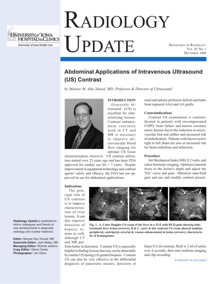
Radiology Update DEPARTMENT OF RADIOLOGY VOL. 20, No. 1 DECEMBER 2008 Abdominal Applications of Intravenous Ultrasound (US) Contrast by Monzer M. Abu-Yousef, MD, Professor & Director of Ultrasound INTRODUCTION Grayscale ultrasound (US) is excellent for characterizing lesions. Contrast enhancement routinely used in CT and MR is necessary to improve microvascular blood flow imaging for optimal US tissue characterization, however. US contrast utilization started over 23 years ago and has been FDA approved for cardiac use for > 7 years. Despite improvement in equipment technology and contrast agents’ safety and efficacy, the FDA has not approved its use for abdominal applications. Radiology Update is published to inform colleagues and friends of new developments in diagnostic radiology and nuclear medicine. Editor: Monzer Abu‑Yousef, MD Associate Editor: Joan Maley, MD Managing Editor: Nichole Jenkins Copy Editor: Glena Clarke Photographer: Jim Olson renal and splenic perfusion defects and leaks from ruptured AAA and AA grafts. Contraindications Contrast US examination is contraindicated in patients with uncompensated COPD, heart failure and known coronary artery disease due to the reduction in microvascular bed and caliber and increased risk of embolization. Patients with known active right to left shunt are also at increased risk for brain embolism and infarction. Procedure Set Mechanical Index (MI): 0.2 watts, and select harmonic imaging. Optimize transmit focus to the lesion’s depth and adjust the TGC curve and gain. Minimize near-field gain and use and modify contrast presets. Indications The principal role of US contrast is to improve characterization of liver lesions. It can also improve detection of Fig. 1. A, Color Doppler US exam of the liver in a 33-F with RUQ pain showing indeh e p a t i c l e - terminate liver lesion (arrows); B & C, early & late contrast US exam showed nodular, sions as well, peripheral, centripetal, arterial & venous enhancement in lesion (arrows), characteristic of hemangioma. although CT and MR perInject 0.2 ml contrast, flush w 3 ml of saline form better in detection. Contrast US is especially over 4 seconds, then start realtime imaging helpful in finding lesions that may not be detectable and clip recording. by routine US during US-guided biopsies. Contrast US can also be very effective in the differential (continued on next page) diagnosis of pancreatic masses, detection of December 2008Radiology Update Abdominal Applications, continued from previous page Potential complications & side effects Cardiac and respiratory arrest, which may be fatal, are serious but rare potential complications for US contrast exams. Less serious complications include hypersensitivity reactions, such as skin rash, but can be serious when patients develop anaphylactic reaction. Side effects are more common and include headache and back pain. Patient monitoring As with CT, patients undergoing contrast US exams are given a contrast explanation sheet. In addition, the vital signs should be monitored before, during and after US contrast exam and should be evaluated for all potential complications and side effects. US contrast nature, properties & common agents US contrast agents are made of microbubbles that are a size smaller than the RBCs (2-8 µm) to enable them to pass through the capillary circulation. They are administered intravenously so they should be typically nontoxic, easily eliminated, physically stable and acoustically responsive. Each bubble is made of a shell composed of either protein, lipid or polymer, and a gas that is either air or heavy gas. The components of these bubbles determine its degree of stability, acoustic responsiveness and safety. While air is more safe, it is less stable and less responsive compared to heavy gas. Similarly, albumen shell is less stable than a shell made of lipid or polymer. The list of US contrast agents is long, but the most commonly used ones include Optison made of albumin, air and PFP (Mallinckrodt); Definity made of lipid, air and PFP (Dupont); Sonovu made of surfactant, powder, air and SF6 (Bracco); Levovist made of Galactose, Palmitate and air (Schering/Berlex); and Echogen made of surfactant and PFC (Sonus). US contrast physics The purpose of US contrast agents is to enhance small blood vessel visualization. Using agents decreases tissue/vascular contrast, however. Different techniques have been used to enhance contrast detection including harmonic imaging, filtration and pulse inversion. There are 3 different ways by which microbubbles interact with US. Linear resonance results in fundamental enhancement, and nonlinear resonance results in harmonic enhancement. Both occur at low US intensity. Transient scattering results from bubble disruption at higher US intensity levels. Harmonic resonance results from the particle oscillating or reflecting US waves at multiples of the frequencies of the incoming US waves. Tissue and blood reflect US waves at the fundamental frequency, while microbubbles reflect at both fundamental and harmonic frequencies. When the equipment is put in the CPS mode during the ultrasound contrast exam, the reflected waves undergo a filtration process that selectively affects the waves at the fundamental frequency. Alternatively, the equipment may send in waves at the same fundamental frequency but Fig. 2. A, suspicious hypoechoic left lobe liver lesion in 60-F with breast ca; B & C, contrast US showed a large central feeding vessel (arrowhead) with centrifugal arterial hyper-enhancement (arrows), which persisted throughout the venous & sinusoidal phases (not shown). Fig. 3 A, US exam shows poorly-defined liver lesion (arrows) in a 55-male with HCV & indeterminate lesion on; B & C, contrast US shows large liver lesion (arrows) with early arterial hyper-enhancement persisting into sinusoidal phase. K = kidney. Fig. 4. A, US with poorly seen RL liver in 71-male with pancreatic ca. & CT with liver lesion; B & C, contrast US shows arterial non-enhancing lesion (arrows) with delayed peripheral hypo-enhancement. at the opposite phase to cancel the waves reflected at this frequency in a process called pulse inversion. Either of these processes leads to prompt enhancement of the harmonic signals coming from the contrast microbubbles. Liver lesions contrast enhancing characteristics On US contrast exams, the various liver lesions demonstrate similar, though slightly different enhancement characteristics compared to that seen on CT and MR contrast exams. Three phases of enhancement are demonstrated by US contrast: arterial (early), portal venous (delayed) and sinusoidal (late) phases. Hemangiomas show peripheral arterial enhancement with delayed centripetal filling (Fig. 1). Focal nodular hyperplasia shows centrifugal hyperintense arterial enhancement with continued portal venous and sinusoidal enhancement (Fig. 2). Adenomas show centripetal arterial hyperintense arterial enhancement with portal venous and (continued on page 5) 2 Radiology Update December 2008 N otes from the Chair Welcome to the latest issue “Radiology Update”. On behalf of the faculty and staff of the University of Iowa, Department of Radiology, I am pleased to highlight recent events and activities within our department. We are excited to let you know about our efforts to be a leader in biomedical imaging research and biomedical imaging informatics, radiological education, and exceptional patient care. In this edition, you will read about new areas of research development and new research grants, a new University of Iowa research institute for biomedical imaging, and the latest accomplishments and kudos of our dedicated faculty and staff. New 2008 grants and contracts for research include: A Merging Multi-scale Model for Simulations of Crystallization/Solidification of Nano-structured Materials on Large-scale Parallel Computing Systems; National Science Foundation, Emerging Models and Technologies; $240,000; PI: Ni, Jun Abnormality Manipulation for Tomographic Imaging Perception Research; US Department of Health & Laurie L. Fajardo, MD, MBA Human Services, National Institutes of Health; $1,010,915.00; PI: Madsen, Mark T Professor & Head, Carotid Revascularization with ev3 Arterial Technology Evolution Post Approval Study (CREAT PAS); Department of Radiology ev3 Endovascular, Inc.; $51,000.00; PI: Chaloupka, John Data Collection for Development and Testing of a Mammography CAD System; VuCOMP, Inc.; 7,500.00; PI: Fajardo, Laurie L Development for RSNA Personal Learning Portfolio; Radiological Society of North America; $20,000.00; PI: D'Alessandro, Michael Diffusion MRI of the Human Brain; University of Wisconsin-Madison; $30,000.00; PI: Kim, Jinsuh Excised Lung Project VPR; VIDA Diagnostics, Inc.; $10,121.00; PI: Hoffman, Eric Genetic Epidemiology of COPD; National Jewish Medical & Research Center; $137,090.00; PI: Hoffman, Eric Genotype-Phenotype Interactions in Severe Asthma Health Study; Wake Forest University; $20,114.00; PI: Hoffman, Eric Imaging Effector Cell Trafficking in Rituximab Therapy of Follicular Lymphoma; Dana Foundation; $100,000.00; PI: Juweid, Malik Inflammation, Myofibroblasts and Distal Lung Disease in Severe Asthma; Washington University in St. Louis; $55,908.00; PI: Hoffman, Eric IPA for Vincent Magnotta; US Department of Veterans Affairs, Iowa City Veterans Affairs Medical Center; $11,000.00; PI: Magnotta, Vincent Large-Scale Computing and Visualization for Cardiopulmonary Imaging; US Department of Health & Human Services, National Institutes of Health; $473,636.00; PI: Lin, Ching-long Multicenter Retrospective Chart Review of the Wingspan Stent System with Gateway PTA Balloon Catheter; Boston Scientific Corporation; $15,000.00; PI: Chaloupka, John Phase 2, Multicenter, Open-Label, Two-Stage Study to Evaluate the Safety and Efficacy of Intra-Arterial Catheter-directed Alfimeprase for Restoration of Neurologic Function and Rapid Opening of Arteries in Stroke (CARNEROS-1); Nuvelo, Inc.; $435,000.00; PI: Chaloupka, John Stenting and Angioplasty with Protection in Patients at High Risk for Endarterectomy (SAPPHIRE WW); Cordis Corporation; $48,205.00; PI: Chaloupka, John The NexStent Carotid Stent System: A Post Market Approval Evaluation Study in Conjunction with the FilterWire EZ Embolic Protection System; Boston Scientific Corporation; $25,625.00; PI: Chaloupka, John Vertical Low Tesla Broadband MRI; US Department of Health & Human Services, National Institutes of Health; $500,000.00; PI: Hoffman, Eric Congratulations to these individuals on their many successes with research! It is a distinct pleasure to recognize our “best doctors” for 2008. The “best doctors” database (http://www.bestdoctors.com) included the following UI radiologists in its latest release: Monzer M. Abu Yousef, Thomas Barloon, Bruce P. Brown, John C. Chaloupka, Georges Y. El-Khoury, Laurie Fajardo, Edmund A. Franken, Jr., Minako Hayakawa, David Kuehn, Yutaka Sato, Wendy Smoker, Alan Stolpen, David Bushnell, Michael Graham, Daniel Kahn, and Yusef Menda. These individuals comprise over 1/3 of our physician faculty. It is also a pleasure to inform you of a new research institute at the University of Iowa that will lead advances in medical imaging. The UI Carver College of Medicine and the College of Engineering established the Iowa Institute for Biomedical Imaging (IIBI) in October 2007. Biomedical imaging and image analysis play critical roles in modern medicine, both in the diagnosis and the treatment of disease. The IIBI aims to foster multi-disciplinary and cross-college research and discovery in biomedical imaging and to improve training and education. A primary objective of the institute is to translate the advances in imaging research to the clinic, improving healthcare for patients. The collaborative nature of the institute ensures that insight from the “bedside” informs and helps direct fundamental imaging research at the “bench”. The institute will reside in the Iowa Institute of Biomedical Discovery building, to be completed in 2011. Nearly 20,000 square feet will be devoted to biomedical imaging and image analysis activities. Radiological research projects will find dedicated space for image acquisition, testing of innovative small animal and human imaging technology, and quantitative imaging analysis. The mission of the Department of Radiology at The University of Iowa is to provide the highest quality, accessible and patient-friendly radiological care while contributing to national efforts to advance training and research in medical imaging. Throughout our various missions, we remain dedicated to progress in medical imaging and patient-centered care. 3 December 2008Radiology Update Research Update Education Update by Kevin S. Berbaum, PhD, Professor, Perceptual Research by Joan Maley, MD, Clinical Associate Professor, Director, Diagnostic Radiology Residency Program Radiologists now read more studies, each containing more images placing greater demands on their vision. Although certain levels of visual fatigue existed with film-viewing, our preliminary data suggests that it is worse with digital displays because they do not provide the same stimulus for oculomotor control as film. Eyestrain, which is known clinically as asthenopia (Ebenholtz, 2001; MacKenzie, 1843), is a consequence of increased image volume and display conditions. With non-medical computer displays, just four hours of near viewing is sufficient to produce asthenopia (Sanchez-Roman, et al., 1996) and prolonged computer use may induce myopia (Komiushina, 2000; Mutti & Zadnik, 1996). Oculomotor fatigue caused by close work with digital displays may add to the effects of extended workdays and aging eyes (Heron, Charman & Gray, 1999). With our colleague Elizabeth Krupinski, a professor in the Radiology Department at the University of Arizona, our Perception Lab will soon be studying the affect of visual fatigue on image interpretation (Eyestrain in Radiologists, NIH Grant R01 EB004987, principal investigator: Elizabeth Krupinski). Preliminary studies suggests that radiologists report increasingly severe symptoms of eyestrain, including blurred vision and difficulty focusing, as they read more imaging studies. Our goal in this research project is to determine whether the detrimental effects of extended inspection of digital displays in radiology go beyond tired eyes and slower reading. We will attempt to assess eyestrain by measuring the changes in accommodation. The lens of the eye is used to alter the refractive index of light entering the eye to focus images on the retina. It is covered by an elastic capsule whose function is to mold the shape of the lens – varying its flatness and therefore its optical power. This variation in optical power is known as accommodation. We will be measuring accommodation using an autorefractor (ours is the so-called WAM-5500 Auto Refkeratometer from Grand Seiko). The device will measure accommodation as the observer looks though a window at an x-ray image presented at a typical viewing distance. The autorefractor will capture the observer’s focus on the screen many times a second for a few seconds. We expect focus to wander in front of and beyond the display screen when the observer’s eyes are tired. We will study how this affects the time needed to interpret images and whether it causes subtle lesions to The Diagnostic Radiology Residency Program at The University of Iowa Hospitals and Clinics continues to move forward and adapt to the ever-changing, post-graduate training requirements. The core competencies (professionalism, patient care, medical knowledge, systems-based practice, practice based learning and communication) have now been with us for seven years. We have completed the first two phases of their implementation, teaching and assessment, and are entering phase three, outcome measurements. In January 2008, the site visitor from the Accreditation Council for Graduate Medical Education reviewed our program and we received notice of reaccreditation for four additional years. We constantly evaluate the curriculum and program to keep pace with the evolving landscape of graduate medical education. A Technology/Systems rotation has been added. This allows the residents the opportunity to shadow the technologists in the individual areas of radiology. The residents will gain a better understanding of the requirements necessary to obtain a diagnostic exam and gain an appreciation of the patients’ experience. Additional rotations in Body MRI will help the residents gain experience in this ever-expanding discipline and increased resident elective time allows the residents to tailor their residency to their educational needs. Currently, the ethics curriculum is being redesigned. Radiology research has always been a cornerstone of our residency and the residents continue to spend dedicated time on research projects. Last year nine different residents presented their research at four different national meetings; the American Society of Head and Neck Radiology (ASHNR), the American Society of Neuroradiology (ASNR), the Radiological Society of North America (RSNA) and the American Institute of Ultrasound in Medicine (AIUM). Dr. Bao Do won the resident research training prize at RSNA for his research on Feedback Natural Language Processing of Fractures in Unstructured Reports of Emergency Department Studies. We had 33 radiology residents in training as of July 1, 2008. Our residents come to train with us from all over the country and we are always looking to add diversity to the classes. In the match just completed March 20, 2008, we matched four women into the eight positions to begin training July 2009. Last year all of our graduating residents successfully completed the oral board exam. Department Researchers Prepare to Study Eyestrain in Radiologists Diagnostic Radiology Residency Program (continued on page 6) 4 Radiology Update December 2008 Department of Radiology at the University of Iowa, allowing us to interact closely with and learn from experienced radiologists who train and teach in systems that are half a world away from ours. Over the past three years the body section has hosted four visiting professors. Drs. Akihiro Nishie and Yoshiki Asayama, were from Kyushu University, Fukuoka, Japan. Drs. Jae Young Lee and Jeong Min Lee were both from Seoul National University College of Medicine in Seoul, South Korea. Dr. Jung Hoon Kim has recently begun a year’s sabbatical with us from Soonchunhyang University Hospital, Seoul, South Korea. Dr. Tom Barloon is the primary teacher of fluoroscopy, passing on the knowledge of classic radiology to the digital generation. Dr. Monzer Abu-Yousef continues to head up the ultrasound division, most recently working to introduce the use of ultrasound contrast agents to our clinical practice. The Body MRI division remains under the direction of Dr. Alan Stolpen who has been helping to guide an ongoing update of those facilities and scanners. When that is completed by early spring, the center will host one 3T and four 1.5T scanners. Dr. Stolpen has also headed up the development of our breast and cardiac MR services. Sectional Update Body Imaging Section by David Kuehn, MD, Clinical Associate Professor, Interim Director of Body Imaging As with the rest of the department, the Body Imaging section has been experiencing a continual evolution of the technology and workflow in radiology. With the number of detector rows in the workhorse CT scanners growing to ever larger multiples of 4, the downstream interpretation technology has also had to adapt. Evolving into a department that is filmless, paperless, using voice transcription, and distributing images widely on the desktop information system, we have also noticed a decrease in our encounters with clinicians. Not only do we see fewer clinical teams dropping by to review images in consultation, we also have fewer telephone interactions with clinicians. An unfortunate byproduct of this ‘de-personalization’ of radiology, both here at the university and in the broader practice of radiology throughout the country, is the risk of becoming just a commodity. Once the end point of a radiology exam—the report—has been reduced in the minds of referring clinicians to a product that is just as easily obtained from Sidney, Minneapolis or Hawaii, it becomes difficult to justify the utility of maintaining your viewbox here in Iowa. Simple numbers such as price and time become the measure of the value of a report. The body imaging section has addressed this trend by remaining very active in participating in multidisciplinary conferences and tumor boards to develop and maintain the relationships that demonstrate the added value of having a discussion with a radiologist. It seems more than coincidence that the clinicians we regularly interact with in these multidisciplinary settings are the same ones who consult us most frequently for an informal image review or consult. The Body section has been adjusting to the retirement of Dr. Bruce Brown last year after 31 years as a physician at The University of Iowa. Board certified in internal medicine before becoming a radiologist, Dr. Brown was involved in the early introduction of ERCP at Iowa and most recently has been instrumental in developing a CT colonography program. He will be greatly missed for the enthusiasm and energy he brings to teaching and we wish him well. There have been four additions to the section in the past year. Former residents and fellows who have joined us include Dr. Maheen Rajput, Dr. Eve Clark, and Dr. Wei Fang. Dr. Fang is taking over for Dr. Brown in coordinating our CTC program. Dr. Rajput is also staffing in breast imaging. Trained outside of Iowa, Dr. Brooke Breen is a graduate of Tufts with a fellowship in MR, who is directing the body fellowship. There has been a long tradition of visiting professors in the Abdominal Applications, continued from page 2 sinusoidal enhancement. Hepatocellular carcinoma typically shows hyperintense arterial enhancement with portal venous and sinusoidal washout (Fig. 3). Metastases show delayed peripheral hypo-enhancement (Fig. 4). Focal fatty infiltration shows early and late iso-enhancement. Other masses may show heterogenous early enhancement with irregular vascularity. Infarcts in kidneys and spleens show perfusion defects. Conclusions US contrast consists of tiny microbubbles that enhance US visualization of microcirculation using harmonic resonance, selective beam filtration, pulse inversion and bubble bursting techniques. US contrast in the liver assists in making a specific diagnosis, confirming a diagnosis, or narrowing the differential diagnosis of the various lesions. Correlation with patient history and laboratory and US findings is important. Although approved for cardiac use, US contrast is still awaiting FDA approval for its abdominal use. Patients’ vital signs should be closely monitored before, during and after the procedure. There should also be careful patient selection, excluding those with known active heart or pulmonary disease, especially coronary artery disease, arrhythmias, COPD and right to left cardiac shunts. References 1. Burns PN, Wilson SR. Focal liver masses: enhancement patterns on contrast-enhanced images--concordance of US scans with CT scans and MR images. Radiology. 2007 Jan;242(1):162-74. 2. Cosgrove D, Blomley M. Liver tumors: evaluation with contrast-enhanced ultrasound. Abdom Imaging. 2004 JulAug;29(4):446-54. 5 December 2008Radiology Update I first met Jason when he was a humor and was always willing to take a medical student. There he stood in chance, try something new. My retireDr. Jason Ross Martin my doorway with a wide grin. He sat ment party was near the time of Jason’s 1/29/70 - 2/25/08 and listened to an idea I had for demfellowship graduation. It was held out onstrating liver anatomy with threein the country around Iowa City; the A Remembrance dimensional reconstructions. Each of green, growing rolling Iowa country us only had a few minutes, but as the side in June -- just about the closest conversation went on, Jason flooded me to heaven anybody could imagine. with questions, most of which I couldn’t Jason was kind enough to be there and answer. So the minutes turned into an shake my hand. That meant a lot to me. hour or so, a research plan, and in less Later, as the party was winding down, than a month he had answered most of I looked up trying to spot an airplane his own questions, completed a literaflying low over the hills, but I never saw ture review, as well as the project that the plane, and it passed. he deftly presented at RSNA that year. There were still a few days left That was Jason: curious, respectful, for me at work, and as I was cleaning hardworking, sometimes stubborn, but out my office, Jason came in, plopped always smiling and on the go. So as well down in the only chair not full of files as being a new physician, he was also and boxes and showed me an aerial a licensed pilot and an award-winning photo of green Iowa fields, a tent, some photographer. people far below. It was a picture he After becoming board-certified in had taken of the retirement party from radiology at the University of Nebraska, that low-flying plane we had heard. Jason spent time in private practice, but One hand on the wheel, one hand he wanted to know more about academic radiology and over his shoulder, snapping a photo of my retirement party. came to UIHC in 2006-7 for a body imaging fellowship. That was Jason, taking a chance, flying low over the green Initially he had difficulty getting used to our dictation fields to present us with a new view of things -- going back and reporting system. He and I had many long and frank to get more information in a new fellowship to better prepare discussions about this. At his age, with his past wide exfor helping people. perience as a private practice radiologist, he could have I will never forget Jason. I’m sure he’s near us right now simply disregarded our suggestions--but he didn’t. He took trying to find a new way of thinking about things. When he his additional training and our comments very seriously, finds it you can be sure that somehow, while we’re trying to staying after hours, working on weekends, asking quesmake sense of cleaning out our office of thirty years, putting tions, making suggestions for our own program. During his up a grandchild’s swing, moving to a retirement home, he fellowship, under the direction of Dr. Monzer Abu-Yousef, will walk into our lives, smile, and show us something we Jason completed a second presentation on the safety of have never seen before. thyroid biopsies, which he presented at the 2007 RSNA. Our deepest condolences go to his family at his death. We So with this give-and-take process, through his hard work, thank them for his life. we all learned something. It was an honor to present him his fellowship diploma. He had earned it. Bruce P. Brown, MD, Associate Professor Emeritus Jason was fun to be around. He had a quirky sense of Research Update, continued from page 4 be missed that were called when the oculomotor system was not fatigued. Preliminary before- and after-workday measurements on a few radiologists show significant degradation in the ability to focus on both near and far objects after a day of image interpretation, although the degradation is more severe for near vision. of visual fatigue during work involving visual stress. Vestnik Oftalmologii 2000;116:33-36. 4. MacKenzie W. On asthenopia or weak-sightedness, Edinburgh J Med & Surg 1843;60:73-103. 5. Mutti DO, Zadnik K. Is computer use a risk factor for myopia? J Am Optometric Assn 1996; 67:521-530. 6. Sanchez-Roman FR, Perez-Lucio C, Juarez-Ruiz C, VelezZamora NM, Jimenez-Villarruel M. Risk factors for asthenopia among computer terminal operators. Salud Publica de Mexico, 1996;38:189-196. References 1. Ebenholtz SM. Oculomotor systems and perception. Cam- bridge University Press, New York, NY, 2001. 2. Heron G, Charman WN, Gray LS. Accommodation responses and ageing. Invest Ophthal Visual Science 1999;40:2872-2883. 3. Komiushina TA. Physiological mechanisms of the etiology 6 Radiology Update December 2008 Welcome New Faculty! Eve D. Clark, MD, joined the Department of Radiology as a Clinical Assistant Professor. Dr. Clark received her medical training from the University of Iowa College of Medicine. She completed her residency in diagnostic radiology, as well as a fellowship in body imaging at University of Iowa Hospitals & Clinics. Dr. Clark joins the faculty of the Body Imaging section. Wei Fang, MD, joined the Department of Radiology as a Clinical Assistant Professor. He received his medical training at the University of Iowa College of Medicine and later went on to complete his diagnostic radiology residency at University of Iowa Hospitals & Clinics. Dr. Fang also completed a fellowship in body imaging at UIHC and recently joined the faculty of the Body Imaging section. Clark Jung Hoon Kim, MD, Visiting Assistant Professor, joined the Body Imaging section of the Department of Radiology. Dr. Kim completed his medical education at College of Medicine, Hanyang University in Seoul, Korea. He completed his residency at Asan Medical Center, University of Ulsan College of Medicine in Seoul. Dr. Kim also completed two fellowships in abdominal radiology, one at Ulsan College of Medicine, and the other at Soonchunhyang University Hospital in Seoul, where he also currently serves as an Assistant Professor. Jun Ni, PhD, joined the Department of Radiology as an Associate Professor. Dr. Ni also has secondary faculty appointments in the Departments of Mechanical & Industrial Engineering and Biomedical Engineering in the College of Engineering at the UI. Dr. Ni also serves as the director of ITS Research Services and is director for the following University of Iowa laboratories: Medical Imaging HPC and Informatics Research Lab (MIHPC Lab), Department of Radiology; Hawkeye Radiology Informatics, Department of Radiology; and the HPC Nanotechnology Research Lab (HPCNano Lab), Department of Mechanical Engineering. Dr. Ni is also Honorable Co-Director of the High-performance Computing Laboratory in the School of Biomedical Engineering & Sciences at Virginia Tech. Fang Kim Ni (continued on next page) In addition to our new faculty, we would also like to welcome our new fellows: Body Imaging Bradley King, MD, Fellow-Associate Scott Greenley, MD, Fellow-Associate Breast Imaging Archana Laroia, MD, Fellow-Associate Chest Stephen Burke, MD, Fellow-Associate Musculoskeletal Glenda Holzman, MD, Fellow-Associate Amol Katkar, MD, Fellow David Watkins, MD, Fellow-Associate Hani Al-Ali, MD, Fellow Nuclear Medicine Neurointerventional Pediatric R. Charles Callison, MD, Jr., Fellow-Assoc Wei Liu, MD, Fellow-Associate John Terry, MD, Fellow-Associate Neuroradiology Aristides Capizzano, MD, Fellow Sandeep Laroia, MD, Fellow Christopher McKinney, MD, Fellow Aaron Settler, MD, Fellow 7 Ravi Sood, MD, Fellow-Associate Achint Singh, MD, Fellow Vascular Interventional Anish Bansal, MD, Fellow Lokesh Khanna, MD, Fellow December 2008Radiology Update New Faculty, continued from previous page Maheen Rajput, MD, Clinical Assitant Professor, joined the Department of Radiology as member of the Body Imaging section. Dr. Rajput completed her medical training at the University of Illinois College of Medicine, Urbana and Peoria, IL. She later went on to complete her residency in diagnostic radiology at the University of Iowa Hospitals and Clinics. Prior to her appointment, Dr. Rajput completed fellowships in musculoskeletal radiology and body imaging at UIHC. Rajput John Sunderland, PhD, MBA, joined the Nuclear Medicine/PET section as an Associate Professor. Prior to his appointment at UIHC, Dr. Sunderland was an instructor at Louisiana State University - Shreveport, LA, in the Department of Chemistry and Physics. He also served as Vice President of PET Operations at the Biomedical Research Foundation in Shreveport. Dr. Sunderland received his PhD in Medical Physics from the University of Wisconsin, Madison, WI, and an MBA from Centenary College, Frost School of Business, Shreveport, LA. He currently serves as the Technical Director for the PET Imaging Center at UIHC and PET Expert Consultant for Lawrence Livermore National Laboratory for the Establishment of a Cyclotron/PET Facility in the Urals region of Russia as part of the US Department of Energy Nuclear Cities Initiative. Sunderland New Residents Diagnostic Radiology Saad Ali, MBBS, Aga Khan University, Pakistan Tamer Ghosheh, MD, University of Iowa Mohammed Sarawan, MBBS, University of Jordan, Jordan David De Bruin, MD, University of Chicago M. Cristian Dobre, MD, University of Minnesota Robert Heninger, MD, University of North Dakota Warren Spencer, MD, Medical College of Wisconsin Nuclear Medicine Jeffrey Meier, MD, University of Pennsylvania Kamaljit Puri, MBBS, Indira Gandhi Medical College, Shimia HP, India James Stecher, MD, University of Iowa Harnoor Singh, MD, Government Medical College, Punjab, India Honors & Awards D. Lee Bennett, MD Appointed Associate Diagnostic Radiology Residency Program Director Michael M. Graham, MD Vice-President Elect, Society of Nuclear Medicine, 2008 Appointed Examiner of the American Board of Radiology, June 2008 8 Geetika Khanna, MD, MS Certificate of Appreciation, Radiol ogical Society of North America, 2007 Radiology Update Joan Maley, MD Selected as one of 30 candidates to participate in the 2008 AUR-Agfa Radiology Management Program during the AUR’s annual meeting Toshio Moritani, MD Appointed to the Electronic Educa- tion and Internet Committee for the American Society of Neuro- radiology, 2006-2010 Listed in Who’s Who in Medicine and Healthcare, 2006-2007 Reviewer for Postgraduate Medi- cine, European Neurology, Neurology India, Cancer Research, Journal of Neurotrauma Brian Mullan, MD One of 3 radiologists chosen to par- ticipate in a 14-day trip to Uganda for an RSNA-sponsored Interna- tional Visiting Professor Program to teach residents new skills using the resources available to them. Jun Ni, PhD Recipient of one of 6 summer faculty fellowships at the University December 2008 Laurie L. Fajardo, MD, MBA, Receives 2008 AUR Gold Medal Award Professor and Chair of the Department of Radiology, Laurie L. Fajardo, MD, MBA, was awarded the presitigious Gold Medal Award by the Association of University Radiologists. The Gold Medal is “awarded in recogni‑ tion of unusually distinguished service or contributions to the Association of University Radiologists, academic radiology, or the field of radiology in general.” Dr. Fajardo was honored with the award, along with David C. Levin, MD, at a rec‑ ognition ceremony in March of 2008. Congratulations, Dr. Fajardo! Photo courtesy of the Association of University of Radiologists of Illinois at Urbana-Champaign’s National Center for Supercomputing and Applications, participating in a cyberinfrastructure-based project on petascale computing applications in multidisciplinary domains Awards Received at National Meetings Jeong D, Park JM, Adkins B, Menda Y, Franken EA, Fajardo LL. PET/CT Findings in Breast Cancer; Correlation with Mammography and Ultrasound. American Roentgen Ray Society, Washington DC, April, 2008. BRONZE MEDAL Baima J, Smoker WRK, Gentry, LR, Michel MA, Reede DL: Don’t Talk with your Mouth Full (of your tongue): Causes of Macroglossia. American Society of Head and Neck Radiology Annual Meeting, Seattle WA, September, 2007. THIRD PLACE AWARD Alsheik NH, Gentry LR, Smoker WRK, Reede DL. Maxillofacial trauma presenting to a level I trauma center: Imaging findings, classification, and complications in 1000 consecutive patients. American Society of Head and Neck Radiology 41st Annual Meeting, Seattle, WA, September 2007. RESIDENT AWARD PAPER Lee HK, Smoker WRK, Moritani T, Lee A: Oculomotor nerve: Anatomy and pathology. Radiological Society of North America 92nd Annual Meeting, Chicago, IL, November 2007. CERTIFICATE OF MERIT Dorantes TM, Reede DL, Hwang W, Smoker WRK, Holliday RA, Gentry LR: Buccal space is “in your face.” Radiological Society of North America 92nd Annual Meeting, Chicago, IL, November 2007. CERTIFICATE OF MERIT AWARD 9 President of the Science, Technol- ogy, Engineering and Math Educa- tion Society Listed in Who's Who in Medicine Higher Education (WWMHE) AcademicKeys Achievement Award, The World Academy of Science, World Computer Congress, Las Vegas, NV, 2007 Founding Editor-in-Chief, Journal of Medical Imaging and Radiology Informatics; Journal of Computa- tional Biosciences and Bioinformatics Associate Editor for Science Letters; and Supplemental Issue Editor, BMC Bioinformatics Honorable Editorial Board, Reports in Medical Imaging Editorial Board, Frontiers of Mechanical Engineering in China; Journal of Computational Intelli- gence in Bioinformatics; Journal of Computational and Theoretical Nanoscience (continued on next page) December 2008Radiology Update 2007-2008 RADIOLOGY TEACHING AWARDS MEDICAL STUDENT TEACHING AWARDS Resident Teacher of the Year Todd Ebbert, MD Resident Educator of the Year Ahmad Izard, MD Outstanding Resident Teachers Dean McNaughton, MD Paul Wheeler, MD Outstanding Senior Faculty Teacher of the Year Monzer Abu-Yousef, MD Abu-Yousef Ahn Clark Dogar Ebbert Izard Patel McNaughton Wheeler Outstanding Junior Faculty Teacher of the Year Eve Clark, MD DEPARTMENTAL TEACHING AWARDS Resident Research Award Rakesh Patel, MD Resident Teacher of the Year Mohammad Asif Dogar, MD Resident Award for Outstanding Clinical Service Mohammad Asif Dogar, MD Krabbenhoft Award for Excellence in Teaching Monzer Abu-Yousef, MD Faculty Teacher of the Year Joong Mo Ahn, MD Breast Imaging Center Receives National Recognition Honors, continued from previous page Guest Editor/Associated Guest Editor, Journal of Computa tional and Theoretical Nanotech- nology; Intl. Journal of Computa tional Science and Engineering Wendy R. K. Smoker, MD Team Leader and First Place Award winner for Stump the Stars, South- eastern Neuroradiological Society Freeport, Bahamas, October 2007 Selected as a 2008 Woman of Achieve ment by the American Biographical Institute John Sunderland, PhD Appointed Technical Director, Positron Emission Tomography Edwin R. J. van Beek, MD Reviewer for Academic Radiology; Critical Care Medicine; EuropeanRadiology, Investigative Radiology; Journal of Magnetic Please join me in congratulating the Breast Imaging Clinic for their achievement of National recognition as a “BREAST IMAGING CENTER OF EXCELLENCE.” To gain recognition as a center of excellence, our center has achieved high standards for technologist and physician training and credentialing, imaging quality assurance and quality control, diagnostic accuracy, patient safety and follow-up, practice audit, and utilization review/appropriateness. Thank you Dr. Jeong Mi Park and Deb Havel for your hard work, leadership and determination in reaching this goal! --Dr. Laurie L. Fajardo, Professor & Head Resonance Imaging; Radiology European Society of Thoracic Imaging 2008 2008 Past-Chair, Hyperpolarized noble gas MR study group, Interna- tional Society of Magnetic Reso nance in Medicine. 10 Paul D. Wheeler, MD Selected as one of 35 candidates to participate in the 2008 Siemens-SUR Radiology Resident Academic Development Program during the AUR’s annual meeting. Radiology Update December 2008 The print version of this section contains a listing of contributors to the Department of Radiology for the period of July 1, 2006 through June 30, 2007. If you wish to receive a print copy, please contact Nichole Jenkins at (319) 353-8690. Publications Books/Book Chapters Ahn JM, El-Khoury GY. Stress Injuries. In: Pope T, Morrison WB, Wilson DJ, eds. Imaging of the Musculoskeletal System, Vol. 1. Philadelphia, PA: Saunders/Elsevier; 2008:975-1001. Menda Y, Graham MM. FDG PET Imaging of Head and Neck Cancers. In: Biersack HJ, Freeman LM, eds. Clinical Nuclear Medicine. New York: Springer; 2007. Ni J (Editor), Jin H, Huang S, Demaria J. Proceeding of Third International Conference on Internet Computing in Science and Engineering, IEEE Computer Science Publications, 2008. Schuknecht B, Smoker WRK. Imaging diseases of the pharynx and oral cavity. In: Diseases of the Brain, Head and Neck, and Spine. Hodler J, vonSchulthess GK, Zollikofer ChL, eds. Springer-Verlag Italia, 2008:149-158 Xiao S, Ni J, Wang S. The Bridhing Domain Multiscale Method and its High Performance Computing Implementation. In: Cao J, ed. Cyber- infrastructure Technologies and Applications. Nova Science Publishers, Inc; 2008. Articles Abada HT, Golzarian J. Gelatine sponge particles: handling characteristics for endovascular use. Tech Vasc Interv Radiol. 2007 Dec;10(4):257-60. Abada HT, Golzarian J. Multidetector CT in abdominal aortic aneurysm following endovascular repair: how to consider the value of a delayed phase. Radiology. 2007 Nov;245(2):610; author reply 610-1. (continued on next page) 11 December 2008Radiology Update Publications, continued from previous page Armato SG 3rd, McNitt-Gray MF, Reeves AP, Meyer CR, McLennan G, Aberle DR, Kazerooni EA, MacMahon H, van Beek EJ, Yankelevitz D, Hoffman EA, Henschke CI, Roberts RY, Brown MS, Engelmann RM, Pais RC, Piker CW, Qing D, Kocherginsky M, Croft BY, Clarke LP. The Lung Image Database Consortium (LIDC): an evaluation of radiologist variability in the identification of lung nodules on CT scans. Acad Radiol. 2007 Nov;14(11):1409-21. Armato SG 3rd, Roberts RY, McNitt-Gray MF, Meyer CR, Reeves AP, McLennan G, Engelmann RM, Bland PH, Aberle DR, Kazerooni EA, MacMahon H, van Beek EJ, Yankelevitz D, Croft BY, Clarke LP. The Lung Image Database Consortium (LIDC): ensuring the integrity of expert- defined "truth". Acad Radiol. 2007 Dec;14(12):1455-63. Aysola RS, Hoffman EA, Gierada D, Wenzel S, Cook-Granroth J, Tarsi J, Zheng J, Schechtman KB, Ramkumar TP, Cochran R, Xueping E, Christie C, Newell J, Fain S, Altes TA, Castro M. Research Program (SARP) NHLBI Severe Asthm. Airway remodeling measured by multidetector computed tomography is increased in severe asthma and correlates with pathology. Chest. 2008 Jul 18. [Epub ahead of print] Baker K, Namati E, Hoffman EA, van Beek EJR, Ferguson JS, Thomas KW, Ross AF, McLennan G. Virtual bronchoscopy: new applications for MDCT in pulmonology. Advances in MDCT – Thoracic Imaging 3(4):10-18. Bayouth JE, Menda Y, Graham MM. Utility of positron emission tomography in radiotherapy practice with emphasis on head and neck cancer. Imaging Decisions MRI 2008; 12:14-24. Bonucchi J, Hassan I, Policeni B, Kaboli P. Thyrotoxicosis associated Wernicke's encephalopathy. J Gen Intern Med. 2008 Jan;23(1):106-9. Buck AK, Herrmann K, Büschenfelde CM, Juweid ME, Bischoff M, Glatting G, Weirich G, Möller P, Wester HJ, Scheidhauer K, Dechow T, Peschel C, Schwaiger M, Reske SN. Imaging bone and soft tissue tumors with the proliferation marker [18F]Fluorodeoxythymidine. Clin Cancer Res 14(10):2970-2977, 2008. Burke SJ, Aggarwala G, Stanford W, Mullan B, Thompson B, van Beek EJ. Preablation assessment for the left atrium: comparison of ECG-gated cardiac CT with echocardiography. Acad Radiol. 2008 Jul;15(7):835-43. Burke SJ, van Beek EJR. Pulmonary embolism. Advances in MDCT – Thoracic Imaging 3(4):30-38. Busacker A, Newell JD Jr, Keefe T, Hoffman EA, Cook Granroth J, Castro M, Fain S, Wenzel S. A Multivariate Analysis of Risk Factors for the AirTrapping Asthmatic Phenotype as Measured by Quantitative CT Analysis. Chest. 2008 Aug 8. [Epub ahead of print] Dakin JH, Evans TW, Hansell DM, Hoffman EA. Regional pulmonary blood flow in humans and dogs by 4D computed tomography. Acad Radiol. 2008 Jul;15(7):844-52. D'Alessandro DM, D'Alessandro MP. Delivering biodefense continuing education to military medical providers by allowing a biodefense educational curriculum to unfold in practice. Mil Med. 2007 Dec;172(12):1293-9. Deng J, Yu H, Ni J, Wang L, Wang G. Parallelism of Iterative CT Algorithm based on Local Reconstruction. Journal of Supercomputing. 2008 March 29. [Epub ahead of print] DeVries N, Gassman EE, Kallemeyn NA, Shivanna KH, Adams BD, Magnotta VA, Grosland NM. Validation of Phalanx Bone 3D Surface Segmenttion from CT Images Using Laser Scanning. Skeletal Radiol. 37(1):35-42, 2008. Donta TS, Smoker WRK: Carcinoma of Unknown Primary. Topics in Magnetic Reson Imag 18(4):281-292, 2007. Dornfeld K, Hopkins S, Simmons J, Spitz DR, Menda Y, Graham M, Smith R, Funk G, Karnell L, Karnell M, Dornfeld M, Yao M, Buatti J. Posttreatment FDG-PET uptake in the supraglottic and glottic larynx correlates with decreased quality of life after chemoradiotherapy. Int J Radiat Oncol Biol Phys 2008; 71(2):386-392. Fajardo LL, Park JM, Rosene B. Digital’s Demands. Enterprising Imaging. Therapeutic Radiology Management. Jan 2008. 33-37. Fuld MK, Easley RB, Saba OI, Chon D, Reinhardt JM, Hoffman EA, Simon BA. CT-measured regional specific volume change reflects regional ventilation in supine sheep. J Appl Physiol. 2008 Apr;104(4):1177-84. Fuld MK, van Beek EJR. Post-processing for pulmonary MDCT. Advances in MDCT –Thoracic Imaging 3 (4): 39-46. Gassman EE, Powell SM, Kallemeyn NA, Devries NA, Shivanna KH, Magnotta VA, Ramme AJ, Adams BD, Grosland NM. Automated bony re gion identification using artificial neural networks: reliability and validation measurements. Skeletal Radiol. 2008 Apr;37(4):313-9. Gu F, Chen H, Ni J, Huang Z. Protein structural class prediction based on an improved statistical strategy. BMC Bioinformatics 2008, 9(Suppl 6):S10. Hillis SL, Berbaum KS, Metz CE. Recent developments in the Dorfman-Berbaum-Metz procedure for multireader ROC study analysis. Acad Radiol. 2008 May;15(5):647-61. Jagasia DH, van Beek EJR. Multidisciplinary collaboration for cardiopulmonary MDCT. Advances in MDCT–Thoracic Imaging 3(4):1-9. Joong MA, El-Khoury GY. Radiologic evaluation of chronic foot pain. Am Fam Physician. 2007 Oct 1;76(7):975-83. Juweid ME. 18F-FDG PET as a routine test for posttherapy assessment of Hodgkin’s disease and aggressive non-Hodgkin’s lymphoma: where is the evidence? J Nucl Med 49(1);9-12, 2008. 12 Radiology Update December 2008 Khanna G, Bushnell D, O'Dorisio MS. Utility of radiolabeled somatostatin receptor analogues for staging/restaging and treatment of somatostatin receptor-positive pediatric tumors. Oncologist. 2008 Apr;13(4):382-9. Khanna G, El-Khoury G. Partial tear of the quadriceps tendon in a child. Pediatr Radiol. 2008 Jun;38(6):706-8. Khanna G, O'Dorisio MS, Menda Y, Glasier C, Deyoung B, Smith BJ, Graham M, Juweid M. Somatostatin receptor scintigraphy in surveillance of pediatric brain malignancies. Pediatr Blood Cancer. 2008 Mar;50(3):561-6. Khanna G, O'Dorisio SM, Menda Y, Kirby P, Kao S, Sato Y. Gastroenteropancreatic neuroendocrine tumors in children and young adults. Pediatr Radiol. 2008 Mar;38(3):251-9, quiz 358-9. Kim BS, Kim JH, Choi GM, Kim SH, Park JK, Song BC, Kang W. Comparison of three free-breathing T2-weighted MRI sequences in the evaluation of focal liver lesions. AJR Am J Roentgenol. 2008 Jan;190(1):W19-27. Kim JH, Choi DH, Won JH et al. Hepatocyte-Like Cells from Human Mesenchymal Stem Cells Engrafted in Regenerating Rat Liver Tracked with in vivo Magnetic Resonance Imaging. Tissue Engeneering 2008;14(1):15-23. Kopans DB, Pisano ED, Acharyya S, Hendrick RE, Yaffe MJ, Conant EF, Fajardo LL, Bassett LW, Baum JK, Gatsonis CA. DMIST results: technologic or observer variability? Radiology. 2008 Aug;248(2):703; author reply 703. Kronmüller KT, Pantel J, Götz B, Köhler S, Victor D, Mundt C, Magnotta VA, Giesel F, Essig M, Schröder J. Life events and hippocampal volume in first-episode major depression. J Affect Disord. 2008 Oct;110(3):241-7. Kronmüller KT, Pantel J, Köhler S, Victor D, Giesel F, Magnotta VA, Mundt C, Essig M, Schröder J. Hippocampal volume and 2-year outcome in depression. Br J Psychiatry. 2008 Jun;192(6):472-3. Kuhn MJ, Chen N, Sahani DV, Reimer D, van Beek EJ, Heiken JP, So GJ. The PREDICT study: a randomized double-blind comparison of contrast induced nephropathy after low- or isoosmolar contrast agent exposure. AJR Am J Roentgenol. 2008 Jul;191(1):151-7. Kunii N, Abe T, Kawamo M, Tanioka D, Izumiyama H, Moritani T. Rathke's cleft cysts: differentiation from other cystic lesions in the pituitary fossa by use of single-shot fast spin-echo diffusion-weighted MR imaging. Acta Neurochirgica 149(8):756-769, 2007. Lang EV, Berbaum KS, Pauker SG, Faintuch S, Salazar GM, Lutgendorf S, Laser E, Logan H, Spiegel D. Beneficial effects of hypnosis and adverse effects of empathic attention during percutaneous tumor treatment: when being nice does not suffice. J Vasc Interv Radiol. 2008 Jun;19(6):897-905. Lee HK, Park SJ, Yi BH, Yeon EK, Kim JH, Hong HS. Portal vein thrombosis: CT features. Abdom Imaging. 2008 Jan-Feb;33(1):72-9. Li C, Grobmyer SR, Chen L, Zhang Q, Fajardo LL, Jiang H. Multispectral diffuse optical tomography with absorption and scattering spectral constraints. Appl Opt. 2007 Dec 1;46(34):8229-36. Li C, Grobmyer SR, Massol N, Liang X, Zhang Q, Chen L, Fajardo LL, Jiang H. Noninvasive in vivo tomographic optical imaging of cellular morphology in the breast: possible convergence of microscopic pathology and macroscopic radiology. Med Phys. 2008 Jun;35(6):2493-501. Liang X, Zhang Q, Li C, Grobmyer SR, Fajardo LL, Jiang H. Phase-contrast diffuse optical tomography pilot results in the breast. Acad Radiol. 2008 Jul;15(7):859-66. Lockhart ME, Fielding JR, Richter HE, Brubaker L, Salomon CG, Ye W, Hakim CM, Wai CY, Stolpen AH, Weber AM. Reproducibility of Dynamic MR Imaging Pelvic Measurements: A Multi-institutional Study. Radiology. 2008 Nov;249(2):534-40. Lu G, Ni J, Casavant T, Athey B. Highlighting computations in bioscience and bioinformatics: review of the Symposium of Computations in Bioinformatics and Bioscience. BMC Bioinformatics 2008, 9(Suppl 6):S1. Lu X, Nadvoretskiy V, Bu L, Stolpen A, Ayres N, Pignatelli RH, Kovalchin JP, Grenier M, Klas B, Ge S. Accuracy and reproducibility of real-time three-dimensional echocardiography for assessment of right ventricular volumes and ejection fraction in children. J Am Soc Echocardiogr. 2008 Jan;21(1):84-9. Epub 2007 Jul 12. Magnotta VA, Adix ML, Caprahan A, Lim K, Gollub R, Andreasen NC. Investigating connectivity between the cerebellum and thalamus in schizo- phrenia using diffusion tensor tractography: a pilot study. Psychiatry Res. 2008 Aug 30;163(3):193-200. McEwan AJ, Atcher RW, Graham MM. Communication – a process of continuous improvement. J Nucl Med 49(5):25N, 2008. McNitt-Gray MF, Armato SG 3rd, Meyer CR, Reeves AP, McLennan G, Pais RC, Freymann J, Brown MS, Engelmann RM, Bland PH, Laderach GE, Piker C, Guo J, Towfic Z, Qing DP, Yankelevitz DF, Aberle DR, van Beek EJ, MacMahon H, Kazerooni EA, Croft BY, Clarke LP. The Lung Image Database Consortium (LIDC) data collection process for nodule detection and annotation. Acad Radiol. 2007 Dec;14(12):1464-74. Mishima H, Lidral AC, Ni J. Application of the Linux cluster for exhaustive window haplotype analysis using the FBAT and Unphased programs. BMC Bioinformatics. 2008 May 28;9 Suppl 6:S10. Moritani T, Kim J, Sato Y, Bonthius D, Smoker WR. Abnormal hypermyelination in a neonate with Sturge-Weber syndrome demonstrated on diffusion-tensor imaging. J Magn Reson Imaging. 2008 Mar;27(3):617-20. Moritani T. Routine work of radiologists in the USA. The Japanese College of Radiology 164, 2008. (continued on next page) 13 December 2008Radiology Update Publications, continued from previous page Namati E, Thiesse J, de Ryk J, McLennan G. Alveolar dynamics during respiration: are the pores of Kohn a pathway to recruitment? Am J Respir Cell Mol Biol. 2008 May;38(5):572-8. Ni J, Deng J, Yu H, He T, Wang G. Analytical Model for Performance Evaluation of Parallel Katsevich Algorithm for 3-D CT Image Reconstruction. Int. Journal on Computational Science and Engineering, Interscience Publisher, 2008. O'Dorisio MS, Khanna G, Bushnell D. Combining anatomic and molecularly targeted imaging in the diagnosis and surveillance of embryonal tumors of the nervous and endocrine systems in children. Cancer Metastasis Rev. 2008 Dec;27(4):665-77. Oudkerk M, Stillman AE, Halliburton SS, Kalender WA, Möhlenkamp S, McCollough CH, Vliegenthart R, Shaw LJ, Stanford W, Taylor AJ,van Ooijen PM, Wexler L, Raggi P. Coronary artery calcium screening: current status and recommendations from the European Society of Cardiac Radiology and North American Society for Cardiovascular Imaging. Int J Cardiovasc Imaging. 2008 Aug;24(6):645-71. Pan H, Han Q, Yin G, Zhang W, Li J, Ni J. A ROI-Based Mining Method with Medical Domain Knowledge Guidance. The Proceedings of ICICSE|08, IEEE Computer Society Press, Los Alamitos, CA, 2008, pp. 91-97. Paradiso S, Turner BM, Paulsen JS, Jorge R, Ponto LL, Robinson RG. Neural bases of dysphoria in early Huntington’s disease. Psychiatry Res, 2008; 162:73-87. Park JM, Franken EA Jr, Garg M, Fajardo LL, Niklason LT. Breast Tomosynthesis: Present Considerations and Future Applications RadioGraphics. 2007 27(Special Issue):S231-S240. Park JM, Franken EA Jr. Triangulation of breast lesions: review and clinical applications. Curr Probl Diagn Radiol. 2008 Jan-Feb;37(1):1-14. Peterson LM, Mankoff DA, Lawton T, Yagle K, Schubert EK, Stekhova S, Gown A, Link JM, Tewson T, Krohn KA. Quantitative imaging of estrogen receptor expression in breast cancer with PET and 18F-Fluoroestradiol. J Nucl Med, 49(3):367-374, 2008. Pisano ED, Hendrick RE, Yaffe MJ, Baum JK, Acharyya S, Cormack JB, Hanna LA, Conant EF, Fajardo LL, Bassett LW, D'Orsi CJ, Jong RA, Rebner M, Tosteson AN, Gatsonis CA; DMIST Investigators Group. Diagnostic accuracy of digital versus film mammography: exploratory analysis of selected population subgroups in DMIST. Radiology. 2008 Feb;246(2):376-83. Policeni BA, Smoker WR. Pathologic conditions of the lower cranial nerves IX, X, XI, and XII. Neuroimaging Clin N Am. 2008 May;18(2):347-68, xi. Policeni BA, Smoker WRK: The lower cranial nerves. Neuroimag Clin N Am 18:347-368, 2008. Rahmouni K, Fath MA, Seo S, Thedens DR, Berry CJ, Weiss R, Nishimura DY, Sheffield VC. Leptin resistance contributes to obesity and hypertension in mouse models of Bardet-Biedl syndrome. J Clin Invest. 2008 Apr;118(4):1458-67. Reede DL, Garcon E, Smoker WR, Kardon R. Horner's syndrome: clinical and radiographic evaluation. Neuroimaging Clin N Am. 2008 May;18(2):36985, xi. Reeves AP, Biancardi AM, Apanasovich TV, Meyer CR, MacMahon H, van Beek EJ, Kazerooni EA, Yankelevitz D, McNitt-Gray MF, McLennan G, Armato SG 3rd, Henschke CI, Aberle DR, Croft BY, Clarke LP. The Lung Image Database Consortium (LIDC): a comparison of different size metrics for pulmonary nodule measurements. Acad Radiol. 2007 Dec;14(12):1475-85. Reinhardt JM, Ding K, Cao K, Christensen GE, Hoffman EA, Bodas SV. Registration-based estimates of local lung tissue expansion compared to xenon CT measures of specific ventilation. Med Image Anal. 2008 Apr 12. [Epub ahead of print] Rogers CS, Abraham WM, Brogden KA, Engelhardt JF, Fisher JT, McCray PB Jr, McLennan G, Meyerholz DK, Namati E, Ostedgaard LS, Prather RS, Sabater JR, Stoltz DA, Zabner J, Welsh MJ. The porcine lung as a potential model for cystic fibrosis. Am J Physiol Lung Cell Mol Physiol. 2008 Aug;295(2):L240-63. Ruprecht, A.. Oral and maxillofacial Radiology: Then and Now. J Am Dent Assoc. 139, Suppl: 5S-6S, June 2008. [http://jada.ada.org/cgi/content/ full/139/suppl_3/5S] Schweitzer ME, Daffner RH, Weissman BN, Bennett DL, Blebea JS, Jacobson JA, Morrison WB, Resnik CS, Roberts CC, Rubin DA, Seeger LL, Taljanovic M, Wise JN, Payne WK. ACR Appropriateness Criteria on suspected osteomyelitis in patients with diabetes mellitus. J Am Coll Radiol. 2008 Aug;5(8):881-6. Sharafuddin MJ, Hoballah JJ, Kresowik TF, Sharp WJ, Golzarian J, Sun S, Corson JD. Long-term outcome following stent reconstruction of the aortic bifurcation and the role of geometric determinants. Ann Vasc Surg. 2008 May-Jun;22(3):346-57. Sheehan FH, Ge S, Vick GW 3rd, Urnes K, Kerwin WS, Bolson EL, Chung T, Kovalchin JP, Sahn DJ, Jerosch-Herold M, Stolpen AH. Threedimensional shape analysis of right ventricular remodeling in repaired tetralogy of Fallot. Am J Cardiol. 2008 Jan 1;101(1):107-13. Shroff R, Kerchner A, Maifeld M, Van Beek EJ, Jagasia D, Dokras A. Young obese women with polycystic ovary syndrome have evidence of early coronary atherosclerosis. J Clin Endocrinol Metab. 2007 Dec;92(12):4609-14. Siddiq F, Vazquez G, Memon MZ, Suri MF, Taylor RA, Wojak JC, Chaloupka JC, Qureshi AI. Comparison of primary angioplasty with stent placement for treating symptomatic intracranial atherosclerotic diseases: a multicenter study. Stroke. 2008 Sep;39(9):2505-10. 14 Radiology Update December 2008 Simons AL, Fath MA, Mattson DM, Smith BJ, Walsh SA, Graham MM, Hichwa RD, Buatti JM, Dornfeld K, Spitz DR. Enhanced response of human head and neck cancer xenograft tumors to cisplatin combined with 2-deoxy-D-glucose correlates with increased 18F-FDG uptake as determined by PET imaging. Int J Radiat Oncol Biol Phys. 2007 Nov 15;69(4):1222-30. Skelly LR, Calhoun VD, Meda SA, Kim J, Mathalon DH, Pearlson GD. Diffusion tensor imaging in schizophrenia: relationship to symptoms. Schizophrenia Res 2008;98 (1):157-162. Smith RB, Robinson RA, Hoffman HT, Graham MM. Preoperative FDG-PET imaging to assess the malignant potential of follicular neoplasms of the thyroid. Otolaryngol Head Neck Surg 138(1):101-106, 2008. Smith RB, Wayne, S, Maley, JE. Adult with Cheek Swelling. Patient of the Month Program – Otolaryngology 2008; 37(5):1-32. Smoker WR, Gentry LR, Yee NK, Reede DL, Nerad JA. Vascular lesions of the orbit: more than meets the eye. Radiographics. 2008 JanFeb;28(1):185-204. Smoker WR, Khanna G. Imaging the craniocervical junction. Childs Nerv Syst. 2008 May 7. [Epub ahead of print] Smoker WR, Reede DL. Denervation atrophy of motor cranial nerves. Neuroimaging Clin N Am. 2008 May;18(2):387-411, xi. Sparacia G, Cannizzaro F, D'Alessandro DM, D'Alessandro MP, Caruso G, Lagalla R. Initial Experiences in Radiology E-Learning. Radiographics 2007, 27(2):573-581. Suter MJ, Reinhardt JM, McLennan G. Integrated CT/bronchoscopy in the central airways: preliminary results. Acad Radiol. 2008 Jun;15(6):78698. Taylor RA, Siddiq F, Suri MF, Martin CO, Hayakawa M, Chaloupka JC. Risk factors for in-stent restenosis after vertebral ostium stenting. J Endovasc Ther. 2008 Apr;15(2):203-12. Thomas CP, Erlandson JC, Edghill EL, Hattersley AT, Stolpen AH. A genetic syndrome of chronic renal failure with multiple renal cysts and early onset diabetes. Kidney Int. 2008 Oct;74(8):1094-9. Thomas SM, Goodacre SW, Sampson FC, van Beek EJ. Diagnostic value of CT for deep vein thrombosis: results of a systematic review and meta-analysis. Clin Radiol. 2008 Mar;63(3):299-304. Epub 2007 Dec 20. Tosteson AN, Stout NK, Fryback DG, Acharyya S, Herman BA, Hannah LG, Pisano ED. DMIST Investigators. Cost-effectiveness of digital mammography breast cancer screening. Ann Intern Med. 2008 Jan 1;148(1):1-10. van Beek EJ, Hoffman EA. Functional imaging: CT and MRI. Clin Chest Med. 2008 Mar;29(1):195-216, vii. van Beek EJ, Mullan B, Thompson B. Evaluation of a real-time interactive pulmonary nodule analysis system on chest digital radiographic images: a prospective study. Acad Radiol. 2008 May;15(5):571-5. Van Beek EJ, Thompson B. Large field-of-view chest reconstruction on cardiac CT – worth the trouble. J Cardiovasc CT 2008;2:64. Washko GR, Criner GJ, Mohsenifar Z, Sciurba FC, Sharafkhaneh A, Make BJ, Hoffman EA, Reilly JJ. Computed tomographic-based quantification of emphysema and correlation to pulmonary function and mechanics. COPD. 2008 Jun;5(3):177-86. Weldon DT, Burke SJ, Sun S, Mimura H, Golzarian J. Interventional management of lower gastrointestinal bleeding. Eur Radiol. 2008 May;18(5):85767. Williams MB, Raghunathan P, More MJ, Seibert JA, Kwan A, Lo JY, Samei E, Ranger NT, Fajardo LL, McGruder A, McGruder SM, Maidment AD, Yaffe MJ, Bloomquist A, Mawdsley GE. Optimization of exposure parameters in full field digital mammography. Med Phys. 2008 Jun;35(6):2414-23. Xia Y, Song G, Zheng Y, Ni J, Zhu M. A Small World Overlay P2P Transfer System with Role-based and Reputation-based Access Control Policies. The Proceedings of ICICSE|08, IEEE Computer Society Press, Los Alamitos, CA, 2008, pp. 517-520 Xiao S, Ni J, Shaowen Wang S. The Bridging Domain Multiscale Method and its High Performance Computing Implementation. Journal of Computational and Theoretical Nanosciences, Vol. 5, pp. 1-10, 2008. Xiao S, Wang S, Ni J, Briggs R, Rysz M. Reliability Analysis of Carbon Nanotubes Using Molecular Dynamics with the Aid of Grid Computing. Journal of Computational and Theoretical Nanosciences. Vol. 5, pp. 528-534, 2008. Yarlagadda R, Menda Y, Graham MM. Tc-99m red blood cell imaging in a patient with blue rubber bleb nevus syndrome. Clin Nucl Med 2008; 33(5):374-376 Zaidat OO, Klucznik R, Alexander MJ, Chaloupka J, Lutsep H, Barnwell S, Mawad M, Lane B, Lynn MJ, Chimowitz M; NIH Multi-center Wingspan Intracranial Stent Registry Study Group. The NIH registry on use of the Wingspan stent for symptomatic 70-99% intracranial arterial stenosis. Neurology. 2008 Apr 22;70(17):1518-24. Zhang D, Donovan M, Fajardo LL, Archer A, Wu X, Liu H. Preliminary feasibility study of an in-line phase contrast X-ray imaging prototype. IEEE Trans Biomed Eng. 2008 Sep;55(9):2249-57. 15 December 2008Radiology Update Scientific Presentations Abu-Yousef MM. Martin, J, MM, Peterson, G, Larotia, A, Wu, A. Do we need to with-hold anticoagulants in patients undergoing US-guided aspirations of thyroid & other neck masses? Society of Radiologists in Ultrasound Fellows Annual meeting, Chicago, IL, October 25-28, 2007. Baker KM, Gabe L, Hoffman E, van Beek E, Look D, McLennan G, Hunninghake G. Comparison of bronchoalveolar lavage using different volumes. American Thoracic Society International Conference, Toronto, Canada, May 16, 2008. Bo Qiang, Jun Ni, and Laurie Fajardo, Parallel Monte Carlo Algorithm for Simulation of Photon Migration inside Biological Tissues. RSNA|07, Chicago, IL, November 25-30, 2007. Fuld MK, Simon BA, van Beek EJ, Hudson M, Sieren J, Hoffman EA. Transitioning from the laboratory to the clinic: adapting the Xe-CT method for human scanning. ATS 2008 Goh JPN, Chawla A, Chuah KL, Tan TY, Smoker WRK, Gentry LR Imaging of extranodal lymphoma in the extracranial head and neck American Society of Head and Neck Radiology Annual Meeting, Seattle WA, September 2007. Gurol ME, Adams H, Moritani T, Maiseri H, Davis PH. Association of Blood Glucose Levels with the Extent of Tissue Injury in the Hyperacute Phase of Ischemic Stroke . 60th Annual Meeting of the American Academy of Jun 7-10, 2008. Hudson M, Saba O, Sieren J, van Beek EJR, Hoffman EA. Functional imaging of the lung via dual energy CT: requirements and limitations. American Thoracic Society International Conference, Toronoto, Ontario, May 16-21, 2008. Ingalhalikar M, Kim J, Magnotta VA, Alexander AL. A comparative study of diffusion tensor field transformations. ISMRM 16th Scientific Meeting, Toronto, #1855, 2008. Ingalhalikar M, Magnotta V, Kim J, Alexander A. Comparison of Tensor Rotation Methods. International Society for Magnetic Resonance in Medicine: ISMRM Sixteenth Scientific Meeting, Toronto, Canda, May, 2008. Jeong D, Park JM, Adkins B, Menda Y, Franken EA, Fajardo LL. PET/CT Findings in Breast Cancer; Correlation with Mammography and Ultrasound. 108th Annual Meeting of the American Roentgen Ray Society, April 13-18, 2008. (CME-assigned and Selected for a presentation for the “Power Hour” program) Juweid ME, Buck AK, Baranowska-Kortylewicz J. Can radiolabeled thymidine analogs (RTAs) be used as in vivo probes of DNA repair synthesis? Presented at the Society of Nuclear Medicine Annual Meeting, New Orleans, LA, June 14-18, 2008. Kim J, Alexander A, Ingalhalikar M, Magnotta V. A simple method for ODF reorientation after deformable imaging registration. International Society for Magnetic Resonance in Medicine: ISMRM Sixteenth Scientific Meeting, Toronto, Canda, May, 2008. Kim J, Ingalhalikar M, Magnotta VA, Alexander AL. A simple method for ODF reorientation after deformable image registration. ISMRM 16th Scientific Meeting, Toronto, #3327, 2008. Laroia AT, Stanford W, Mullan BF, Thompson B, van Beek EJR. Radiologic manifestations of pulmonary papillomatosis. Society of Thoracic Radiology Postgraduate Course, Fort Myers, FL. March 2008. Laroia AT, Stanford W, Mullan BF, Thompson B, van Beek EJR. Unexpected findings on coronary CTA – worth the trouble? Laroia AT, Thompson B, Mullan B, Stanford W, van Beek EJR. Chest X-ray CAD system: clinical experience in cancer follow-up patients. Society of Thoracic Radiology Postgraduate Course, Fort Myers, FL. March 2008. Lee HK, Smoker WRK, Moritani T, Kim J. Large Meckel’s Cave: Clinical presentations and MR features. Radiological Society of North America 92nd Annual Meeting. Chicago, IL. Nov-Dec 2007. Magnotta V, Harris G, Ingalhalikar M, Tao X. GTRACT: An Open Source Diffusion Tensor and Fiber Tracking Toolkit. MICCAI 2008 Workshop on Computational Diffusion MRI. New York, NY, September 2008 Magnotta VA, Li W, Grosland NM. Comparison of Displacement-Based and Force-Based Mapped Meshing. MICCAI Computational Biome- chanics for Medicine III. New York, NY, September 2008. Martin J, Abu-Yousef MM, Peterson G, Laroia A, Wu A. US-guided aspirations of thyroid & other neck masses are safe to perform in anticoagu lated patients. Scientific Assembly and Annual meeting of the Radiological Society of North America, Chicago, IL, November 25-30, 2007. Moritani T, Kademian J, Smoker WRK, Policeni BA, Lee H, Kim J, Sato Y. Diffusion-weighted imaging of scalp and skull lesions. American Society of Neuroradiology annual meeting. New Orleans, LA, June 2008. Moritani T, Kirby P, Greenlee JDW, Sato Y. Inflammatory Myofibroblastic Tumor with Extensive Orbital, Paranasal Sinus, Intracranial, and Systemic Skeletal Involvements. 46th American Society of Neuroradiology. New Orleans, LA, May-Jun 2008. Moritani T, Sato TS, Hitchon P, Sato Y. Intradiploic Cerebrospinal Fluid Pseudocyst: Unusual Complication of the Ventriculoperitoneal Shunt. 46th American Society of Neuroradiology. New Orleans, LA, May-Jun 2008. Moritani T, Sato TS, Kirby P, Buatti J. Papillary Tumor of the Pineal Region. 46th American Society of Neuroradiology. New Orleans, LA, May-Jun 2008. 16 Radiology Update December 2008 Parekh NN, Smoker WRK, Menezes AH. MR and CT imaging findings of calcium pyrophosphate dihydrate deposition disease at the cranio- vertebral junction. American Society of Neuroradiology annual meeting. New Orleans, LA, June 2008 Park JM, Franken EA, Fajardo LL. Minimally Invasive Percutaneous Surgical Device for Breast Lesions. 108th Annual Meeting of the American Roentgen Ray Society, April 13-18, 2008. Park JM, Franken EA, Garg M, Fajardo L. Breast Tomosynthesis: Preliminary results and future applications (AMA PRA Category I) Radiologi cal Society of North America, Chicago, IL. November, 2006. (Invited by RadioGraphics and published) And The University of Iowa Review of the 2007 San Antonio Breast Cancer Symposium. Iowa City, IA. February 29, 2008 Policeni BA, Smoker WRK, Policeni FC, Gentry LR, Reede DL. Lower Cranial nerve pathology. A segmental approach. American Society of Neuroradiology Annual Meeting, New Orleans, LA, June 2008. Qiang B, Ni J, Fajardo LL. Parallel monte carlo algorithm for Simulation of Photon Migration inside Biological Tissues. Scientific Assembly and Annual meeting of the Radiological Society of North America, Chicago, IL, November 25-30, 2007. Ramme A, Magnotta V, Grosland NM. Comparison of the Expectation Maximization Algorithm and an Artificial Neural Network for Automated Bone Segmentation. 54th Annual Meeting of the Orthopaedic Research Society, San Francisco, California, March, 2008. Schultz MK, McAlister D, Tewson TJ, Harvey TJ, Horwitz P. Evaluation of a new Ge-68/Ga-68 generator for preparing high-purity radio- pharmaceuticals for PET imaging. Presented at the Society of Nuclear Medicine Annual Meeting, New Orleans, LA, June 2008. Schultz MK, Watkins GL, Tewson TJ. Removal of a [C-11]-carbonic acid impurity arising in the synthesis of 1-[C-11]-acetate using the captive solvent method. Presented at the Society of Nuclear Medicine Annual Meeting, New Orleans, LA, June 14-18, 2008. Shivanna K, Kallemeyn N, Tadepalli S, DeVries N, Magnotta V, Grosland N. IA-FeMesh: An Interactive All Hexahedral Mesh Generator For Discrete Anatomic Closed Surfaces. 2008 Summer Bioengineering Conference, Marco Island, FL, June, 2008. Soebbing S, Tewson T, Franklin S. Incorporation of histidine-rich metal-binding sites onto small protein scaffolds: toward developing direct Tc- labeling of proteins. Society of Nuclear Medicine Annual Meeting, New Orleans, LA, June 2008. Surapaneni K, Reede DL, Smoker WRK, Holliday RA, Gentry LR. Head and neck infections: Patterns of spread and complications. American Society of Neuroradiology annual meeting. New Orleans, LA, June 2008. Surapaneni K, Reede LR, Smoker WRK, Holliday RA, Gentry LR. Road rules: Imaging evaluation of lesions in the thoracic inlet. American Society of Neuroradiology annual meeting. New Orleans, LA, June 2008. Tadepalli SC, Shivanna KH, Magnotta VA, Grosland NM, Patient –Specific Hexahedral Mesh Generation for Orthopaedic Surgical Planning, 16th Annual Pre-ORS meeting, San Francisco, CA, 2008. Thomas D, Bushnell D. Comparison of I-123 pre-therapy scintigraphy to post-therapy I-131 scintigraphy in postsurgical thyroid cancer patients. Presented at the Society of Nuclear Medicine Annual Meeting, New Orleans, LA, June 14-18, 2008. Voria PR, Destian S, Reede DL, Smoker WRK. All that jazz: The sellar region: A review of anatomy and pathology. American Society of Neuroradiology annual meeting. New Orleans, LA, June 2008. Watkins GL, Thede-Reynolds KR. [F-18]Sodium fluoride “on demand”: an answer to reliance on technetium diphosphonates. Society of Nuclear Medicine Annual Meeting, New Orleans, LA, June 14-18, 2008. Weldon DT, Policeni BA, Donta TS, Smoker WRK: Intraosseous lipomas of the skull base: CT and MRI features a differentiation from other pa- thologies. American Society of Head and Neck Radiology 41st Annual Meeting. Seattle, WA, September 2007. Woodhouse N, Wild JM, Hoggard N, van Beek EJR, Taylor CJ. Hyperpolarised Helium-3 MRI pre and post physiotherapy in cystic fibrosis: preliminary experiences. American Thoracic Society International Conference, Toronoto, Ontario, May 16-21, 2008. Wu A, Martin J, Abu-Yousef MM, Peterson G, Laroia A. US-guided aspirations of thyroid & other neck masses are safe to perform in anticoagu- lated patients. Annual A.R.R.S. meeting, Washington, DC, April 13-18, 2008. Invited Speakers Abu-Yousef, MM. 1) A primer in obstetrical ultrasound; 2) A primer in gynecologic ultrasound; 3) Mock Boards in abdominal ultrasound. Univ- ersity of Indiana, Indianapolis, IN, September 10, 2007. [Visiting Professor] Abu-Yousef MM. 1) Role of US Imaging in the diagnosis of thyroid gland disease; 2) Role of intravascular bubble contrast in ultrasound imaging. Iowa Radiologic Technology Annual Meeting, Cedar Rapids, IA, May 3, 2008. Abu-Yousef MM. 1) Ultrasound of extratesticular masses; 2) Contribution of Doppler ultrasound to diagnosis of bowel disease; 3) Ultrasound role in diagnosis of thyroid disease; 4) Role of liver Doppler in diagnosis of hepatic and extra hepatic diseases. First Arab Radiological Conference, ARC, Dead Sea, Jordan, April 25-29, 2007. Abu-Yousef MM. Intravascular contrast in ultrasound. Iowa Radiological Society Annual Meeting. Iowa City, Iowa, November 2-3, 2007. (continued on next page) 17 December 2008Radiology Update Invited Speakers, continued from previous page Abu-Yousef, MM. Advances in renal duplex Doppler ultrasound imaging. Nephrology Division; Department of Internal Medicine, The University of Iowa, College of Medicine, Iowa City, IA, May 2008. Abu-Yousef, MM. Introduction to Liver duplex Doppler ultrasound imaging. Gastroenterology & Hepatology Division; Department of Internal Medicine, The University of Iowa, College of Medicine, Iowa City, IA, January, 2008. Abu-Yousef, MM. Abdominal aortic aneurysm ultrasound screening program: logistics & organization. American Institute of Ultrasound in Med- icine Annual meeting, San Diego, CA, March 13-16, 2008. D'Alessandro MP. Collaboration: Learning in a Pediatric and Radiology Community of Practice on the Internet Using Social Networking Software. Japanese Society of Pediatric Radiology 2008 Annual Meeting, Tokyo, Japan, June 20-21, 2008. El-Khoury GY. Pelvic Ring and Acetabulum. American Roentgen Ray Society Annual Meeting, Washington, DC, April 13-18, 2008. Fajardo LLF. Imaging Studies for Local-Regional and Systemic Assessment. 61st Annual Cancer Symposium, Chicago, IL, March 13, 2008. Fajardo LLF. Diagnostic Radiology Grand Rounds, “Digital Mammography.” University of Maryland, Feb. 20, 2008. Fajardo LLF. Co-Chair Department of Defense, Breast Cancer Research Program Meeting, Era of Hope, “Concurrent Symposia Sessions III”. Baltimore, MD, June 25-28, 2008. Graham MM. Demonstrating Value of PET/CT Imaging (moderator) at the 29th Annual High Country Nuclear Medicine Conference, Vail, CO, February 29-March 5, 2008. Graham MM. Head and Neck PET/CT: Normal Structures, Common Pathology at the Pacific Northwest Chapter of the Society of Nuclear Medicine Annual Meeting, Portland, OR, March 15-16, 2008. Graham MM. PET/CT – Head and Neck Cancer at the 53rd Annual Meeting of the Southwestern Chapter of the Society of Nuclear Medicine, Little Rock, AR, March 28-30, 2008. Graham MM. Update on IRAT (Imaging Response Assessment Team) at the 29th Annual High Country Nuclear Medicine Conference, Vail, CO, February 29-March 5, 2008. Jun Ni. Cyber infrastructure-enabled Computations for the Discovery of Innovative Technology in Medical Imaging through Nanotechnology. HPCNano07, in IEEE/ACM Supercomputing, Reno, NV, 2007. Jun Ni. Cyber-enabled HPC for Accelerating Medical Imaging & Informatics Research. National Center for Supercomputing Applications (NCSA), Summer Faculty Fellowship, Opening, June 5, 2008. Jun Ni. HPC-enabling Technology for Medical Imaging and Informatics in Integrated Medical Image Informatics Systems. First Annual Microsoft HPC Summit, in IEEE/ACM SC07, Reno, NV, 2007. Jun Ni. Modeling of biotransport in biophysics systems using petascale computing and medical imaging. NSF invited workshop on Biological Communications and Molecular Communications, Feb. 20-21, 2008. Jun Ni. Parallel Monte Carlo Algorithm for Simulation of Photon Migration inside Biological Tissues. The Iowa Institute of Biomedical Imaging, Seminar, May 1, 2008. Jun Ni. Vision of Cyberinfrastructure for Biomedical Imaging. ECE Graduate Seminar and the Iowa Institute of Biomedical Imaging, Feb. 7, 2008. Juweid ME. Application of targeted imaging to cancer care. At the "Molecular and Translational Oncology Workshop,” organized bythe Cancer Education Consortium (CEC), Reston, VA, April 2008. Juweid ME. Beyond FDG-PET: FLT-PET for assessment of lymphoma. Yale University School of Medicine, Department of Radiology, New Haven, CT, February 2008. Juweid ME. FDG-PET for Management of Lymphoma. Department of Nuclear Medicine Technische Universitat Munchen, Munchen, Germany, April 2008. Juweid ME. Proposed response criteria for interim PET/CT in lymphoma. 10th International Conference on Malignant Lymphoma, Lugano, Switzerland, June 2008. Juweid ME. Radioimmunotherapy of Lymphoma. The 31st Annual Ted Bloch Memorial Lecture. Society of Nuclear Medicine Southwestern Chapter Annual Meeting, Little Rock, AK, March 2008. Juweid ME. Role of PET for Lymphoma Management. Society of Nuclear Medicine Southwestern Chapter Annual Meeting, Little Rock, AK, March 2008. Kahn D. CTCA and coronary artery disease. Presentation to primary care providers at the VAMC, December 2007. Kahn D. CTCA: How to perform and in whom. UIHC Division of Cardiovascular Diseases, Fellow Core Conference, March 2008. 18 Radiology Update December 2008 Moritani T. CNS infection. Showa University, School of Medicine, Tokyo, Japan, Jan 18, 2008. Moritani T. Imaging of Gliobalstoma multiforme. St. Luke's International Hospital, Tokyo, Japan, Jan 18, 2008. Moritani T. Invited as presenter and discusser. Productive/successful life in U.S.A for Japanese radiologists. Film reading session: Japan vs. USA. The 5th Eisai Evening Seminar in Chicago, during RSNA. Nov 28, 2007, Chicago IL, 2007. Ruprecht A. 1) Dental Implants: Why CT? 2) Interesting Radiographic Findings for Yonsei’s AGD; 3) Clinical Dental Education at The University of Iowa. Yonsei University, Seoul, March 17-18, 2008. Ruprecht A. CBCT What is it and what can it do for me? University District Dental Society, Iowa City, April 9, 2008. Ruprecht A. Specialty Recognition, Certification and Accreditation in the United States of America. Korean Academy of Oral and Maxillofacial Radiology, Seoul, March 15, 2008. Smoker WRK. CT of Cervical Spine Trauma. Annual Meeting of the Iowa Society of Radiologic Technologists. Iowa City, Iowa, April 2008. Stolpen AH. MR Contrast Policy at the University of Iowa. Nephrogenic Systemic Fibrosis Advisory Board Meeting (GE), Chicago, IL, February 2008. van Beek EJR. 1) Meet the Professor-Assessing lung function using MRI. 2) New developments in lung imaging: CT, PET and MRI. American Thoracic Society, Toronto, ON, May 2008. van Beek EJR. MDCT of coronary arteries-pitfalls and artifacts. European Society of Thoracic Imaging, Nice, France, May 2008. van Beek EJR. PHELINET Course, Sorbonne University, January 2008. [Visiting Professor] Invited / Refresher Course Faculty El-Khoury GY. 1) New Trends in Imaging of Cervical Spine Trauma; 2) New Trends in Imaging of Thoraco-Lumbar Spine Trauma. National Diagnostic Imaging Symposium. (Presented by World Class CME) Lake Buena Vista, FL, December 2-6, 2007. Graham MM. 1) Issues Involving Approval of New Radiopharmaceuticals at the Clinical Nuclear Medicine/PET Postgraduate Course (Harvard); 2) Molecular Imaging in Oncology at the Clinical Nuclear Medicine/PET Postgraduate Course (Harvard). Boston, MA, May 13-16, 2008. Jeong D, Park JM, Adkins B, Menda Y, Franken EA, Fajardo LL. PET/CT Findings in Breast Cancer ; Correlation with Mammography and Ultrasound. American Roentgen Ray Society, Washington DC, April, 2008 (CME-assigned and Selected for a presentation for the “Power Hour” program) Ni J. Modeling of biotransport in biophysics systems using petascale computing and medical imaging. NSF invited workshop on Biological Communications and Molecular Communications, February 20-21, 2008. Ni J. Vision of Cyberinfrastructure for Biomedical Imaging. ECE Graduate Seminar and the Iowa Institute of Biomedical Imaging, February 7, 2008. Smoker WRK. 1) Spinal Neoplasms; 2) Degenerative Spine Disease and Spinal Infections; 3) Vascular and Miscellaneous Spinal Pathology; 4) Suprahyoid Neck I (SS, PPS, CS); 5) Suprahyoid Neck II (MS and P); 6) Suprahyoid Neck III (PMS and oral cavity). Lecturer in Neuroradiology. Armed Forces Institute of Pathology, Bethesda, MD, May 2008. Smoker WRK. Evaluation of the patient with motor cranial neuropathy. American Society of Head and Neck Radiology 42nd Annual Meeting, Toronto, Canada, September, 2008. Smoker WRK. 1) Sinonasal Masses; 2) Lesions of the Pharyngeal Mucosal Space. World Class Radiology, Lake Buena Vista, FL, December 2007. Smoker WRK. 1) Superficial, Parapharyngeal and Carotid Spaces of the Neck; 2) Oral Cavity Pathology; 3) The Pharyngeal Mucosal Space; 4) ENT Symptomatology of Craniovertebral Junction Pathology; 5) The Parotid Space. Los Angeles Radiological Society, Los Angeles, CA, January 2008. Smoker WRK. CT of Cervical Spine Trauma. Annual Meeting of the Iowa Society of Radiologic Technologists, Iowa City, Iowa, April 2008. Smoker WRK. Denervation atrophy of motor cranial nerves. Radiological Society of North America 92nd Annual Meeting, Chicago, IL, Nov-Dec 2007. Smoker WRK. DJD and Infections of the Spine. 2007 Fall Meeting of the Iowa Radiological Society, Iowa City, IA, November 2, 2007. Smoker WRK. Long Island College Hospital, Brooklyn NY, May 2008. [Visiting professor] Smoker WRK. The patient presenting with a motor cranial neuropathy. American Society of Neuroradiology Annual Meeting, New Orleans, La, June 2008. Smoker WRK. 1) Pharyngeal mucosal space and oral cavity pathology; 2) Parapharyngeal Space-the Crossroad; 3) The Craniovertebral Junction; 4) Masticator and parotid spaces. Allegehny General Hospital, Pittsburgh, PA, December 2007. [Visiting Professor] 19 December 2008Radiology Update NON-PROFIT ORGANIZATION U.S. POSTAGE PAID UNIVERSITY OF IOWA Radiology Update Department of Radiology University of Iowa Hospitals and Clinics 200 Hawkins Dr Iowa City, IA 52242 Invited Course Faculty, continued from previous page Smoker WRK. The Pharynx and Oral Cavity. International Diagnostic Course in Davos- 40th Annual Meeting, Davos, Switzerland, April 2008. Smoker WRK. Winthrop Hospital, Mineola, NY, May 2008. [Visiting professor] Exhibits / Posters Goh JPN, Chawla A, Chuah KL, Tan TY, Smoker WRK, Gentry LR. Imaging of extranodal lymphoma in the extracranial head and neck. American Society of Head and Neck Radiology Annual Meeting, Seattle WA. September 2007. Gurol ME, Moritani T. et al. Magnetic Resonance Imaging (MRI) Abnormalities in Status Epilepticus (SE) and Frequent Repetitive Seizures (FRS). 132nd Annual Meeting of the American Neurological Association, Washington DC, Oct 7-10, 2007. Krupinski EA, Johns A, Berbaum KS. Measurement of visual strain in radiologists.(Poster). Annual Meeting of the SPIE – The International Society for Optical Engineering, Medical Imaging 2008: Image Perception and Performance, San Diego, CA, February, 2008. Laroia AT, Stanford W, Mullan B, Thompson B, van Beek EJR. Radiographic manifestations of pulmonary papillomatosis. Society of Thoracic Radiology, Fort Myers, FL, March 2-5, 2008. Laroia AT, Stanford W, Mullan B, Thompson B, van Beek EJR. Unexpected Findings on Coronary CTA – Worth the Trouble. Society of Thoracic Radiology, Fort Myers, FL, March 2-5, 2008. Lee HK, Smoker WRK, Lee AG, Yun J, Moritani T. Oculomotor nerve: Anatomy and pathology. Radiological Society of North America 92nd Annual Meeting, Chicago, IL. Nov 2007. Llewellyn K, Smoker WRK, Lee HK, Michel MA, Gentry LR: Temporomandibular Joint CPPD and its Mimics. American Society of Head and Neck Radiology Annual Meeting, Seattle WA. September 2007. Moritani T, Kirby P, Smoker WRK, Ando K, Kobayashi N, Sato Y. Spectrum of Symmetric Cytotoxic Edema in the Corpus Callosum with or without White Matter Involvement: Central Nervous Syste Cytokinopathy? American Society of Neuroradiology, 46th Annual Meeting, New Orleans, LA, May 31-Jun 5, 2008. Moritani T, Smoker WRK, Lee HK, Donta T, Policeni B, Kademian J, Sato Y. Diffusion-weighted imaging of the scalp and skull lesions. 1) American Society of Head and Neck Radiology Annual Meeting, Seattle WA, September 2007; 2) American Society of Neuroradiology Annual Meeting. New Orleans, LA, June 2008. 20

