Full text
advertisement
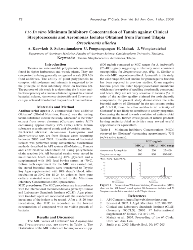
Proceedings 7th Chula. Univ. Vet. Sci. Ann. Con., 1 May, 2008. 72 P16 In vitro Minimum Inhibitory Concentration of Tannin against Clinical Streptococcosis and Aeromonas Isolates Obtained from Farmed Tilapia Oreochromis nilotica A. Kaewkoh S. Sukwattanakorn U. Pongsangangan H. Maisak J. Wongtavatchai Department of Veterinary Medicine, Faculty of Veterinary Science, Chulalongkorn University. Thailand. Keywords: Tannin, Streptococcosis, Aeromonas, Tilapia Introduction Tannins are water-soluble polyphenols commonly found in higher herbaceous and woody plants. They are categorized as being generally recognized as safe (GRAS) food additives. The ability of plant polyphenols to complex with polymers and minerals is suggested to be the principle of their inhibitory effect on bacteria (2). The purpose of this study is to determine the in vitro antibacterial potency of a tannin substance against the clinical bacterial isolates, Aeromonas hydrophila and Streptococcus spp, obtained from farmed tilapia Oreochromis nilotica. Materials and Method Antibacterial agent: A commercial feed additive Globatan® (Global Nutrition, France) was the testing tannin substance used in the study. Globatan® is the water extract from sweet chestnut (Castanea sativa Mill) containing approximately 75% (w/w) active tannin substance as a mixture of esteric and glycosidic tannins. Bacterial strains: Aeromonas hydrophila and Streptococcus spp. are from disease cases occurring between 2005 and 2007. Identification of bacterial isolates was performed using conventional biochemical methods described in API system (BioMerieux, France) and confirmative identification using polymerase chain reaction (4). All bacterial strains were stored in maintenance broth containing 40% glycerol and a supplemented with 10% fetal bovine serum, at -70oC. Before each experiment for the MIC was carried out, the stored bacterial strains were transferred to Tryptic Soy Agar supplemented with 10% sheep’s blood. After incubation at 30 oC for 18-24 hr, colonies from pure culture material were transferred to the Minimum Inhibitory Concentration (MIC) procedures. MIC procedures: The MIC procedures are in accordance with the international recommendations given by Clinical and Laboratory Standards Institute (CLSI). Agar plates with two-fold agent are inoculated with the standardized inoculums of the isolate to be tested. After a 18-20 hour incubation, the MIC is recorded as the lowest concentration of compound with no visible growth of bacteria. Results and Discussion (800 +g/ml) compared to MIC ranges for A. hydrophila (25-400 +g/ml) suggesting a relatively more consistent susceptibility for Streptococcus spp. (Fig. 1). Similar to the wide MIC range observed for A. hydrophila in this study, the wide range MICs of tannins for gram negative bacteria has been reported in previous studies. Gram negative bacteria poses the outer lipopolysaccharide membrane which may be capable of repelling the phenolic compound, and hence, they are not very sensitive to tannins (5). In spite of the acidity quality claimed for polyphenolic compounds, the present study demonstrates the antibacterial activity of Globatan® in the test system posing pH 6.5-7.0, thus, in vitro antibacterial activity of Globatan® is not likely to contribute to acidity properties. Concerning the trend towards evolution of antimicrobial resistant strains, further investigation of natural products having antimicrobial activities may reveal useful applications for aquaculture. Table 1 Minimum Inhibitory Concentrations (MICs) observed for Globatan® (containing approximately 75% (w/w) active tannin). MIC (+g/ml) MIC 50 Aeromonas hydrophila Streptococcus spp. 1 200 800 MIC 902 MIC range 200 800 25-400 800-800 MIC 50% 2MIC 90% Streptococcus Aeromonas Figure 1 Frequencies of Minimum Inhibitory Concentrations (MICs) observed for Globatan® tested against 20 Aeromonas isolates and 20 Streptococcus isolates associated with Tilapia disease. References 1. 2. 3. 4. ® The MIC values of Globatan for A.hydrophila and Streptococcus spp. are shown in Table 1. The Distribution of the MIC values are for Streptococcus spp. 1 5. API Company. https://apiweb.biomerieux.com Bossi et al. 2007. J. Appl. Microbiol. 102: 787-795. Clinical and Laboratory Standards Institute (CLSI) (formerly NCCLS). 2002. 9 th Informational Supplement-6th Edition. 19(1): 91-93. Maisak et al., 2007. Proceeding of the 6th Chula. Univ. Vet. Ann. Con. Smith et al. 2005. Microb. Ecol. 50: 197-205.
