Functional Anatomy - PE Studies Revision Seminars
advertisement
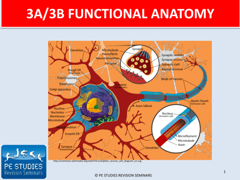
3A/3B FUNCTIONAL ANATOMY http://commons.wikimedia.org/wiki/File:Complete_neuron_cell_diagram_en.svg © PE STUDIES REVISION SEMINARS 1 CONTENTS SLIDE Structure of a skeletal muscle • How do we produce movement? • Structure of a sarcomere • Sliding filament theory 3 10 17 19 Nervous control of muscular contractions • Spinal cord • Motor neuron • Motor unit 22 24 25 26 Types of muscle fibres • Type IIa • Type IIb • Type I 28 29 30 31 Mechanical characteristics of a muscle • Force - velocity • Force - length • Force - time 35 36 45 46 References 47 © PE STUDIES REVISION SEMINARS 2 STRUCTURE OF A SKELETAL MUSCLE This diagram represents the various levels of skeletal musculature 3 Home © PE STUDIES REVISION SEMINARS http://commons.wikimedia.org/wiki/File:Skeletal_muscle_diagram.jpg HOW DO WE PRODUCE MOVEMENT? The human body is made up of over 600 muscles, working together to provide both conscious and subconscious movements for the human body. One of the major functions of the muscular system is to produce movement; PRODUCE MOVEMENT • • • Skeletal muscles which are consciously controlled (voluntary) are attached to bones When we want to produce movement e.g. walking, swimming, the CNS sends a message from the brain to the relevant muscle to contract, resulting in “pulling the bone” causing movement to occur. This enables the human body to respond quickly to changes in the external environment e.g. changing direction in a game of sport © PE STUDIES REVISION SEMINARS www.flickr.com/photos/jimmyharris/116283470/ 4 STRUCTURE OF A SARCOMERE The diagram below shows the part a myofibril called a sarcomere. This is the smallest unit of skeletal muscle that can contract. Each myofibril is made up of many sarcomeres joined end to end which are separated by their Z – lines. ACTIN FILAMENT MYOSIN FILAMENT SARCOMERE Z LINE CROSSBRIDGES H ZONE Z LINE 5 Home © PE STUDIES REVISION SEMINARS SLIDING FILAMENT THEORY • Biomechanists use sliding filament theory to explain the shortening of the sarcomere (concentric contraction) and the resulting contraction of the muscle. – When there is a neurochemical stimulation, calcium is released into the muscle prompting a reaction in each muscle fibre between the myosin and the actin filaments. – Myosin filaments contain crossbridges at regular intervals. These cross bridges attach and reattach at different times along the actin pulling on them to create movement and maintain tension – This causes the actin to move into the centre of the sarcomere, shortening the myofibril and causing the actin and myosin filaments to be almost fully overlapped when in a fully contracted position – As each sarcomere shortens, so does the total length of each muscle fibre – When the contraction finishes, the myosin and actin filaments return to a relaxed position © PE STUDIES REVISION SEMINARS Home 6 SLIDING FILAMENT THEORY The myosin cross bridges attach and reattach at different times along the actin pulling on them to create movement and maintain tension http://commons.wikimedia.org/wiki/File:Querbr%C3%BCckenzyklus_1.png © PE STUDIES REVISION SEMINARS Home 7 NERVOUS CONTROL OF MUSCULAR CONTRACTION • Understand the function of the nerve, impulse, spinal chord, motor unit (dendrite, axon, neurone) • Describe the relationship between muscle contraction and nerve function. Curriculum Council of Western Australia. Physical Education Studies Support Document 2009. The Motor Neuron http://commons.wikimedia.org/wiki/File:Neuron.svg © PE STUDIES REVISION SEMINARS 8 MOTOR NEURON Dendrites Cell body MOTOR NEURON • Refers to neurons located in the CNS that project their axons outside the CNS and directly or indirectly control muscles. • It is responsible for carrying impulses away from the spinal cord and brain to the muscles or glands • It consists of; Axon – A cell body that directs the neuron’s activities – Dendrites that act as branches to pick up the impulse – An axon that transmits the message to the muscle and away from the cell body – Motor end plates which attach to the muscle fibres Home © PE STUDIES REVISION SEMINARS Motor end plates 9 COMPARISON OF FORCE DEVELOPMENT OVER TIME Type IIb fibres Type IIa fibres F O R C E Type 1 fibres TIME 10 © PE STUDIES REVISION SEMINARS Fibre Type Slow Twitch (Type 1) Fast Twitch (Type 2A) Fast Twitch (Type 2B) Contraction Time Slow Fast Very Fast Size of Motor Neuron Small Large Very Large Resistance to Fatigue High Medium Low Activity used for Aerobic Long term Anaerobic Short term Anaerobic Force Production Low High Very High Capillary Density High Intermediate Low Oxidative Density High Glycolitic Capacity Low High High Major Fuel Source Triglycerides and glycogen Creatine phosphate and glycogen Creatine phosphate and glycogen Home Moderate Low 11 MECHANICAL CHARACTERISTICS OF A MUSCLE FORCE 1. FORCE – VELOCITY During isometric contraction, force generated does not result in change of muscle length During eccentric muscle contraction (lengthening) , max force achieved during max velocity LENGTHENING VELOCITY Home During concentric muscle contraction (shortening), max force achieved during minimum velocity 0 SHORTENING VELOCITY © PE STUDIES REVISION SEMINARS 12 REFERENCES • • • • • • • • • • Home Curriculum Council. (2009). Physical Education Studies Scope and Sequence Year 11 2010, Year 12 2011: Osborne Park, WA. Dawson, T., Dawson, P. (2007). E – Teaching Physical Education compact disc 1. Gaugers, R. (2009). Physical Education Studies: A Resource for Units 2A & 2B. Cottesloe, WA: Impact Publishing. Heberle, M., Middleton, C. (2007). Physical Education Studies: A Resource for Units 3A & 3B. Cottesloe, WA: Impact Publishing. McArdle, W., Katch, F., Katch, V. (2001). Exercise Physiology (5th ed.). Energy, Nutrition and Human Performance. Baltimore, Maryland: Lippincott Williams & Wilkins. McPartland, D., Pree, A., Malpeli, R., Telford, A. (2010). Physical Education Studies for 3A,3B. South Melbourne, VIC: Cengage Learning Australia Pty Limited. Smyth, D., Brown, H., Judge, W., McCallum, C., Pritchard, R. (2006). Live it up 2 (2nd ed.). Milton, QLD: John Wiley & Sons Australia, Ltd. Smyth, D., Brown, H., Judge, W., McCallum, C., Wright, P. (2006). Live it up 1 (2nd ed.). Milton, QLD: John Wiley & Sons Australia, Ltd. Whipp, P., Elliot, B., Guelfi, K., Dimmock, J., Lay, B., Landers, G., Alderson, J. (2010). 3A-3B Physical Education Studies: A Textbook for Teachers and Students. Crawley, WA: UWA Publishing. Wilmore, J., Costill, D. (1994). Physiology of Sport and Exercise. Champaign, IL: Human Kinetics. ©PE Studies Revision Seminars 13
