...where molecules become realTM
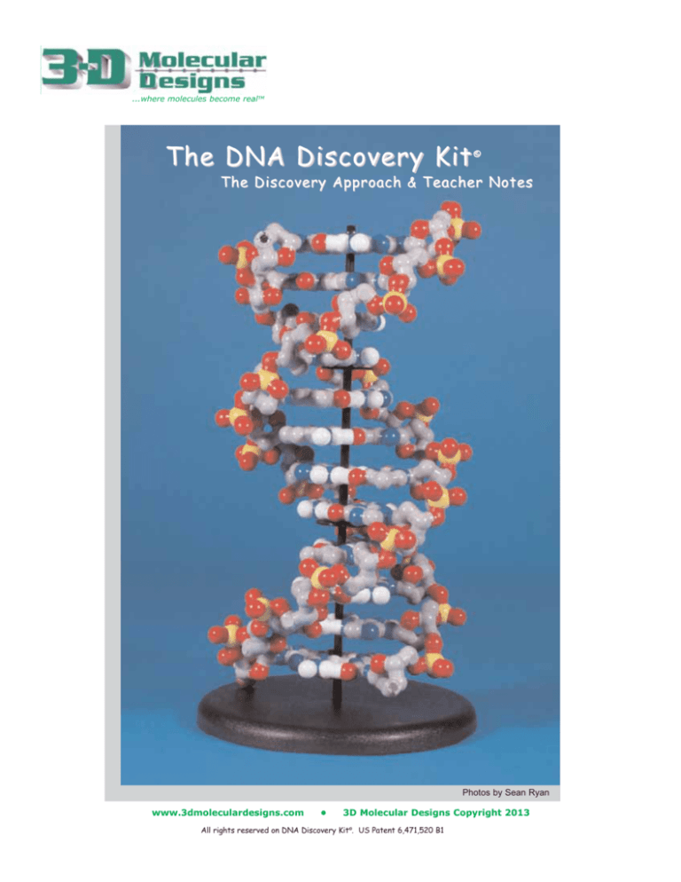
The DNA Discovery Kit
©
The Discovery Approach & Teacher Notes
Photos by Sean Ryan
www.3dmoleculardesigns.com
3D Molecular Designs Copyright 2013
All rights reserved on DNA Discovery Kit©. US Patent 6,471,520 B1
The DNA Discovery Kit ©
Contents of The Discovery Approach
Contents of the DNA Discovery Kit©
12 Base Pair Kit . . . . . . . . . . . . . . . . . . . .2
Contents of the DNA Discovery Kit©
2 Base Pair Kit . . . . . . . . . . . . . . . . . . . . .2
Assembly Instructions . . . . . . . . . . . . . . . . .3
The Discovery Approach . . . . . . . . . . . . . .6
Optional Questions and Activities . . . . . . .8
Reviewing Bonds . . . . . . . . . . . . . . . . . . .11
Mini-Toober DNA . . . . . . . . . . . . . . . . . . .12
Three Frequently Asked Questions . . . . .14
The Discovery of the Structure of DNA . .17
Student Handout . . . . . . . . . . . . . . . . . . . .18
Transparency Templates
Chemical Structures . . . . . . . . . . . . . . .20
Information Available to
Watson & Crick in 1953 . . . . . . . . . . .21
Watson and Crick DNA Papers . . . . . . . .11
Contents of The DNA Discovery Kit© 12 Base Pair Kit
6 each of Adenosine, Thymine, Guanine
and Cytosine Nucleotides
Black Rod
48 Nucleotide Labels
Assembly Instructions
Black Base
2 Mini-Toobers
Black Helix Guide
Contents of The DNA Discovery Kit© 2 Base Pair Kit
1 Each: Adenosine, Guanine, Thymine and
Cytosine Nucleotides
Assembly Instructions
8 Nucleotide Labels
Contents of Online Resources found at 3dmoleculardesigns.com/resources.php
Contents & Introduction PDFs
DNA Resource Information
©
DNA Discovery Kit Introduction
Read Me First
DNA Contents & Assembly Directions
Teacher Notes
DNA Activities & Teacher Notes PDFs
The Discovery Approach
The Guided Discovery Approach
Student Handout
Student Worksheet
Student Answer Sheet
DNA Websites
Additional DNA Resources
Three Frequently Asked Questions
Watson & Crick Papers PDFs
Watson & Crick April 1953
Watson & Crick May 1953
Annotated Version of Watson & Crick
Paper
2
The Discovery Approach
3D Molecular Designs Copyright 2013
The DNA Discovery Kit ©
DNA Discovery Kit© Assembly Instructions
Nucleotides Assembled
The nucleotides are preassembled.
You have the option of using labeled or
unlabeled nucleotides. To label a
nucleotide, peel a letter from its protective
backing and press it into the depression
on a corresponding base. After placing
the label on one side, flip the base over
and repeat with another label. Use the
photo to correctly place the labels on the
nucleotides. (Labels only fit inside the
larger depression on the Adenosine and
Guanine nucleotides.)
Phosphodiester Bond
Magnets Simulate Bonding
Magnets
The nucleotide models have
magnets embedded in them to
simulate the spontaneous
bonding that occurs between
complementary base pairs
(hydrogen bonds) and between
the phosphate group of one
nucleotide to the deoxyribose of
another nucleotide
(phosphodiester bonds).
Hydrogen Bonds
Arrows in the photo above point
to the magnet(s) in each piece.
You can break the hydrogen
bonds by pulling apart the G-C
and A-T base pairs. When examining the deoxyribose and phosphate groups, you will see
the single magnet embedded in the deoxyribose group and one embedded in the phosphate group.
3D Molecular Designs Copyright 2013
The Discovery Approach
3
The DNA Discovery Kit ©
Nucleotides Separate Into Component Parts
Each nucleotide separates into its three component parts
the nitrogenous base, deoxyribose group and phosphate
group.
To separate the pieces, pull the three pieces apart as
shown in the photos. Be sure to pull the pieces apart with a
straight motion. The attachment posts can break if a
twisting or bending motion is used.
Three Ways to Display DNA Discovery Kit ©
We encourage you to
leave the DNA
Discovery Kit© pieces
out on a table for your
students to explore in
their free time.
You can also easily
display or store the fully
assembled double helix
by setting up the black
base and black rod that are included in
the 12 Base Pair DNA Discovery Kit©.
Or you can hang the double helix
from a ceiling by threading a strong cord
through the eyelet at the top of the rod.
Do not use the black base when hanging
the DNA.
4
The Discovery Approach
3D Molecular Designs Copyright 2013
The DNA Discovery Kit ©
Setting Up Base & Rod to Displaying DNA
Eyelet for Hanging DNA on Rod
Push the bottom of the rod
(end without the eyelet) into the
black base. Press down
firmly so it rests securely in the
base. The lowest disk will rest
about 3/4 inch above the base.
Before correctly placing
the Guanine - Cytosine base pairs
around the rod, look carefully at the models. You will see that
the Guanine model has two hydrogen that are longer than the third hydrogen. (See photo at right
and refer to the page 2 photo labeled, Hydrogen Bonds.) The Cytosine model has one hydrogen
that is longer and two shorter hydrogen. Adenosine and Thymine each have one longer hydrogen
and one shorter hydrogen.
The Guanine - Cytosine base pair should be placed so that the rod is between the longer and
the shorter hydrogen. (See the above photos.) The Adenosine - Thymine base pair should be
placed so that the rod is between the two hydrogen (one is longer and one is shorter. As each
base pair is placed on the rod, rotate it until it forms the phosphodiester bond with the previous
base pair. Four base pairs fit above each disk.
Making Mini -Toober DNA
Black Helix Guide
Place two Mini-Toobers side-byside, so the red end cap on one is
even with the blue end cap on the
other. Next, line them up with the
first two grooves in the black helix
guide. Begin wrapping the MiniToobers around the guide following the
grooves.
Once the Mini-Toobers are wound around
the guide, you can remove them by twisting
the guide as though you are unscrewing it
from the Mini-Toobers. Then loosen and
separate the coils by gently unwinding and
pulling them apart.
(See the Activities and Teacher Notes at
3dmoleculardesigns.com/resources.php for
information on this activity.)
3D Molecular Designs Copyright 2013
The Discovery Approach
5
The Discovery Approach Teacher Notes
The Discovery Approach to investigating the structure of DNA allows students to
discover the double helix in much the same way as Watson and Crick discerned it in 1953.
In addition to gaining an understanding of the structure, this approach enables your students
to experience science as a dynamic, creative process, in which groups of people work
together or in competition to construct an understanding of the natural world. The story
of the discovery of DNAs double helical structure is a compelling example of real science.
For this approach, evenly divide the bases, deoxyribose sugars, and phosphate groups
among your students. Then pass out copies of the Student Handout, which contains the
information that was available to James Watson and Francis Crick in 1953. This includes the
polymeric nature of DNA, Chargaff's rules, and the crystallographic data of Maurice Wilkins
and Rosalind Franklin. Then challenge your students to develop a model for the structure of
DNA that satisfies the information available in 1953. Your students will quickly see:
How the phosphate groups, the deoxyribose groups and the bases join together to form
four nucleotides,
How the complementary bases go together via hydrogen bonding to form the AT and
GC base pairs
How the base pairs stack upon each other to create the elegant double-helical structure
of DNA that is described in Watson and Crick's 1953 publication in Nature.
The Student Handout begins on page 17. The Information Available to Watson and Crick in
1953 appears in the blue box on page 2 of the Student Handout (page 18 in this document).
(A summary of key points of the Information Available to Watson and Crick in 1953, (page
21), can be printed on transparency film for overhead use. The Student Handout is also
available as a separate document on the website.)
Phosphates
Deoxyribose
Nitrogenous Bases
Hand out the 12 DNA pieces shown above (four phosphate groups, four deoxyribose groups, and
the four nitrogenous bases, A,G,T and C), to each small group of students. Once each group of
your students has the pieces, you may want them to refer to their last page of the Student
Handout (see page 19) which identifies: the phosphate groups, deoxyribose groups, nitrogenous
bases and the coloring scheme. Then tell your students to play with the pieces until they
discover how all fit together in a compact structure.
6
The Discovery Approach
3D Molecular Designs Copyright 2013
The Discovery Approach Teacher Notes
1
2
One of the many possible ways to put together
two base pairs of DNA (a GC and an AT base
pair) is shown:
Photo 1: The phosphates are joined to the
deoxyribose groups.
Photo 2: Sugar-phosphate backbone of DNA is discovered. This polymer contains
no information. How do we know that it contains no information?
3
Photos 3, 4, and 5: The bases (A,G,T and C)
are added to the backbone.
4
5
6
Photo 6: Tell your students to unassemble the
single-stranded DNA chain, so that they have
four nucleotide units.
3D Molecular Designs Copyright 2013
The Discovery Approach
7
The Discovery Approach Teacher Notes
Tell your students to play
with the four nucleotides
until they discover how The
AT and GC base pairs fit
together.
Once each student group has put
together their AT and GC base pairs,
the base pairs can be combined to
form the 12 base-pair double helix
(if you purchased the
12 Base-Pair DNA Discovery Kit©).
The DNA can be assembled
horizontally on a table or in DNAs more
iconic vertical orientation.
For the vertical assembly,
you will need the black base and rod that
are provided with the 12 Base-Pair DNA
Discovery Kit©. (Please refer to the printed
base and rod assembly directions,
on pages 4 and 5.)
Correct Pairing for Forming
Double Helix
Optional Questions & Activities
You can use the questions in the yellow boxes on the next three pages to guide your
students, if they have difficulty building an accurate DNA double helix. Alternatively, you can use
the following questions to prompt discussion after your students have correctly assembled the
double helix. If you dont use the questions on the next three pages, skip ahead to the green box
entitled, Watson & Crick DNA Papers on page 11.
Please also see The Guided Discovery Approach on the website for additional
information and ways to use the DNA Discovery Kit©.
Background Information on DNA is available at 3dmoleculardesigns.com/resources.php.
8
The Discovery Approach
3D Molecular Designs Copyright 2013
The Discovery Approach Teacher Notes
Use your models to build the individual nucleotides (base + deoxyribose + phosphate).
What you can discover about how the nucleotides might interact with each other?
What general feature of these nucleotide pairs do you see?
Remember that the crystallographic data indicated DNA was a helix made up of two strands.
Using what we learned previously about the primary structure of DNA, and what youve just
discovered about pairs of nucleotides, can you build a helix made up of two strands of DNA?
Students who build the correct A - T and G - C
base pairs should also discover that they can
build the double helix by forming the correct
bonds between the 5' phosphates and the 3'
hydroxyl groups.
Incorrect Pairing for Forming Double Helix
They may also find they can form incorrect
C - C and G - G base pairs using only two of
the three hydrogen bonds, or A - A and T - T
base pairs. (Note photo on right.)
When the phosphate-deoxyribose is added to
the incorrectly paired bases, your students will not be able to add them to correctly paired A - T
or G - C base pairs to form a double helix. (Note photo below. Both of the phosphate-sugar
groups have a downward orientation. Now note the similar photo on the previous page) Ask
your students if they can discover alternative base pairing that will allow them to put two base
pairs together. You may want to explain that in a natural environment, nitrogenous bases can
form incorrect bonds, but the bonds
Incorrect Pairing for Forming Double Helix
will be unstable and break apart
since they wont be able to bond
with correctly paired A - T or G - C
base pairs and form the stable
double helix structure.
After students explore base pairing, have them disassemble
the nucleotides and then form a two-nucleotide chain as was
done in the first exercise. Then ask the students to use what
they learned about base pairing to add two nucleotides to the
structure so that they build a helix with two strands as was
suggested by the crystallographic data. The students should
discover that the only way to build a double helix is by
following the A - T, G - C base pairing rule.
3D Molecular Designs Copyright 2013
The Discovery Approach
9
The Discovery Approach Teacher Notes
The 5' phosphate of one strand is opposite the 3' hydroxyl of the opposite strand, and vice versa.
Note that the two DNA strands run in the opposite directions. As a result, one strand is oriented in
the 5' - 3' direction, while the opposite strand is orientated in the 3' - 5'. The two strands are said to
be anti-parallel due to the different directions.
Looking at your double helix model of DNA, can you identify the 5' phosphate and 3' hydroxyl of
each strand?
Where are they in relation to each other on the two strands?
The DNA sequence is always read in the 5' - 3' direction. Can you write the sequence of each
strand in your DNA model?
Check to make sure the sequences are read in the 5' - 3' direction.
At this point, it would be useful for your students to assemble all
12 base pairs into the double helix.
Lets examine the full DNA model to see if we satisfied all of the
experimental predictions.
Is it a polymer?
Yes.
Does it form a double stranded helix with 10 residues per turn
as predicted from the x-ray crystallography?
Yes. Starting with the 5' phosphate at the top of the model, count
down 10 residues. The tenth 5' phosphate will line up directly
under the first 5' phosphate.
What do you notice about the location of the phosphate
molecules and the bases?
The phosphate molecules, which are negatively charged, are on the outside where they interact
with the aqueous environment. The bases are stacked on top of one another with the planes of
the bases nearly perpendicular to the helix axis.
How does the base paring in the model explain Chargaffs Rules?
While the overall nucleotide composition (percentage of G - C pairs and percentage of A - T pairs)
of the DNA of different organisms can vary, the concentration of A always equals the concentration
of T, and G is always equal to C. This is a direct consequence of Chargaffs Rules, which state
that A is always paired with T, and G is always paired with C.
10
The Discovery Approach
3D Molecular Designs Copyright 2013
The Discovery Approach Teacher Notes
How does this organization of DNA's bases, deoxyriboses and phosphates make DNA a stable molecule?
In other words, what forces make this a stable structure?
The ribose subunits in the backbone are connected by covalent phosphodiester bonds.
The backbone is connected to the nitrogenous bases by covalent bonds.
The nucleotide base pairs are formed by hydrogen bonding. A-T base pairs form two hydrogen
bonds and are less stable than G-C base pairs, which form three hydrogen bonds.
Hydrophobic interactions between the base pairs provide additional stability to the double helix.
Reviewing Bonds
A covalent bond forms when two atoms share two electrons. A covalent bond is an intramolecular bond within one molecule. Covalent bonds can be either polar (which have partially
charged atoms) or non-polar (without charged atoms).
A hydrogen bond is an intermolecular force between the two molecules where a positively
charged hydrogen atom interacts with a negatively charged fluorine, nitrogen, or oxygen atom
in a second molecule.
An ionic bond is the complete transfer of an electron between two atoms resulting in one
positively and one negatively charged atom. Ionic bonds are intra-molecular bonds within one
molecule.
Ions are charged atoms that have gained or lost electrons as a result of an ionic bond.
Watson and Crick DNA Papers
After your students discover the structure of DNA by putting the model together, they should read
the classic paper published by Watson and Crick in Nature, April 23, 1953. You can download the
PDF at 3dmoleculardesigns.com/resources.php. (an annotated version of the paper is included
as a teacher resource.) Watson and Crick published a second paper in the next issue of Nature
that expanded on the significance of their proposed structure. This paper provides an interesting
description of what was known and unknown at that time, and sets the stage for the construction
of the Central Dogma of Molecular Biology, which was developed in the following years.
One interesting feature of the DNA structure that is addressed in Watson and Cricks second
paper concerns the way in which the two strands of DNA wrap around each other. Watson and
Crick clearly understood the topological problem this structure presents, even though they did not
understand at that time how the cell would deal with it. To help your students better understand
that the two strands of DNA are intertwined and to appreciate the problem this intertwining poses,
we have included the supplies to create a Mini-Toober model of double-stranded DNA,with the 12
Base-Pair DNA Discovery Kit©.
Instructions and photos follow on the next two pages.
3D Molecular Designs Copyright 2013
The Discovery Approach
11
The Discovery Approach Teacher Notes
Making a Mini-Toober Model of DNA
Place two MiniToobers side-by-side,
so the red end cap on
one is even with the
blue end cap on the
other. Next, line them up with the first two grooves in the
black helix guide. Begin wrapping the Mini-Toobers
around the guide following the grooves.
You just made a right-handed double
helix DNA model. How do you know
if your helix is right-handed?
Imagine the helix is a spiral staircase.
As you walk up, one of your hands
rests on the outside rail of the staircase.
If it is your right hand, then you are
walking up a right-handed helix.
The structure of DNA is always a
Once the Mini-Toobers are completely wound around
right-handed double helix.
the guide, you can remove the Mini-Toobers by twisting
the guide, as though you are unscrewing it from the
Mini-Toobers. Then loosen and separate the coils by gently unwinding
and pulling them apart.
Carefully position each strand of DNA to show the major and minor grooves
(right photo). Then, holding the DNA horizontally by only one strand,
demonstrate that the two strands are wrapped around each other(above
photo). (The term used to describe this property of DNA is plectonemeic.)
Another way to show that that DNA is plectonemeic is to separate the two
strands of Mini-Toober DNA, by unwinding one strand from the other
(Photo next page top). Separate the Mini-Toobers before class. Once
class starts ask one of your students to put the two strands together in the
12 The Discovery Approach
3D Molecular Designs Copyright 2013
The Discovery Approach Teacher Notes
same way that the double stranded DNA fits together. After a few false starts your student will
probably try winding one of the strands into the second strand.
How do you think the cell separates the two strands of DNA for replication and transcription,
when they are wound around each other?
Watson and Crick realized
the problem intertwined
DNA poses for DNA
replication and RNA
transcription. In their May
1953 paper, published in
Nature (see Watson &
Crick PDFs online), they
wrote,Since the two chains
in our model are
intertwined, it is essential
for them to untwist if they
are to separate. As they make one complete turn around each other in 34 A, there will be about
150 turns per million molecular weight, so that whatever the precise structure of the chromosome, a
considerable amount of uncoiling would be necessary. It is well known from microscopic
observation that much coiling and uncoiling occurs during mitosis, and though this is on a much
larger scale it probably reflects similar processes on a molecular level. Although it is difficult at the
moment to see how these processes occur without everything getting tangled, we do not feel that
this objection will be insuperable.
We now know that the solution is provided by a family of proteins known as topoisomerases.
These enzymes function to unwind or wind double-stranded DNA by:
Cleaving a phosphodiester bond in the backbone of one strand of DNA
Effectively unwinding the free end one turn around the other DNA strand
Re-forming the phosphodiester bond.
In this way DNA can be unwound one turn at a time. You can simulate the result but not the
mechanism of this localized unwinding by grasping the toober model with both hands spaced
about 6 inches apart. Then unwind the double-helix to form the replication bubble shown above.
Discussion Opportunity Compare the DNA Discovery Kits plastic model with its Mini-Toober DNA model. What
are the advantages and disadvantages of each? Compare them to textbook drawings and illustrations of DNA.
(Drawings appear on the next page. Larger images that can be printed on transparency film for overhead use
appear on page 20.)
3D Molecular Designs Copyright 2013
The Discovery Approach
13
Three Frequently Asked Questions
How do these models compare with the chemical drawings of nucleotides in my textbook?
As your students become familiar with DNAs phosphate groups, deoxyribose groups and bases, by
handling the models, the 2-D drawings of DNAs chemical structure will be more meaningful.
When your students compare the models with the chemical drawings in textbooks, it is important that
they understand that most of the hydrogen atoms have been eliminated from the models in order to
more clearly reveal the underlying structure. A direct comparison of the physical models with a typical
chemical drawings of the nucleotide structures is provided below.
G
C
A
14
The Discovery Approach
T
3D Molecular Designs Copyright 2013
Three Frequently Asked Questions
How does the model show that the two strands of DNA are anti-parallel?
One powerful feature of this model is that it clearly demonstrates that the two strands of DNA are
running in opposite directions. Look at the photo shown below, and focus on the red oxygen atom
found in the two deoxyribose groups. Notice how the oxygen of the deoxyribose on the left is below
the plane of the base pair, while the oxygen of the deoxyribose on the right is above the plane. This
is a clear indication that the polarity of the nucleotides in the two strands are opposite each other.
5
3
3
5
Now focus your students attention on the phosphate groups
from each nucleotide. Again, one of these phosphates will be
below the plane of the base pair while the other will be above
the plane. And since the phosphate group is attached to the
5carbon of the deoxyribose group, the DNA chain on the right
of the double helix shown in the photo above is said to run
5 to 3 from the top of the photo to the bottom while to other
strand is running 5 to 3, from the bottom to the top.
3D Molecular Designs Copyright 2013
The Discovery Approach
15
Three Frequently Asked Questions
Can incorrect base pairs be formed with the model pieces?
Yes, non-standard base pairs (other than the A - T and G - C that form the double
helix) can be formed by this model just as these base pairs can form in solution
with real nucleotides.
Four of these non-standard base pairs are shown below. However, these
non-standard base pairs are not compatible with double helical DNA, for two reasons.
Base pairs formed with two purines or two pyrimidines will have a different diameter
than standard A - T and G - C base pairs that consist of one purine paired with one
pyrimidine. Therefore, the non-standard base pairs shown below cannot be assembled
into a double helical model with a uniform diameter.
Encourage your students to discover these non-standard base pairs -- and then
determine for themselves why these base pairs are not consistent with the model
proposed by Watson and Crick.
For the non-standard hydrogen bonded base pairs to form, the
polarity of the two strands of DNA must be parallel, not anti-parallel.
Therefore, notwithstanding the problem with the diameter of these
non-standard base pairs (see above paragraph), it is not possible
to accommodate these parallel base pairs in the Watson-Crick
model of DNA.
16
The Discovery Approach
3D Molecular Designs Copyright 2013
...where molecules become realTM
The Discovery of the Structure of DNA
On April 25, 1953, a one-page paper entitled, A
Structure for Deoxyribonucleic Acid, appeared in the
British journal, Nature. The authors of this paper were
James Watson, a young American post-doctoral
candidate who had recently received a Ph.D. from the
University of Illinois, and Francis Crick, a physicist who
was completing his doctoral dissertation at Cambridge
University, England. The paper began; "We wish to
suggest a structure for the salt of deoxyribose nucleic
acid (D. N. A.). This structure has novel features which
are of considerable biological interest."
This initial description of the structure of DNA
marked a major milestone in the development of molecular biology. In addition to reporting the
correct structure of DNA, the paper also contained their classic understatement in scientific
literature: "It has not escaped our notice that the specific pairing we have postulated immediately
suggests a possible copying mechanism for the genetic material." Their paper serves as an
excellent example of what has become a recurring theme in the molecular biosciences Forms
Follows Function. That is, the structure of a macromolecule often explains the macromolecules
function (how the macromolecule) works.
Watson and Crick's achievement is notable in several ways, including the fact that they determined
the structure of DNA without performing a single experiment. They used the information from
numerous other scientists who were investigating various properties of DNA.
Modeling was the major approach Watson and Crick used. Using paper cut-outs
of the shapes of the four nitrogenous bases (A,T, G and C), they were able to
combine all of the different facts that had accumulated to that date into a
plausible model for the structure of DNA.
...The structure has two helical chains coiled around the same axis (see
diagram). We have made the usual chemical assumptions, namely, that each
chain consists of phosphate diester groups joining B-D-deoxyribofuranose
residues with 3',5' linkages. The two chains (but not their bases) are related
by a dyad perpendicular to the fibre axis. Both chains follow right-handed
helices, but owing to the dyad the sequences of the atoms in the two chains
run in opposite direction.
Watson, J.D. and Crick, F.H.C., Nature, 171, 737-738 (1953)
(Page 1 of Student Handout)
3D Molecular Designs Copyright 2013
The Discovery Approach
17
Student Handout
The DNA Student Challenge
Your challenge today is to see if you can discover the correct structure of double-stranded DNA, just
as Watson and Crick did over 50 years ago.
Your model should satisfy all of the pieces of experimental information that was known in 1953, as
noted in the blue box below. Rather than using paper cut-outs to represent the DNA bases, you will
use plastic models of the four deoxyribonucleotides whose 3D structures are based on known
atomic coordinates of the B-form DNA. In these nucleotide models, magnets are used to represent
both:
the phosphodiester bonds that link the nucleotide units together into a long, linear polymer
the hydrogen bonds that bond one base to another.
Information Available to Watson and Crick in 1953
DNA is a Polymer: Previous studies identified DNA as the genetic material of cells,
and that DNA was a polymer consisting of three components:
A nitrogenous base
A pentose (5-carbon) sugar called deoxyribose
A phosphate group.
Moreover, experiments suggested that the DNA molecule was unbelievably large, with
molecular weights ranging from 25 x 106 to 3 x 10⁹ daltons. (Since each nucleotide
has a mass of 330 daltons, DNA molecules were believed to be composed of between
76,000 and 9,000,000 nucleotides.)
DNA is more dense than protein. At a density of 1.6 gm/cm3, DNA was known to
be more dense than protein (1.3gm/cm3). This suggested that DNA was a densely
packed structure.
Chargaff's Rules: In 1947, Erwin Chargaff demonstrated that while the four
nucleotides were not present in equal amounts in the DNA from different organisms,
the amount of adenine was the same as thymine, and the amount of guanine was the
same as cytosine. This became known as Chargaff's Rules:
The proportion of A always equals that of T, and the proportion of
G always equals that of C. Thus, A = T and G = C.
X-ray Crystallography Data: In the laboratory of Maurice Wilkins, Rosalind
Franklin used X-ray diffraction to analyze fibers of DNA. The pattern of spots on the
X-ray diffraction pattern suggested that:
Phosphate was on the outside, nitrogenous bases were on the inside.
DNA was a double helix, made up of two strands.
The two strands of DNA run in opposite directions (anti-parallel).
There are 10 base pairs per turn of the double helix.
(Page 2 of Student Handout)
18
The Discovery Approach
3D Molecular Designs Copyright 2013
Background information for students
Phosphates
Deoxyribose
Nitrogenous Bases
Each group of students should have physical models of the four nucleotides, separated
into their component parts. These include:
Phosphate group which is negatively charged
Deoxyribose group which is a cyclic ring structure
Four nitrogenous bases (A, G, C and T)
Each component of the nucleotides is color coded according to atom type, following the
standard CPK coloring scheme:
Oxygen is RED
Carbon is GRAY
Nitrogen is BLUE
Phosphorus is YELLOW
Hydrogen is WHITE
(Page 3 of Student Handout)
3D Molecular Designs Copyright 2013
The Discovery Approach
19
G
C
A
T
Transparency Template of Comparison of Models to Textbook Drawings of Nucleotides. See page 14.
20 The Discovery Approach
3D Molecular Designs Copyright 2013
Information Available to Watson and Crick in 1953
DNA is the genetic information of cells
DNA is a Polymer
A nitrogenous base
A pentose (5-carbon) sugar called deoxyribose
A phosphate group.
DNA molecule is unbelievably large
DNA is more dense than protein
Chargaffs Rules
The proportion of A always equals that of T
The proportion of G always equals that of C
A = T and G = C
X-ray Crystallography Data
The phosphate is on the outside; nitrogenous base is on the inside.
DNA is a double helix, made up of two strands.
The two strands of DNA run in opposite directions (anti-parallel).
There are 10 base pairs per turn of the double helix.
Transparency Template of Information Available to Watson & Crick. See pages 6 and 18.
3D Molecular Designs Copyright --
The Discovery Approach
21


