Micrococcus species Staphylococcus aureus
advertisement
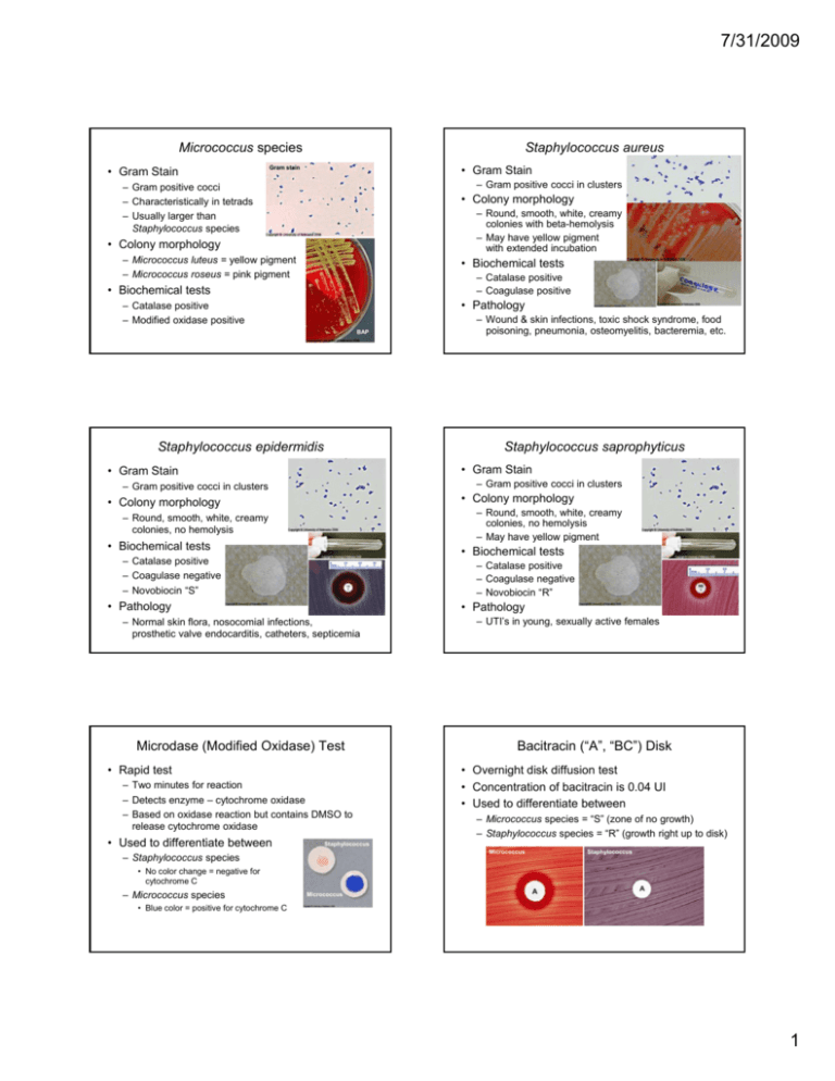
7/31/2009 Micrococcus species • Gram Stain Staphylococcus aureus • Gram Stain Gram stain – Gram positive cocci in clusters – Gram positive cocci – Characteristically in tetrads – Usually larger than Staphylococcus species • Colony morphology – Round, smooth, white, creamy colonies with beta-hemolysis – May have yellow pigment with extended incubation • Colony C l morphology h l – Micrococcus luteus = yellow pigment – Micrococcus roseus = pink pigment • Biochemical tests – Catalase positive – Coagulase positive • Biochemical tests • Pathology – Catalase positive – Modified oxidase positive BAP Staphylococcus epidermidis Staphylococcus saprophyticus • Gram Stain • Gram Stain – Gram positive cocci in clusters – Gram positive cocci in clusters • Colony morphology • Colony morphology – Round, smooth, white, creamy colonies, no hemolysis – May have yellow pigment – Round, smooth, white, creamy colonies, no hemolysis • Biochemical tests • Biochemical tests – Catalase positive – Coagulase negative – Novobiocin “S” – Catalase positive – Coagulase negative – Novobiocin “R” • Pathology • Pathology – Normal skin flora, nosocomial infections, prosthetic valve endocarditis, catheters, septicemia Microdase (Modified Oxidase) Test • Rapid test – Two minutes for reaction – Detects enzyme – cytochrome oxidase – Based on oxidase reaction but contains DMSO to release cytochrome oxidase • Used U d tto diff differentiate ti t b between t – Wound & skin infections, toxic shock syndrome, food poisoning, pneumonia, osteomyelitis, bacteremia, etc. – UTI’s in young, sexually active females Bacitracin (“A”, “BC”) Disk • Overnight disk diffusion test • Concentration of bacitracin is 0.04 UI • Used to differentiate between – Micrococcus species = “S” (zone of no growth) – Staphylococcus p y species p = “R” (g (growth right g up p to disk)) Staphylococcus Micrococcus – Staphylococcus species Staphylococcus • No color change = negative for cytochrome C – Micrococcus species Micrococcus • Blue color = positive for cytochrome C 1 7/31/2009 Coagulase Tube Test Mannitol Salt Agar • Tests for free coagulase – an extracellular enzyme that causes a clot to form when bacterial cells are incubated in rabbit plasma • Used to differentiate between – Staphylococcus aureus = positive (clot at 4 hours) • Some strains of Staph. aureus may lyse the clot (due to staphylokinase) so it is important to check reaction every ½ hour for the first 4 hours) – Other Staphylococcus species = negative (no clot at 24 hours) Staphylococcus aureus – Selective for Staphylococcus species (↑ NaCl) – Differential for the fermentation of mannitol, the sole carbohydrate in the media • Mannitol fermented ↓ pH indicator turns pink to yellow • Staphylococcus p y aureus = p positive,, yellow colonies surrounded by yellow zone DNase Agar • Other Staphylococcus species & Micrococcus species = negative, reddish colonies – no color change of media aureus • Disk diffusion test using novobiocin (antibiotic) at a concentration of 5 ųg for the presumptive identification of Staphylococcus saprophyticus in urine cultures • Staphylococcus saprophyticus = “R” – Organism O i grows up to t the th disk di k or a zone <16 mm Staphylococcus saprophyticus • Other Staphylococcus species = “S” – Organism’s growth is inhibited by antibiotic with a zone > 16 mm Staphylococcus species, not aureus Hemolysis • Beta (β) hemolysis – Complete lysis of RBC’s surrounding colony causing a clear zone or clearing of blood from the agar • Alpha (α) hemolysis – Partial lysis of RBC’s surrounding colony causing a greenish i h di discoloration l ti iin media di – Also known as Gamma hemolysis (δ) – No lysing of RBC’s and no color change of the medium surrounding colony Staphylococcus species, not aureus Novobiocin Disk • Media classified as differential • Inoculum is a dime-sized area of growth • Used to detect the production of an active DNase exoenzyme by aerobic bacteria • Used to differentiate Staphylococcus • Non-hemolytic Staphylococcus aureus – Note: some coag neg Staph can be + Coag neg Staph sp. • Report as “Coagulase negative Staphylococcus species” – Staphylococcus aureus = positive, pink color around growth – Other Staphylococcus species = negative, no color change • Media classified as selective and differential Staphylococcus species, not saprophyticus Bacitracin (“A”, “BC) Disk • Overnight disk diffusion test • Concentration of bacitracin is 0.04 UI • Used to differentiate between Group A Strep – Streptococcus pyogenes (Group A Strep) = “S” (any zone of no growth around the disk) • There are a small % of other beta-hemolytic Strep that are “S” – Most other beta-hemolytic Strep = “R” (growth up to edge of disk) Beta Strep not Group A 2 7/31/2009 Hippurate Hydrolysis Test CAMP Test • A rapid test that detects the enzyme hippuricase – Na hippurate benzoic acid + glycine blue/purple color change Grp B Strep hydrolyzes 2 hr, 35°C Ninhydrin Reagent 15 min. – A streptococcal protein that acts synergistically with Staphylococcal beta-hemolysis on sheep RBC’s • Used to differentiate between – Streptococcus p agalactiae g (Group B Strep) = positive, blue/purple color – Other beta-hemolytic Strep = negative, no color change • An overnight test that detects the extracellular CAMP factor • Used to differentiate between – Streptococcus agalactiae (Group B Strep) = positive, “arrow” formation Positive Negative – Other beta-hemolytic Strep = negative, no “arrow” formation Group B Strep Staph aureus Enhanced hemolysis Beta Strep not Group B Optochin (“P”) Disk PYR Hydrolysis Test • Overnight disk diffusion test • Disk contains 5 ųg ethyl-hydrocupreine hydrochloride • Used to differentiate between – Streptococcus pneumoniae = “S” (>14 mm zone of no growth around the disk) – Other Streptococcus species = “R” (growth up to edge of disk) • Rapid test – Enzyme L-pyrroglutamyl-aminopeptidase hydrolyzes the substrate L-pyrrolidonyl-beta-naphthylamide to produce a betanaphthylamine – When the reagent cinnamaldehyde combines with the betanaphthylamine, a bright red color is produced Strep pneumo Negative Bile Esculin Hydrolysis • Certain GPC are able to grow in 6.5% NaCl whereas others are inhibited by this salt concentration • Used to differentiate between Growth No Growth – Streptococcus pyogenes = positive – Enterococcus species = positive – Other Streptococcus species = negative (no color change) Other Strep 6.5% Sodium Chloride Test – Enterococcus species = positive i i ((growth, h turbid) bid) – Group D Streptococcus = negative (no growth, clear) – Streptococcus species viridans group = negative Positive • Used U d tto diff differentiate ti t • Certain GPC are able to grow in the presence of bile and hydrolyze esculin to form a blackening of media, whereas others are inhibited by the bile • Used to differentiate between – Enterococcus species = positive (black) – Group D Streptococcus = positive (black) – Other Streptococcus species = negative (no color change) Positive Negative 3 7/31/2009 Streptococcus pyogenes (Group A) • Gram Stain – Gram positive cocci in pairs/chains • Colony morphology – Small, transparent, smooth colony with well-defined zone beta-hemolysis • Biochemical tests – Catalase negative – Bacitracin (A) “S” – PYR positive • Pathology – Pharyngitis, skin infections, necrotizing fasciitis, toxic shock syndrome, post-streptococcal sequelae Streptococcus pneumoniae • Gram Stain – Gram positive cocci in pairs (lancet-shaped) • Colony morphology – Small, round, glistening, mucoid, dome-shaped colony with alpha-hemolysis – Autolytic changes (center collapses) when older • Biochemical tests – Catalase negative – Optochin (P) “S” – Bile solubility positive • Pathology Streptococcus agalactiae (Group B) • Gram Stain – Gram positive cocci in pairs/chains • Colony morphology – Small, grayish-white mucoid colony with small zone beta-hemolysis • Biochemical tests – – – – Catalase negative Bacitracin (A) “R” Hippurate positive CAMP positive • Pathology – Neonate meningitis, endometritis, wound infections Streptococcus species viridans group • Gram Stain – Gram positive cocci in pairs (lancet-shaped) • Colony morphology – Small, round, glistening, mucoid, dome-shaped colony with alpha-hemolysis – Autolytic changes (center collapses) when older • Biochemical tests – Catalase negative – Optochin (P) “R” – Bile esculin negative • Pathology – Normal flora, opportunistic, subacute bacterial endocarditis – Pneumonia, sinusitis, otitis media, bacteremia, meningitis Enterococcus species MacConkey Agar • Classified as a selective and differential media – Selective for Gram negative rods (Gram positive organisms are inhibited by bile salts) – Differential for lactose fermentation • Media contains lactose as its only carbohydrate • Indicator is neutral red (agar is pink) • Lactose Fermenter – Pink colonies Non”F” ”F” • Non-lactose Fermenter – Clear colonies – Note: Proteus will not swarm on this media Uniniculated 4 7/31/2009 Hektoen-Enteric (HE) Agar • Classified as a selective and differential media Eosin Methylene Blue (EMB) Agar • Classified as a selective and differential media – Selective for Gram negative rods (Gram positive organisms are inhibited by bile salts) – Differential for lactose & sucrose fermentation – Selective for Gram negative rods (Gram positive organisms are inhibited by eosin dye) – Differential for lactose fermentation • Media contains lactose and sucrose • Indicator is bromthymol blue (agar is green) • Lactose and/or Sucrose “F” (Yellow colonies) • Lactose & Sucrose Non-“F” (Bluish-green colonies) • H2S production = black precipitate • Media contains lactose as its only carbohydrate • Indicators is eosin & methylene blue • Lactose Fermenter – Dark-pink-centered colonies • Lowers pH which precipitates dye Lactose &/or Sucrose ”F” Lactose & Sucrose Non-”F” Lactose & Sucrose Non-”F” with H2S + Xylose Lysine Desoxycholate (XLD) Agar • Classified as a selective and differential media – Selective for Gram negative rods (Gram positive organisms are inhibited by bile salts) – Differential for lactose, sucrose and xylose fermentation • Media contains lactose, sucrose, and xylose • Indicator is phenol red (turns yellow when pH decreases) – Differential for lysine decarboxylation (LDC) • Media contains lysine • • • • Yellow colonies = Fermenter of carbohydrates Yellow colonies with black centers = “F” & H2S + Colorless/red colonies = Non “F” Red colonies with black centers = LDC + & H2S + Oxidase Test • Enzyme cytochrome oxidase in the presence of the reagent tetra-methyl- (or dimethyl-) paraphenylene-diamine-dihydrochloride to form a blue color within 30 seconds – Positive = blue color development within 30 seconds – Negative g = no color development p • Must use a platinum loop or sterile wooden stick, because nichrome loops can cause a false positive Positive Negative • Tetra-methyl reagent is more sensitive than dimethyl reagent – Note: E. coli = metallic green sheen • Non-lactose Fermenter – Clear colonies Escherichia coli Salmonella-Shigella (SS) Agar • Classified as a selective and differential media – Selective for pathogenic Enterics (Gram positive organisms are inhibited by bile salts) – Differential for lactose fermentation • Media contains lactose as its only carbohydrate • Indicator is neutral red (agar is pink) • Lactose “F” (Red/Pink colonies) • Lactose Non-“F” (Colorless colonies) • H2S production = black precipitate Glucose OF Media • Fermentative (“F”) organism will produce an acid reaction (yellow) throughout tube • Oxidative (“O”) organism will produce an acid reaction in the upper pp 1/3 of media • Non-fermenter & Nonoxidizer (Non-“F” & Non“O”) will produce an alkaline reaction (blue) in upper portion of tube due to peptone metabolism. Non-“F” Uninoculated Non-“O” “O” “F” with gas 5 7/31/2009 Kligler’s Iron Agar (KIA) Lysine Iron Agar (LIA) • Detects deamination (LDA) and decarboxylation (LDC) of the amino acid lysine • Slant • Lactose Fermenter, Glucose Fermenter – Acid (yellow) slant & butt (A/A) • Lactose Non-Fermenter, Glucose Fermenter – LDA positive = wine-red – LDA negative = purple (alkaline) – Alkaline (red) slant & acid butt (K/A) • Butt – LDC positive = purple (alkaline) – LDC negative = yellow (acid) • Glucose Non-Fermenter – Alkaline (red) slant & butt (K/K) • H2 S • H2 S LF GF – Positive = Black precipitate – Negative = No precipitate LNF GF H2S neg LNF GF H2S pos GNF – Positive = Black precipitate – Negative = No precipitate • Note: This test is invalid if the organism is unable to ferment glucose! Ornithine Decarboxylase Media • Detects decarboxylation (ODC) of the amino acid ornithine • Top 1/3 of media always purple • Butt K/K Red/A K/A LDA – LDA – LDA + LDC – LDC + LDC – H2 S + H2 S – K/K LDA – LDC + H2 S – Indole Test • Detects the enzyme Tryptophanase Tryptophane ---------- Indole ----------------------- Cherry Red color tryptophanase ODC – ODC + – ODC positive = purple – ODC negative = yellow (lower 2/3 or tube) Para-dimethylamino-benzaldehyde Kovac’s Reagent • Indole positive – Cherry red color after ft adding ddi Kovac’s K ’ Reagent • Indole negative – No color reaction after adding Kovac’s Reagent • Note: This test is invalid if the organism is unable to ferment glucose! Motility S Media Positive Negative Nitrate Reduction Test • Detects organism’s ability to reduce nitrate (NO3) to nitrite (NO2) or nitrogen gas (N2↑) • Add 2 reagents to the Motility S tube • Motile – Diffuse clouding away from the line of inoculation – Alpha-naphthylamine & sulfanilic acid • NO3 NO2 • Non-motile –G Growth th only l along l the line of inoculation Uninoculated – Add reagents g = Red color • NO3 Non-motile Motile – Add reagents = No color reaction – Add zinc dust = Red color Pos Neg • NO3 N2↑ – Add reagents = No color reaction – Add zinc dust = No color reaction 6 7/31/2009 Simmon’s Citrate Media Christiansen’s Urea Media • Detects organism’s ability to use citrate as its sole source of carbon • Positive – Blue color – alkaline end products,, bromothymole p y blue indicator turns blue when pH increases – Growth on slant – with or without color change • Detects the enzyme urease Urea + H2O -------- ammonium carbonate urease Negative g Positive • Negative – No color change and no growth on slant • If the organism can hydrolyze urea, there will be an increase in pH due to the alkaline end product. The indicator, phenol red, will turn a bright pink color. • Positive = pink color • Negative = no color change Positive Gelatin • Detects proteolytic enzymes which hydrolyze large protein molecules into smaller peptides and peptones • Reagent = acidic mercuric solution • Positive Strong Positive Negative Klebsiella pneumonia • Growth on sheep blood agar is very mucoid – Clear zone adjacent to line of growth after addition of reagent • Negative – A milky color in area adjacent to growth after addition of reagent Escherichia coli • Growth on sheep blood agar is very often betahemolytic Proteus mirabilis • Growth on sheep blood agar swarms across the entire plate One colony swarms to edge of plate 7 7/31/2009 Oxidase Test • Enzyme cytochrome oxidase in the presence of the reagent tetra-methyl- (or dimethyl-) paraphenylene-diamine-dihydrochloride to form a blue color within 30 seconds – Positive = blue color development within 30 seconds – Negative g = no color development p CTA Sugars for Neisseria ID • Detects oxidation of carbohydrates (CHO) • Indicator is phenol red • CHO oxidized – Decrease in pH, indicator turns yellow Thayer-Martin (TM) Media – Colistin – inhibits Gram negative rods – Vancomycin – inhibits Gram positive organisms – Nystatin – inhibits yeast • Modified TM agar – Contains antibiotic trimethoprim/ sulfamethoxazole to inhibit Proteus species – Pyoverdin – yellow-green / yellow-brown – Pyocyanin – blue – Combination of pyoverdin & pyocyanin – green – Pyorubin – red – Pyomelanin – brown / black Organism Glucose Maltose Lactose Sucrose DNase Nitrate N. gonorrhoeae + - - - - - N. meningitidis + + - - - - N. lactamica + + + - - - M. catarrhalis - - - - + + Moeller’s Decarboxylase Tests • Detects enzymatic ability of organism to decarboxylate or hydrolyze an amino acid to form an amine • pH increases (alkaline) and changes indicator to purple • Amino acids tested include: – Arginine, Lysine and Ornithine • Positive – Purple color • Negative – Yellow/purple or green/purple • Note: this media works with glucose “F”, glucose “O”, and glucose Non-“F” Non-“O” Pigment Production for Pseudomonas • Use Trypticase Soy Agar (TSA slant) to view pigmentation • Pigments Negative – No pH change, change indicator stays red • Must use a platinum loop or sterile wooden stick, because nichrome loops can cause a false positive Positive Negative • Tetra-methyl reagent is more sensitive than dimethyl reagent • A selective agar for Neisseria gonorrheae, N. meningitidis, and N. lactamica (normal flora) • Agar contains antibiotics Positive • CHO not utilized Positive Negative Pseudomonas aeruginosa • Gram negative rod • BAP – Beta-hemolytic – Flat, spreading colony – Fruity, grape-like smell • • • • • • • • • • • Oxidase positive Glucose “O” A i i dih Arginine dihydrolase d l positive iti Lysine decarboxylase negative Ornithine decarboxylase negative Nitrate variable Esculin hydrolysis negative “O” Pigment Gelatin variable DNase negative Growth at 42°C Sometimes produces green or red pigment ADH+ ODC- LDC- 8 7/31/2009 X & V Quad Plate Stenotrophomonas maltophilia • • • • • • • • • • • • Gram negative rod Oxidase negative Glucose “O” Arginine dihydrolase negative Lysine decarboxylase positive Ornithine decarboxylase negative Nitrate variable Esculin hydrolysis variable Gelatin positive DNase positive No growth at 42°C Sometimes can develop a lavender-green to light purple pigment • Used for the speciation of Haemophilus • Determines requirement of X and V factors and hemolysis on horse or rabbit blood • X Factor – Hemin, Hemin heat stable • V Factor – NAD, heat labile – – – – Haemophilus species I contains X factor II contains V factor III contains X & V factors IV contains horse blood I • Gram negative rod BAP – Small (can be coccoid), can exhibit bipolar staining – Will not grow on sheep blood agar – Must use Chocolate agar – Requires CO2 • Porphyrin test – Delta Delta-aminolevulinic aminolevulinic acid (ALA) is substrate – Determines if organism has enzyme required to produce hemin (X factor) – If positive, organism does not require X factor Requires X Requires V Hemolysis on horse blood ALA-Porphyrin test H. Influenzae Positive Positive None Negative H. Parainfluenzae Negative Positive None Positive H. Hemolyticus Positive Positive Beta Negative H. parahemolyticus Negative Positive beta Positive • No growth on MAC • Oxidase positive • Glucose “F” weak (apple green color) • Nitrate positive • ODC positive • Indole negative • Urease negative • “S” to 2 units of Penicillin Campylobacter jejuni • Curved Gram negative rod • Grows at both 37°C & 42°C in 10% CO2 (microaerophilic) • Oxidase positive • Darting motility on wet prep • Hippurate hydrolysis positive • Nalidixic Acid “S” • Cephalothin “R” II Pasteurella multocida • Gram negative coccobacilli • Fastidious Organism IV III • Quadrants Gram Stain OF Glucose MAC Aeromonas hydrophila • Gram negative rods • Beta-hemolytic on BAP • Grows on MAC BAP – Lactose variable • • • • • • • • • Oxidase positive Glucose “F” LDC variable ODC negative Indole positive Gelatin positive VP positive Esculin positive DNase positive MAC Campy BAP 9 7/31/2009 Acinetobacter species • Gram negative coccobacilli • Oxidase negative • Acinetobacter baumannii – Glucose “O” – Lactose “F” – Grows at 42°C BAP Acinetobacter baumannii MAC • Acinetobacter lwoffii – Glucose Non-“F”, Non-“O” – Lactose Non-“F” – No growth at 42°C • Nitrate negative • Non-motile Acid Fast Stain / Ziehl-Neelsen Stain • Mycobacteria are readily stained with 1° stain, which binds to mycolic acid in their lipid-rich cell wall. This stain cannot be removed (decolorized) and organism is referred to as being acid-fast. • Primary Stain is carbo-fuschin • Decolorizer is acid alcohol • Counterstain is methylene blue • Acid Fast Bacilli (AFB) – Red branching rods against a blue background Kirby Bauer Susceptibility E-Test Susceptibility MIC Bacteroides fragilis group • Most common anaerobic organism isolated in the clinical laboratory • Pleomorphic Gram negative Gram stain rod • Grows in 20% bile • Hydrolyzes esculin • Penicillin “R” AnaBAP • Beta-lactamase positive 10 7/31/2009 Clostridium perfringens Bacillus species Gram stain • Anaerobic organism • Large Gram positive rods • Large Gram positive rods – Blunt, rounded ends (“boxcar” shaped) – May see spores Double zone beta-hemolysis • BAP – double zone of beta-hemolysis • Catalase negative • Lecithinase positive on egg yolk agar • Causes Gram stain – With age can appear Gram variable or Gram negative – Can see spores (if organism is stressed) • Blood Agar – Large colonies – Rough edges – Beta-hemolytic – Gas gangrene – Food poisoning BAP • Motile • Considered a contaminant Gas in Thio broth Egg yolk agar Bacillus anthrasis Listeria monocytogenes • Large Gram positive rods – Subterminal spores (not at end or in the middle) • Blood Agar – Non-hemolytic • Small Gram positive rods (can be coccobacilli) • Blood agar BAP – Grayish white colony – Small zone beta-hemolysis BAP • Non-motile • Causes anthrax • Catalase p positive – Demonstrated on T-soy agar slant – tube catalase • Esculin hydrolysis positive • Hippurate hydrolysis positive • Motility Hippurate Esculin – Wet prep = tumbling motility – 25°C semisolid media = inverted umbrella Motility Gram stain CTA Sugars for Corynebacterium ID • Detects oxidation of carbohydrates (CHO) • Indicator is phenol red • CHO oxidized Corynebacterium diphtheriae • Pleomorphic Gram positive rods – Often palisading – Can be club-shaped – Decrease in pH, indicator turns yellow – May have a very small zone beta-hemolysis • CHO not utilized – No pH change, change indicator stays red Positive Negative Organism Glucose Maltose Sucrose Urease Nitrate + C. diphtheriae + + - - C. jeikeium + +/- - - - +/- +/- +/- +/- +/- Diphtheroids Gram stain • BAP • • • • • • • Catalase positive Nitrate positive Glucose positive Maltose positive Sucrose negative Urease negative Note: must test for the diphtheria toxin 11 7/31/2009 Diphtheroids Corynebacterium jeikeium • Pleomorphic Gram positive rods – Often palisading – Can be club-shaped • Pleomorphic Gram positive rods – Often palisading – Can be club-shaped Gram stain • BAP Gram stain • BAP – Non-hemolytic – Opaque colonies – Non-hemolytic – Opaque colonies • • • • • • • Catalase positive Nitrate negative Glucose positive Maltose variable Sucrose negative Urease negative Key to identification – resistant to most antibiotics used to treat Gram-positive infections. It is “S” to vancomycin • Causes infections in immunocompromised persons Albert’s Stain • Methylene blue stain • Stains Corynebacterium species irregularly, giving a beaded appearance • Metachromatic areas of cell stain more intensely (dark purple/black) than rest of cell and are referred to as Babès-Ernst granules (or metachromatic granules) • • • • • • • Catalase positive Nitrate variable Glucose variable Maltose variable Sucrose variable Urease variable Considered normal skin flora or contaminant Cystine-Tellurite Blood Agar (CTBA) • Classified as a selective and differential media • Selective for Corynebacterium – Potassium tellurite inhibit many non-coryneform bacteria • Differential for tellurite reduction – Corynebacterium species (also Staph & Strep) • Colonies appear gray or black – Corynebacterium diphtheriae, Corynebacterium ulcerans, & Corynebacterium pseudotuberculosis • Brown halo surrounding colony due to cystinase activity 12

