Nervous tissue - PEER - Texas A&M University
Histo 07
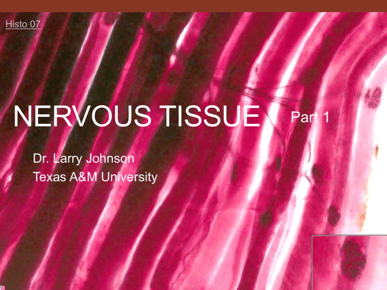
NERVOUS TISSUE
Dr. Larry Johnson
Texas A&M University
Part 1
Objectives
Part 1
• Describe the components of a neuron.
• Distinguish between neurons and glial cells.
• Identify the components of the meninges, grey and white matter.
Part 2
• Recognize peripheral nerves and identity their structural components (in longitudinal and transverse sections); understand the structural and functional differences between myelinated and unmyelinated fibers with both light microscopy and electron micrographs.
From: Douglas P. Dohrman and TAMHSC Faculty 2012 Structure and Function of Human Organ
Systems, Histology Laboratory Manual
Four Basic Types of Tissues in the Body
-----------------------------------------------
Epithelium
(90% of tumors)
Connective Tissue
Muscular Tissue
Four Basic Types of Tissues in the Body
-----------------------------------------------
Epithelium
(90% of tumors)
Connective Tissue
Muscular Tissue Nervous Tissue
Nerve is
like
Epithelium
Origin of nerve is
ectoderm
like epidermis
(epithelium) of skin
Nervous Tissue
Functions: specialized for
the transmission , reception ,
and integration of
electrical impulses
Distinguishing features:
Neurons – very large excitable cells with long processes called axons and dendrites. The axons make contact with other neurons or muscle cells at a specialization called a synapse where the impulses are either electrically or chemically transmitted to other neurons or various target cells (e.g., muscle).
Others secrete hormones.
Nervous tissue
Distribution: comprise the central nervous system.
Individual peripheral nerves are found throughout the body. Individual neurons and clusters of neurons (called ganglia ) are found in most organs.
Function of the Nervous System is
Communication
Dependent upon
special signaling
properties of neurons
Long processes of neurons (e.g.,
1 meter
motor neuraxon)
Function of the Nervous System is
Communication
Characteristics of neurons
Irritability - protoplasm capable to react to various physical and chemical agents
Conductivity - ability to transmit the resulting excitation from one locality to another
Activity of the Nervous System
Information
• Receive : receptors afferent pathway
• Process : CNS ( centralization is paramount)
• Transmit : efferent pathways effect
Voluntary (conscious ) = somatic
Involuntary = autonomic
• Sympathetic - fight or flight
• Parasympathetic - vegetative
CNS & PNS divisions
• Central Nervous System ( CNS )
• Brain and spinal cord
• Grey matter (neuron’s somas and some processes)
• White matter (myelinated and unmyelinated axons)
• Myelination is from oligodendrocytes
• Glial cells:
• Astrocytes
• Microglial cells
Part of the blood-brain barrier (BBB)
• Peripheral Nervous System ( PNS )
• Nerves, sensory ganglia, and autonomic ganglia (or plexus)
• Schwann cells : myelin producing glial cells of the PNS
Axon hillock
Nervous system & neuron
Copyright McGraw-Hill Companies
All images copyright McGraw-Hill Companies
Slide 3 : Cerebral cortex (Luxol fast blue)
Grey matter White matter
Neuropil
Neuron with large nucleus containing a large nucleolus
Neuroglia support cell
Slide 3 : Cerebral cortex (Luxol fast blue)
Gray matter White matter
Neuropil = felt-work of a complex and highly ordered meshwork of dendritic, axonal, and glial processes whose entanglements facilitate an ordered activity important in the communications function of the nervous system. The neuropil provides an enormous region for synaptic contact and functional interactions among nerve cell processes.
Neuropil
Slide DHD100
: Fibrous Astrocytes
Cajal staining method
• Used to visualize: the numerous stellate processes of fibrous astrocytes and neurons
McGraw Hill Companies McGraw Hill Companies
INTERMEDIATE FILAMENTS of cells
FIVE CLASSES (not conserved)
KERATIN - INSOLUBLE SUBSTANCE, EPITHELIUM
INTERMEDIATE FILAMENTS of cells
FIVE CLASSES (not conserved)
KERATIN - INSOLUBLE SUBSTANCE, EPITHELIUM
INTERMEDIATE FILAMENTS of cells
FIVE CLASSES (not conserved)
KERATIN - INSOLUBLE SUBSTANCE, EPITHELIUM
DESMIN – CYTOSKELETON IN MUSCLE
VIMENTIN - NUCLEAR ENVELOPE FOR MECHANICAL
SUPPORT AND STABILITY OF ITS LOCATION IN CELL,
MESENCHYMAL CELL
INTERMEDIATE FILAMENTS of cells
FIVE CLASSES (not conserved)
KERATIN - INSOLUBLE SUBSTANCE, EPITHELIUM
DESMIN – CYTOSKELETON IN MUSCLE
INTERMEDIATE FILAMENTS of cells
FIVE CLASSES (not conserved)
KERATIN - INSOLUBLE SUBSTANCE, EPITHELIUM
DESMIN – CYTOSKELETON IN MUSCLE
VIMENTIN - NUCLEAR ENVELOPE FOR MECHANICAL
SUPPORT AND STABILITY OF ITS LOCATION IN CELL,
MESENCHYMAL CELL
INTERMEDIATE FILAMENTS
FIVE CLASSES con’t
NEUROFILAMENTS
• DENDRITES AND AXONS OF NERVE CELLS
• INTERNAL SUPPORT - GELATED STATE OF CYTOPLASM
INTERMEDIATE FILAMENTS
FIVE CLASSES con’t
NEUROFILAMENTS
• DENDRITES AND AXONS OF NERVE CELLS
• INTERNAL SUPPORT - GELATED STATE OF CYTOPLASM
GLIAL FILAMENTS - ASTROCYTES
INTERMEDIATE FILAMENTS
IMMUNOFLUORESCENCE DETECTION - TOOL IN
DISTINGUISHING CELL TYPE OF ORIGIN OF
MALIGNANT TUMORS
Astrocytes
GLIAL FILAMENTS
- ASTROCYTES
Intermediate filaments of GFAP (glial fibrillary acidic proteins) filament is used clinically to identify astrocytes as in the diagnostic classification of a tumor as an astrocytoma
Spinal cord
Copyright McGraw-Hill Companies
Slide 4 , 17 , 18 : Spinal Cord and Meninges
Central “H” shaped grey matter White matter Central canal lined by ependymal cells
The ependymal cells have microvilli and can have cilia. Cerebral spinal fluid (CSF) surrounds the brain and spinal cord and acts as a cushion, protecting the brain and spine from injury. The fluid is clear normally, and the pressure in the spinal fluid is important too.
Slide 4 , 17 , 18 : Spinal Cord and Meninges
Ventral horn
(motor)
Dorsal horn
(sensory)
Glial cells Motor neuron in ventral horn
Nissl bodies
17 Nissl bodies = rough endoplasmic reticulum and free ribosomes(polyribosomes or polysomes for short)
Slide 4 , 17 , 18 : Spinal Cord and Meninges
Grey matter
White matter
White matter
Grey matter
Axonal tracts
Axons surrounded by dissolved myelin sheath
Slide 4 , 17 , 18 : Spinal Cord and Meninges
Dura mater
Subdural space
Arachnoid matter
Spinal nerve
Subarachnoid space
Pia mater
Slide 4 , 17 , 18 : Spinal Cord and Meninges
Histo 18
Dura mater
Subdural space
Arachnoid matter
Spinal nerve
Subarachnoid space
Slide 17 Spinal Cord and dorsal root ganglia
Dorsal root ganglia
Given pseudounipolar neurons found in the dorsal root ganglia reach from the sensory detection to the spinal cord, there are no synapses of these sensory neurons in the dorsal root ganglia.
THE NERVOUS SYSTEM
CENTRAL NERVOUS SYSTEM (CNS)
• BRAIN AND SPINAL CORD
• NEURONS AND SUPPORT
NEUROGLIA
Histo 07
NERVOUS TISSUE
Part 2
Dr. Larry Johnson
Texas A&M University
Objectives
Part 1
• Describe the components of a neuron.
• Distinguish between neurons and glial cells.
• Identify the components of the meninges, grey and white matter.
Part 2
• Recognize peripheral nerves and identity their structural components (in longitudinal and transverse sections); understand the structural and functional differences between myelinated and unmyelinated fibers with both light microscopy and electron micrographs.
THE NERVOUS SYSTEM
PERIPHERAL NERVOUS SYSTEM (PNS)
ALL NERVOUS TISSUE (NEURONS, SUPPORT CELLS,
AND AXONS) OUTSIDE THE BRAIN AND SPINAL CORD
Auerbach’s Plexus (“Myenteric Plexus”) is sandwiched between the two layers of smooth muscle in the muscularis externa that controls gut peristalsis
Ganglia - collections of nerve cell bodies in PNS
Smooth muscle Smooth muscle
32409
Meissner’s plexus
Rat small intestine
Auerbach’s Plexus
Ganglia -collections of nerve cell bodies in organs
nerve cell bodies
29291
Human pancreas Cystic duct of monkey
Peripheral ganglion, monkey
Axons
Satellite cells
Neuron
Nissl bodies
Organization of Peripheral Nerves
Nerve fibers - axons invested by connective tissue
Epineurum - surrounding entire nerve
Perineurum - surrounding fascicles –
constitutes the PNS blood barrier via tight junctions between fibroblasts
Endoneurum - between individual nerve axons
Organization of Peripheral Nerves
Nerve fibers - axons invested by connective tissue
Epineurum - surrounding entire nerve
Perineurum - surrounding fascicles –
constitutes the PNS blood barrier via tight junctions between fibroblasts
Endoneurum - between individual nerve axons
Slide 6 Nerve (cross section; Bodian’s stain/lissamine red)
Bodian’s stain/lissamine red stain –
• axons and nuclei: black
•
• myelin: pink-red connective tissue: brown to black
Fascicle : group of neurons surrounded by perineurium endneurium : surrounds each axon’s myelin sheath
Nerve : group of fascicles bound by epineurium
perineurium
19673
Myelinated axon
Unmyelinated fibers (axons) do not have a myelin sheath covering, but they are still supported by simple folds of Schwann cells. unmyelinated axons
EM 54 and 55: Nerve
Function of Myelin
Increase speed of condition
• 1 meter/sec TO 120 meters/sec
High-resistance low capacitance
• Insulator
Protection of axon
Possible nutritional role
Direct regenerating axons
SCHWANN CELL
STRUCTURE /
FUNCTION
MOST PERIPHERAL NERVES
ARE MYELINATED
1 SCHWANN CELL/1 AXON
FOR LOCATION
SCHWANN CELL
STRUCTURE /
FUNCTION
MOST PERIPHERAL NERVES
ARE MYELINATED
1 SCHWANN CELL/1 AXON
FOR LOCATION
FORMATION OF MYELIN
SHEATH
• NODES OF RANVIER
• SCHMIDT-LANTERMAN
CLEFTS
• NEUROKERATIN NETWORK
NODE OF RANVIER
Slide
5
: Nerve (longitudinal section)
H&E
• Stains axons, Schwann cells (neurolemmocytes), and connective tissue
• Myelin contains lipids, this is not stained and will have dissolved away during tissue preparation
Longitudinal section Cross section
Schwann cell nuclei
Axons
Neurolemma
Node of Ranvier
Fibroblast
Nerve (longitudinal section)
Schwann cells, the principle glia of the
PNS, wraps around axons of motor and sensory neurons of the PNS to form their myelin sheath.
Schwann cell nuclei
Fibroblast
Node of Ranvier
Axons
Neurolemma
Peripheral nerve,
monkey ut 193
Nerve
(cross section)
Fibroblasts of perineurium
Capillaries
Axons
Neurolemma of Schwann cell
29297
Slide 7 : Myoneural junction (gold chloride/formic acid)
• Skeletal muscle fibers still stain purple to black.
Skeletal muscle cells
Slide 7 : Myoneural junction (gold chloride/formic acid)
• Skeletal muscle fibers still stain purple to black.
Skeletal muscle cells
Axons
Slide 7 : Myoneural junction (gold chloride/formic acid)
• Skeletal muscle fibers still stain purple to black.
Skeletal muscle cells
Motor end plates
Axons
Slide 7 : Myoneural junction (gold chloride/formic acid)
• Skeletal muscle fibers still stain purple to black.
Skeletal muscle cells
Motor end plates
Axons
End
feet
(terminal
buttons and end of presynaptic cell)
NEURONAL STRUCTURE / FUNCTION
Axonal transport
Anterograde - toward terminal - kinesin
Retrograde - toward cell body - dynein
• Tetanus toxin
• Neurotropic viruses (herpes and rabies) use path to get to cell body in CNS
Summary of the Physiological Events at the
Synapse
• Arrival of action potential at axon terminal
• Opening Ca++ channels
• Influx of Ca++ into axon terminal
Summary of the Physiological Events at the
Synapse
• Arrival of action potential at axon terminal
• Opening Ca++ channels
• Influx of Ca++ into axon terminal
• Exocytosis of neurotransmitter
• Diffusion of neurotransmitter across synaptic cleft
• Binding of neurotransmitter to receptors on target cell
• Opening of Na+ channels causing depolarization of target cell
• Removal of neurotransmitter
Neurotransmitter ACh vs NE
Acetylcholine (ACh) is the neurotransmitter released in the synaptic cleft of the motor end plate innervating muscle.
However, norepinephrine (NE) is the neurotransmitter released from most sympathetic postganglionic nerves.
Histo 07
Clinical Correlation
When a peripheral nerve is injured/severed, the segment of the axon distal to the injury and its myelin sheath completely degenerate and their remnants are removed by macrophages. However, Schwann cells proliferate and form cellular columns that act as guides for the regrowing axon back to the original muscle.
Macrophages
Figure 6. Astrocytes. Adapted from “Inducing Alignment in astrocyte tissue constructs by surface ligands patterned on biomaterials,” by Meng, F.,
Hlady, V., Tresco, PA., 2012, Biomaterials , 33:5, p. 1328, Copyright 2012 by Elsevier.
Schwann cells
Copyright McGraw-Hill Companies
Many illustrations in these VIBS Histology YouTube videos were modified
•
•
•
•
•
•
•
•
•
•
•
•
•
•
•
• from the following books and sources: Many thanks to original sources!
Bruce Alberts, et al. 1983. Molecular Biology of the Cell. Garland Publishing, Inc., New York, NY.
Bruce Alberts, et al. 1994. Molecular Biology of the Cell. Garland Publishing, Inc., New York, NY.
William J. Banks, 1981. Applied Veterinary Histology. Williams and Wilkins, Los Angeles, CA.
Hans Elias, et al. 1978. Histology and Human Microanatomy. John Wiley and Sons, New York, NY.
Don W. Fawcett. 1986. Bloom and Fawcett. A textbook of histology. W. B. Saunders Company, Philadelphia, PA.
Don W. Fawcett. 1994. Bloom and Fawcett. A textbook of histology. Chapman and Hall, New York, NY.
Arthur W. Ham and David H. Cormack. 1979. Histology. J. S. Lippincott Company, Philadelphia, PA.
Luis C. Junqueira, et al. 1983. Basic Histology. Lange Medical Publications, Los Altos, CA.
L. Carlos Junqueira, et al. 1995. Basic Histology. Appleton and Lange, Norwalk, CT.
L.L. Langley, et al. 1974. Dynamic Anatomy and Physiology. McGraw-Hill Book Company, New York, NY.
W.W. Tuttle and Byron A. Schottelius. 1969. Textbook of Physiology. The C. V. Mosby Company, St. Louis, MO.
Leon Weiss. 1977. Histology Cell and Tissue Biology. Elsevier Biomedical, New York, NY.
Leon Weiss and Roy O. Greep. 1977. Histology. McGraw-Hill Book Company, New York, NY.
Nature (http://www.nature.com), Vol. 414:88,2001.
A.L. Mescher 2013 Junqueira’s Basis Histology text and atlas, 13 th ed. McGraw
Douglas P. Dohrman and TAMHSC Faculty 2012 Structure and Function of Human Organ Systems, Histology Laboratory
Manual - Slide selections were largely based on this manual for first year medical students at TAMHSC


