5.1 Skin Study Guide By Hisrich
advertisement
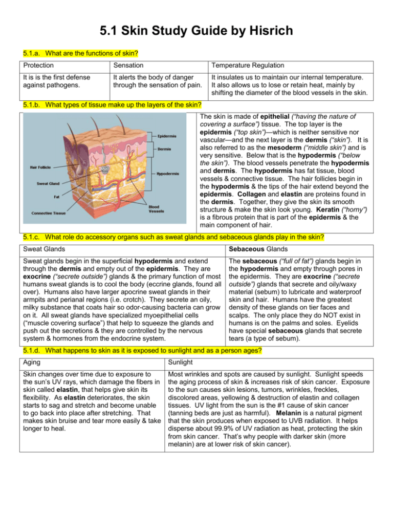
5.1 Skin Study Guide by Hisrich 5.1.a. What are the functions of skin? Protection Sensation Temperature Regulation It is is the first defense against pathogens. It alerts the body of danger through the sensation of pain. It insulates us to maintain our internal temperature. It also allows us to lose or retain heat, mainly by shifting the diameter of the blood vessels in the skin. 5.1.b. What types of tissue make up the layers of the skin? The skin is made of epithelial (“having the nature of covering a surface”) tissue. The top layer is the epidermis (“top skin”)—which is neither sensitive nor vascular—and the next layer is the dermis (“skin”). It is also referred to as the mesoderm (“middle skin”) and is very sensitive. Below that is the hypodermis (“below the skin”). The blood vessels penetrate the hypodermis and dermis. The hypodermis has fat tissue, blood vessels & connective tissue. The hair follicles begin in the hypodermis & the tips of the hair extend beyond the epidermis. Collagen and elastin are proteins found in the dermis. Together, they give the skin its smooth structure & make the skin look young. Keratin (“horny”) is a fibrous protein that is part of the epidermis & the main component of hair. 5.1.c. What role do accessory organs such as sweat glands and sebaceous glands play in the skin? Sweat Glands Sebaceous Glands Sweat glands begin in the superficial hypodermis and extend through the dermis and empty out of the epidermis. They are exocrine (“secrete outside”) glands & the primary function of most humans sweat glands is to cool the body (eccrine glands, found all over). Humans also have larger apocrine sweat glands in their armpits and perianal regions (i.e. crotch). They secrete an oily, milky substance that coats hair so odor-causing bacteria can grow on it. All sweat glands have specialized myoepithelial cells (“muscle covering surface”) that help to squeeze the glands and push out the secretions & they are controlled by the nervous system & hormones from the endocrine system. The sebaceous (“full of fat”) glands begin in the hypodermis and empty through pores in the epidermis. They are exocrine (“secrete outside”) glands that secrete and oily/waxy material (sebum) to lubricate and waterproof skin and hair. Humans have the greatest density of these glands on tier faces and scalps. The only place they do NOT exist in humans is on the palms and soles. Eyelids have special sebaceous glands that secrete tears (a type of sebum). 5.1.d. What happens to skin as it is exposed to sunlight and as a person ages? Aging Sunlight Skin changes over time due to exposure to the sun’s UV rays, which damage the fibers in skin called elastin, that helps give skin its flexibility. As elastin deteriorates, the skin starts to sag and stretch and become unable to go back into place after stretching. That makes skin bruise and tear more easily & take longer to heal. Most wrinkles and spots are caused by sunlight. Sunlight speeds the aging process of skin & increases risk of skin cancer. Exposure to the sun causes skin lesions, tumors, wrinkles, freckles, discolored areas, yellowing & destruction of elastin and collagen tissues. UV light from the sun is the #1 cause of skin cancer (tanning beds are just as harmful). Melanin is a natural pigment that the skin produces when exposed to UVB radiation. It helps disperse about 99.9% of UV radiation as heat, protecting the skin from skin cancer. That’s why people with darker skin (more melanin) are at lower risk of skin cancer). 5.1.e. Which layers of the skin are damaged in different types of burns? (info from http://hospitals.unm.edu/burn/classification.shtml) 1st Degree Burn—damages Epithelium, painful/tender 2nd Degree Burn—damages Epithelium & top of dermis, very painful 3rd Degree Burn—damages epithelium and dermis—little to no pain 5.1.f. How does burn damage in the skin affect other functions in the body? Skeletal Circulatory Muscle Nervous Respiratory Bone marrow works to replace RBCs destroyed by burnt skin—blood transfusions may be needed BP and blood volume drop, decreasing blood flow and oxygenation—can lead to shock/death Metabolism increases and the body starts to consume muscle mass K+ levels become abnormal makes nerve transmissions irregular (faster, slower or not at all) Rate of breathing can increase from higher metabolism & edema—edema of throat can also obstruct the airway Endocrine Lymphatic Immune Digestive Urinary Adrenalin secretions can raise body temperature & increase metabolism System under strain from inflammation (due to damaged tissues) Becomes less effective because 1st line of defense (skin) compromised Intestinal lining increases absorption of nutrients to support metabolism and repair cells Kidneys increase reabsorption to compensate for lost fluid (can damage kidneys) 5.1.h. What role does pain play in the human body? 5.1.i. How does the body interpret and process pain? 5.1.j. Why would the inability to feel pain actually put the human body in danger? Role of Pain Processing of Pain Risk from Lack of Pain The survival purpose of pain is to alert the body to a risk of tissue damage so the person can take action to remove the risk (i.e. touching a hot stove—pull hand away). The higher the risk, the greater the pain. Pain is received by naked nerve endings (dendrites of sensory nerves) & is often experiences as physical discomfort (pricking, throbbing or aching). A person typically responds by taking action to remove the source of the pain. The brain secretes endorphins (a type of hormone made in the pituitary gland) in response to stress and pain. Endorphins can act in a similar way to morphine, reducing perception of pain as well as to feelings of euphoria and an increased immune response. (May be responsible for “runner’s high”) Acute (short term) pain is necessary to survival. If a human felt no risk from pain (i.e. touching a hot stove), he would take no action to remove the threat (i.e. pulling hand away) and would suffer more damage. There is not thought to be any survival benefit to chronic (long term) pain (i.e. cancer pain), however)

