Evaluate a Blood Film
in less than 3 minutes
by Dr. Dennis DeNicola DVM, PhD, DACVP
Frequently Asked Questions
Q: How accurate do you feel manual differentials are?
A: Assuming that the individual performing the manual leukocyte differential can accurately differentiate the different leukocytes present on
the blood film, accuracy of the manual differential is relatively good; however, the basic problem with the manual differential is precision.
When counting only 100 leukocytes, a relatively wide range of results must be acceptable as being correct (or accurate). As more
leukocytes are counted, precision improves but it is highly impractical to count enough leukocytes to come close to the precision of a
five-part leukocyte differential on an advanced automated hematology analyzer assuming the analyzer is performing correctly. These
advanced hematology analyzers count a minimum of 10,000 leukocytes for reporting distributions.
Example:
Total Leukocytes Counted In A Manual Differential
Incidence (%) of leukocyte
in circulation
100
200
500
1,000
5
1 – 12
2 – 10
3–8
3–7
20
12 – 30
14 – 27
16 – 24
17 – 23
50
39 – 61
42 – 58
45 – 55
46 – 54
75
65 – 84
68 – 81
70 – 79
72 - 78
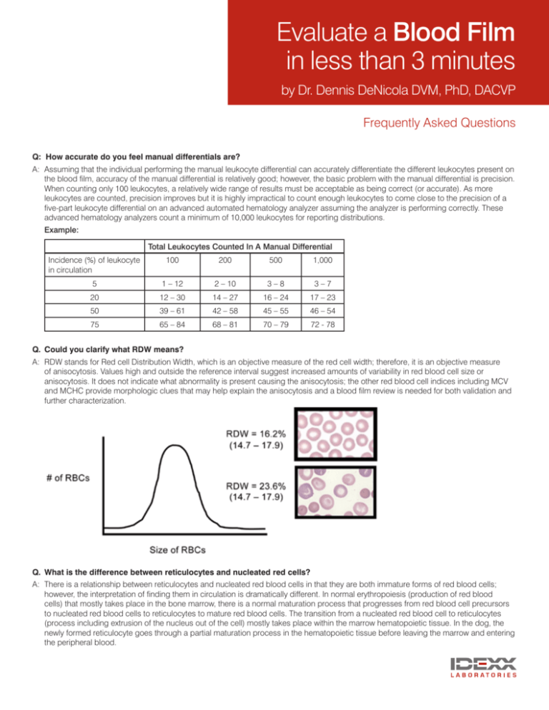
Q. Could you clarify what RDW means?
A: RDW stands for Red cell Distribution Width, which is an objective measure of the red cell width; therefore, it is an objective measure
of anisocytosis. Values high and outside the reference interval suggest increased amounts of variability in red blood cell size or
anisocytosis. It does not indicate what abnormality is present causing the anisocytosis; the other red blood cell indices including MCV
and MCHC provide morphologic clues that may help explain the anisocytosis and a blood film review is needed for both validation and
further characterization.
Q. What is the difference between reticulocytes and nucleated red cells?
A: There is a relationship between reticulocytes and nucleated red blood cells in that they are both immature forms of red blood cells;
however, the interpretation of finding them in circulation is dramatically different. In normal erythropoiesis (production of red blood
cells) that mostly takes place in the bone marrow, there is a normal maturation process that progresses from red blood cell precursors
to nucleated red blood cells to reticulocytes to mature red blood cells. The transition from a nucleated red blood cell to reticulocytes
(process including extrusion of the nucleus out of the cell) mostly takes place within the marrow hematopoietic tissue. In the dog, the
newly formed reticulocyte goes through a partial maturation process in the hematopoietic tissue before leaving the marrow and entering
the peripheral blood.
In the dog, the maturation to a normal mature red blood cell takes approximately
24 hours in the blood. In the cat, there is a slight difference in that the majority of
the reticulocyte maturation takes place in the hematopoietic tissue itself. Punctate
reticulocytes, reticulocytes with only very small amounts of cytoplasmic immaturity,
are released to the peripheral blood and these may stay in circulation for 10, and
potentially, even 14 days before completing the process of maturation to a normal
mature red blood cell. During moderate to severe anemia in the cat, the less mature
reticulocyte identified as an aggregate reticulocyte with moderate cytoplasmic
immaturity is released from the marrow. These aggregate reticulocytes mature to
punctate reticulocytes typically within a 24-hour period.
Q. Are reticulocyte counts accurate in cats on LaserCyte® due to the different types of reticulocytes in cats?
A: This is a great question on a somewhat controversial topic. We know that there are minimally two basic types of reticulocytes in the
cat, punctate and aggregate. Aggregate reticulocytes, when stained with a reticulocyte stain, will have multiple coarse aggregates of
precipitated ribosomal RNA (immature cellular components responsible for hemoglobin production); punctate reticulocytes have only
a few specs of precipitated material with the same stain because they have much less cytoplasmic immaturity. In most veterinary and
veterinary technology curricula, it is commonly stated that only the aggregate reticulocyte count is important since it represents the
count of the most recently released and “younger” reticulocytes and, therefore, represents evidence of a bone marrow response to an
anemia allowing classification of the anemia as regenerative.
Current advanced hematology analyzers, including the IDEXX LaserCyte Analyzer and commercial hematology analyzers used in reference
and academic laboratories, only detect aggregate reticulocytes. These analyzers are not sensitive enough with current methodologies to
detect the punctate reticulocytes. At first glance this all sounds great; however, there is a little bit of key information that is missing, and
if we had a sensitive enough system (in-house or reference laboratory) to detect punctate reticulocytes, I believe that we would be much
better off. The important bit of information to know is that in cats with only mild anemia, where the HCT is greater than 20-24%, punctate
reticulocytes are not easily released from the marrow and we primarily see a punctate reticulocyte response. In these cases, a missed
classification of a mild anemia as a non-regenerative anemia because of the lack of an aggregate reticulocytosis could be made. Only
very few academic laboratories actually report punctate reticulocyte counts. This can be extremely labor-intensive since it requires manual
inspection and identification of the punctate reticulocyte forms and because punctate reticulocyte counts can easily go into the several
hundred thousands or millions per microliter during a marrow response. Manual counting recommends determining the percentage of
non-nucleated red blood cells that are aggregate and punctate reticulocytes during evaluation of 1,000 red blood cells stained with a
reticulocyte stain. Simple inspection and identification of large numbers of punctate reticulocytes without counting is all that is really needed
to indicate that a mild anemia in a cat should still be classified as “regenerative.”
Q. What is the best stain and method?
A: From a hematologist’s perspective, a classic Romanovsky stain such as Wright’s stain, Giemsa stain, Wright’s-Giemsa combination
stain, etc., would be considered a gold standard for hematology. If used properly, the rapid and generally aqueous stains commercially
available to veterinarians should prove completely acceptable. The added advantage of an aqueous stain over some of the more
traditional stains is seen in the minimization of artifacts such as stain precipitates when most rapid stains are used. The key to
successful usage of these stains is to experiment and modify staining procedures as you find that “things don’t look right” when
doing your blood film evaluation. Many technicians seem to produce stained blood films that are too blue in character, which clearly
will interfere with recognition of polychromatophils, identification of neutrophil toxicity, and in some instances, simple leukocyte
differentiation. As with anything, practice makes perfect. One quick tip to follow when using the rapid aqueous stains is to assure that
the first step of the process, the fixation step, is adequate. Leaving a blood film in the fixative for an extended period of time beyond the
manufacturer’s recommendation, or even leaving it in the fixative for several minutes, will only potentially improve the actual staining
process. Practice and modification of timing may be required initially, but the end result is of great value.
Q. What stain do you recommend for viewing RBC inclusions?
A: This is another great question. The presence of any debris on a blood film or stain precipitate actually interferes with accurate
characterization of any sort of red blood cell inclusion. To minimize stain precipitate problems, I believe the rapid aqueous stains prove
to be superior. There have been many times in the past, when examining a blood film stained with a classic Wright’s stain, where I
believed that there was something on the blood film such as Mycoplasma haemofelis organisms on the surface of the red blood cells.
Many of these times, I would return to stain another blood film with one of the rapid aqueous stains to discover that I was only seeing
stain precipitate. I personally prefer a well-stained film using one of the rapid aqueous stains; however, this is one of those questions
where you might find other microscopists with differing opinions/preferences.
Q. What stain do we use for blood film cytology?
A: Since I examine both blood films and various cytologic preparations from other organ systems from a variety of sources, including
stains from different types of automated stainers as well as stained material sent directly to me from veterinarians
in the field, I am used to looking at pretty much everything. There are advantages and disadvantages to essentially
all stains. I feel quite strongly that the commercially available rapid aqueous stains, such as Diff Quik, are great
all-purpose stains for both blood film and other organ cytology specimens. They are simple to use, and if a little time is invested in
developing a protocol for proper staining, all the essential information needed can be obtained from diagnostic specimens.
Q. What, in your opinion, is the best way to perform a manual platelet count in the hospital?
A: I honestly do not recommend performing a manual platelet count. If one is not happy with their hematology analyzer’s performance
related to platelet counts, a manual estimate can be attempted by looking at a well-prepared and well-stained blood film. In the dog and
cat, 8-10 platelets per average 100x oil objective field of view is a minimum number of platelets to be considered “adequate.” An actual
number is not needed if adequate, but an approximate platelet count can be made by multiplying the average number of platelets seen
in ten 100x oil objective fields of view times 20,000 to obtain an estimate of platelets per microliter. Manual platelet counts can still be
performed using a commercially available Unopette system for platelet and leukocyte counting using a haemocytometer; however, one
must perform significant numbers of counts to develop any degree of precision for this count. Additionally, a good microscope with
capability for light focus control and, potentially, a phase contrast microscope may be needed to obtain accurate results. Again, I would
not recommend manual platelet counting. A quick inspection of a blood film can provide all the clinically important information needed
in most cases. If manual platelet counts are performed, be aware that platelet clumping will interfere with accurate manual platelet
counts just as much as they interfere with automated counts, so a peripheral blood film inspection is still essential.
Q. If the hematology analyzers are so accurate (such as our LaserCyte), why does it give us the warning of “differential
algorithms exist” on almost every sample we run?
A: There are two potentially important points surfacing with this question.
First, the “warning” or “flag” or “message code” being reported on almost every sample run should be considered a potential issue
with that particular analyzer unless CBCs on only clinically abnormal patients are performed, which is unlikely. I recommend that you
contact your customer support representative and do some investigation. There might be a very simple problem, such as instrument
overheating. A series of simple questions can be answered (Is there good temperature control in the area of the in-house laboratory? Is
the filter controlling airflow through the analyzer cleaned regularly?, etc.). The various in-house analyzers require appropriate operating
conditions. If these and other questions can be answered by the customer support people, a service event might be considered.
Second, a general comment on warning messages is warranted. In general, both reference laboratory and in-house hematology
analyzers will have a series of checks and balances in place to assure producing the best data possible. Each will have a series of
specific metrics in place, that if met, will release data with no message codes; however, if performance of a particular sample run does
not meet the defined metrics, a message code will be reported. Depending on the severity of the failure in meeting the defined metrics,
data may or may not be reported. If there is a severe deviation from normal operation, data may be completely blocked from reporting,
but if the deviation from normal performance is only minor, results are reported with the message code. These codes are basically
informing the operator that there is an extremely high likelihood that the results reported are correct but validation should be considered.
Most of this validation for the various parameters measured can be accomplished with the 1-3 minute blood film review.
Q. Are we looking at the body of the film and at 100X?
A: Recommendations for the approach to the blood film review are included in the files that could be downloaded at the end of the
presentation. Below is a quick summary:
Feathered edge of film – 10x objective field of view, 10 seconds
• Looking for large objects or cells (abnormal leukocytes, clumps of cells, including platelets, microfilaria, etc.)
Body of the film – 10x objective field of view, 10 seconds
• Looking to characterize red blood cell to red blood cell associations (rouleaux versus agglutination) and to obtain a quick perspective
on overall leukocyte distribution
Monolayer of film – 10x, 20x, 40x and 100x objective fields of view, less than 3 minutes
• Detailed cellular morphologic evaluation, numerical data evaluation, etc.
Q. Would you use a 40x objective if you did not have a 20x objective on your microscope?
A: The primary advantage of using a lower magnification than the 100x objective when examining a blood film is to allow scanning
of a larger area of the slide at a magnification where morphology abnormalities can be seen. With the 10x objective field of view,
morphology, and even leukocyte identification, can be very difficult and at the 100x objective where detailed morphologic evaluation is
possible, too small of a field of view is available and overall scanning of the slide is impaired. The analogy of the forest and the trees
applies well to this situation. At the 100x objective field of view, you can detail specifics about the individual tree, but miss the overall
picture of the forest regarding what different types of trees are present, if there are any general differences in sizes of the trees or even
if there are broken branches seen on the occasional tree. The 20x objective field of view provides this “big picture” that may be missed
by only using the 100x objective. The 40x, which is present on most microscope setups in the veterinary practice, can serve as a
compromise objective to use; however, it is not intended for use without a coverslip. To assure sharp focus on the specimen examined,
a drop of immersion oil can be placed on the stained and dried blood film and a coverslip can be placed on this drop. The oil will
spread between the stained blood film and the coverslip and now the specimen will be in focus using your 40x objective.
Q. Do you ever read the blood film before evaluating data from the hematology analyzer?
A: This is an absolutely fantastic question. With time, I believe this is exactly what should be done. In doing the blood film evaluation
independent of the hematology analyzer data, a great internal quality control program can be developed. This would be the absolute
perfect world to live in, where basically each run on the analyzer would have a rapid independent evaluation. However, if just beginning
with blood film evaluation, I would strongly recommend that without much practice and experience, validation of numerical data from the
hematology analyzer should be done with knowledge of the data themselves. Since morphology comments on red blood cells, white
blood cells and platelets are not included with any automated hematology analyzer, these can be established without the actual data
generated by the analyzers.
Q. Do the “bluish” red blood cells (polychromatophils) always have less central pallor on a typical hematology stain?
A: This is somewhat related to the quality of the slide; however, if a blood film is made properly, many, if not most, of the
polychromatophils, will have increased central pallor compared to mature red blood cells. These younger red blood cells do not
have their normal complement of cytoplasmic content (primarily hemoglobin) and they have excess cytoplasm membrane and the
combination of these two developmental findings makes the polychromatophil more “floppy” with less cytoplasmic content, which gives
these cells an apparent increased central pallor.
Q. Can you comment on the use of “percent reticulocytes” and how this relates to regenerative anemia?
A: The percent reticulocyte count is often difficult to interpret since it is a “relative” number related back to the total number of red blood
cells present in the sample. If the bone marrow is releasing the normal number of reticulocytes for a dog, a maximum of 60,000
reticulocytes per microliter, or 1% of the total number of red blood cells, will be found in the blood. Assume a situation where there is a
sudden severe loss (blood loss or hemolysis) of half of the circulating red blood cell mass and the bone marrow continues to release
only the “normal” number of reticulocytes. In a very short time, the relative number of reticulocytes will change from the maximum 1%
to 2%; however, the absolute number of reticulocytes will still be 60,000 per microliter. Simply looking at the increased relative number
of reticulocytes can give misleading information about the regenerative capacity at a particular point in time. The absolute reticulocyte
count (number of reticulocytes per microliter) is the preferred reticulocyte parameter to use for differentiating regenerative from nonregenerative anemia since it avoids the “concentrating” effect when there is a decreased red blood cell mass.
Q. Regarding band neutrophils, can you tell how to differentiate a band from a mature neutrophil and how to determine their
count without doing a complete differential?
A: There are multiple definitions of a band neutrophil, the precursor to the mature neutrophil. One definition, which I find limiting, is that the
nucleus has no indentation and that the thickness of the nucleus is uniform throughout. Another definition is that the thinnest portion of
the nucleus is greater than one-third the widest portion of the nucleus. In both cases, the nucleus clearly is less indented and also less
dense than that of a mature neutrophil nucleus. Since during preparation of a blood film, the nucleus may be twisted or folded upon
itself, neither “rule” for identifying a band neutrophil may work for all neutrophil forms visualized on the blood film. In most cases, a “less
mature”-appearing neutrophil, which correlates to a “band neutrophil,” can be differentiated from a mature neutrophil even when there
is twisting or folding of the nucleus. Since the recognition of band neutrophils is so important for characterizing a “left shift” to help
properly identify when inflammation is present, I believe that these cells need to be identified and reported. However, because of the
difficulty in accurately identifying them because of nuclear distortion during the blood film preparation and the availability of different
“definitions” of how a band morphology is defined, subjective terms for their presence on the blood film is more valuable than an
actual attempted count. In a specific clinic, the technicians can develop their own definitions for mild, moderate, and marked left shifts.
Knowing the imprecision of a 100 leukocyte differential, these subjective terms are of greater value as long as there is consistency in
how they are used by a specific technician.
Q. How important is it to identify schistocytes (fragmented red blood cells) and is it possible to cause this morphology with
the blood film preparation itself?
A: Schistocytes, or fragmented blood cells, are an important finding; they indicate mechanical fragmentation of the cells. This can be
due to a variety of situations, but some of the more common include fragmentation due to red blood cells traveling through strands
of fibrin (disseminated or localized intravascular coagulopathy) or through abnormal microvascularity as might be present within a
vascular neoplasm. Heavy adult heartworm loads, hear valvular disease, renal glomerular abnormalities, and other conditions can
result in fragmentation also. Unlike other morphologic changes in red blood cells, schistocytes will be typically much less frequent, and
significant findings may be when only one fragment is seen in every five to ten 100x objective fields of view. Significant poikilocytosis
for other morphologic changes is when multiple morphologic abnormalities are seen in every 100x objective field of view. Therefore,
more diligence is required to identify significant schistocytosis. The actual process of making the blood film should not typically cause
fragmentation to occur; however, in cases where there is significant other poikilocytosis (acanthocytosis, oxidant injury changes, etc.),
where the red blood cells are fragile, apparent fragmented cells may be seen. As an isolated artifact of
blood film preparation, this should not be a problem; if seen, there will be many other artifacts on the
blood film also.
idexx.com
© 2008 IDEXX Laboratories, Inc. All rights reserved. • 6219-00
All ®/TM marks are trademarks or registered trademarks of IDEXX Laboratories, Inc. or its affiliates in the United States and/or other countries.


