1. Structure & classification of neurons 2. Role of
advertisement

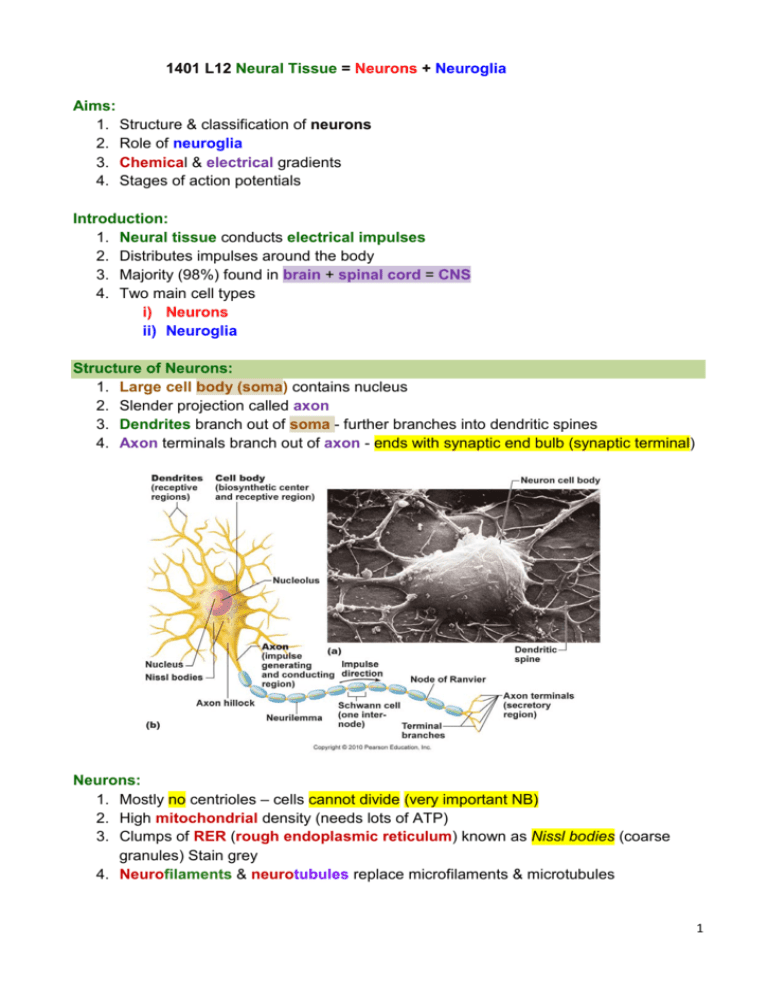
1401 L12 Neural Tissue = Neurons + Neuroglia Aims: 1. Structure & classification of neurons 2. Role of neuroglia 3. Chemical & electrical gradients 4. Stages of action potentials Introduction: 1. Neural tissue conducts electrical impulses 2. Distributes impulses around the body 3. Majority (98%) found in brain + spinal cord = CNS 4. Two main cell types i) Neurons ii) Neuroglia Structure of Neurons: 1. Large cell body (soma) contains nucleus 2. Slender projection called axon 3. Dendrites branch out of soma - further branches into dendritic spines 4. Axon terminals branch out of axon - ends with synaptic end bulb (synaptic terminal) Neurons: 1. Mostly no centrioles – cells cannot divide (very important NB) 2. High mitochondrial density (needs lots of ATP) 3. Clumps of RER (rough endoplasmic reticulum) known as Nissl bodies (coarse granules) Stain grey 4. Neurofilaments & neurotubules replace microfilaments & microtubules 1 Structural Classification of Neurons: 1. Multipolar = Two or more dentrites & one axon 2. Bipolar = Distinct dentritic & axon processes with soma between them 3. Unipolar = Fused continuous axon with soma attached Functional Classification of Neurons: 1. Sensory (afferent) neurons: i) Relay information from sensory (skin/organs) receptors to CNS ii) Most Unipolar iii) Can be very long - Over 1m long from toe to spinal cord 2. Motor (efferent) neurons: i) Transmit impulses from CNS to effectors organs (muscles & organs) ii) Multipolar structure iii) Cell bodies mainly in CNS (brain + spinal cord) 3. Interneurons: i) Situated between motor & sensory neurons ii) Act as messengers for impulse signal iii) Distribute signal through CNS iv) Make up most of neurons in CNS v) Most multipolar Myelin Sheath = Myelinated Axons: 1. Axon covered with Schwann cells 2. Nodes of Ranvier bisect Schwann cells 3. Myelinated axons allow faster (saltatory) conduction compared to non-myelinated axons 2 Conduction Speed: SALTATORY = FASTER = fast bursts between Schwann cells Activation of Neurons: 1. Neurons extremely excitable & respond to stimuli 2. Once activated impulse runs down axon 3. This is known as action potential 4. Process begins with change in resting potential of neuron Electricity Basics: 1. Voltage: i) Measure of potential energy generated by separated charge ii) Measured in volts (V) or millivolts (mV) iii) Always measured between two points iv) Known as potential difference or just potential 2. Current: i) Flow of electrical charge from one point to another ii) Depends on voltage & resistance 3. Resistance: i) Inhibits the flow of current ii) Insulators have a high resistance iii) Conductors have low resistance 3 Ohm’s Law: 1. Relationship between voltage, current & resistance Current = Voltage Resistance Cell Membrane Ion Channels: 1. To fire a neuron need to create (a charge separation) or potential 2. achieved this by arranging sodium & potassium ions in different concentrations 3. Ion channels open & close to control entry & exit of these ions Chemical Gradients: 1. Chemical gradients exist across cell membrane i) Inner (ICF) = high K+ and low Na+ ii) Outer (ECF) = high Na+ and low K+ 2. Concentration gradient exists for sodium (Na+) & potassium (K+) across cell membrane Sodium-Potassium Pump: 1. Maintains gradient of sodium and potassium ions across membrane 2. Requires energy – ATP 3. For each molecule of ATP: i) 3 sodium ions pumped out of cell ii) 2 potassium ions pumped into cell Electrical Gradient: 1. Electrical gradient exist across cell membrane i) Inner membrane slightly negative ii) Outer membrane slightly positive 2. Difference of -70mV i) Can be -40mV to -90mV 3. This is known as the resting membrane potential Resting Membrane Potential: 4 Changes in Membrane Potential: 1. Cell membrane is dynamic (constantly changing in response to environment) 2. Membrane potential changes as result of ion movement across membrane 3. Three different types of membrane channel for ions Membrane Channels: Very important (NB) 1. Leakage channels i) Alternate between open & closed ii) More potassium (K+) channels than sodium (Na+) channels 2. Voltage-gated channels i) Open in response to change in membrane potential 3. Ligand-gated channels i) Open in response to specific chemical stimulus (Ligand = chemical natural/man-made) Cell Membrane Ion Channels Neuron Activation 1. Neurons ‘fire’ when their resting membrane potential is reversed or depolarised 2. This creates an action potential 3. Membrane potential changes from (-70 mV to +30 mV) (100 mV difference) 4. Followed by repolarisation Action Potential: 1. Initiated by stimulation of dendrites 2. Opens voltage-gated Na+ channels at axon hillock 3. ‘All or nothing’ in function 4. A ‘wave’ of depolarisation runs down axon Depolarisation: 1. Change in membrane potential by stimuli activates gated Na+ & K+ channels 2. Na+ channels open faster & so more Na+ enters cell 3. Influx of Na+ causes reduction in –ve charge of inner membrane (-70mV to 0mV) 4. Depolarisation in wave-like flow down axon to synaptic terminals (telodendria) 5. Larger diameter axon - increased flow rate 6. Myelinated axon - fastest flow rate – action potential jumps between Nodes of Ranvier 5 Repolarisation: 1. Reducted Na+ influx due to closure of Na+ gated channels 2. Increase in K+ flow out of cell as K+ channels still open 3. Restores –ve charge of inner membrane 4. Hyperpolarisation i) Increase in –ve charge of inner membrane ii) Caused by slow K+ channel inactivation Action Potential Process: 6 Synapse: 1. Synapse: a junction where neurons communicate with other cells via neurotransmitter or electrical current 2. Presynaptic cell - sends message via synaptic terminals on axon 3. Postsynaptic cell - receives message (usually) across synaptic cleft Neurotransmitters: 1. Chemicals released from neuron that affects another cell’s membrane potential 2. Acetylcholine = Voluntary muscle movement 3. Norepinephrine = Alertness & arousal 4. Dopamine = Pleasure/wanting – associated with addictions Activation Sequence: 1. Action potential reaches axon terminal & causes calcium (Ca2+) channels to open 2. Ca2+ more conc. (concentrated) in ECF (extracellular fluid) 3. Ca2+ enter axon terminal & trigger exocytosis of synaptic vesicles with neurotransmitter 4. Neurotransmitter released across synapse cleft & binds to receptors on postsynaptic cell 5. Receptor channels open to allow ions to enter cell 6. Voltage changes due to ion conc 7. When threshold is reached the post-synaptic cell is activated 7 Neuroglia: 1. Glia – Gr. meaning ‘glue’ 2. Non-neuronal cells in nervous system 3. Provides support, nutritional needs & Myelin 4. Four types of neuroglia: i) Ependymal cells - line central canal (spinal cord) & ventricles around brain, secrete cerebrospinal fluid (CSF) & monitor composition of CSF ii) Astrocytes support endothelial cells in regulating Blood Brain Barrier iii) Oligodendrocytes: contact exposed surfaces of neurons, help form ‘myelin sheath’ along axon, myelinated axons permit faster transmission of info iv) Microglia - help protect CNS via phagocytosis of waste, debris and pathogens Summary: 1. Neurons & neuroglia make up neural tissue 2. Neurons conduct electrical information 3. Convey sensory or motor data to & from brain or distribute information within brain 4. Rely on action potentials to ‘fire’ 8


