Summary and Future
advertisement
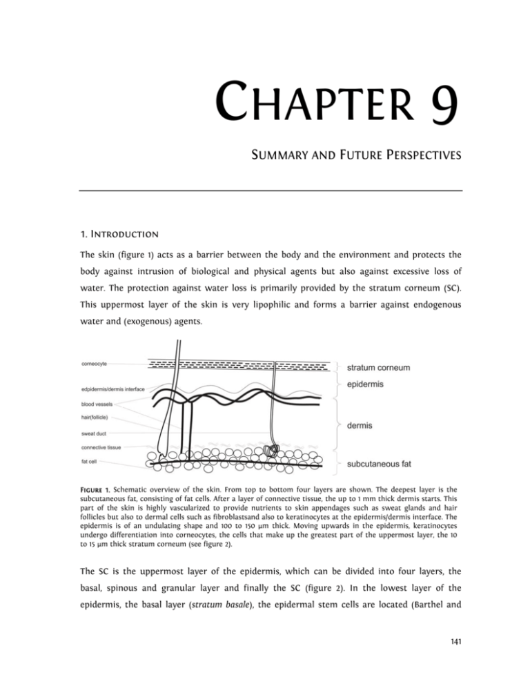
CHAPTER 9 SUMMARY AND FUTURE PERSPECTIVES 1. Introduction The skin (figure 1) acts as a barrier between the body and the environment and protects the body against intrusion of biological and physical agents but also against excessive loss of water. The protection against water loss is primarily provided by the stratum corneum (SC). This uppermost layer of the skin is very lipophilic and forms a barrier against endogenous water and (exogenous) agents. Figure 1. Schematic overview of the skin. From top to bottom four layers are shown. The deepest layer is the subcutaneous fat, consisting of fat cells. After a layer of connective tissue, the up to 1 mm thick dermis starts. This part of the skin is highly vascularized to provide nutrients to skin appendages such as sweat glands and hair follicles but also to dermal cells such as fibroblastsand also to keratinocytes at the epidermis/dermis interface. The epidermis is of an undulating shape and 100 to 150 µm thick. Moving upwards in the epidermis, keratinocytes undergo differentiation into corneocytes, the cells that make up the greatest part of the uppermost layer, the 10 to 15 µm thick stratum corneum (see figure 2). The SC is the uppermost layer of the epidermis, which can be divided into four layers, the basal, spinous and granular layer and finally the SC (figure 2). In the lowest layer of the epidermis, the basal layer (stratum basale), the epidermal stem cells are located (Barthel and 141 Chapter 9: Summary and Future Perspectives Aberdam, 2005). In this layer continuous cell division takes place. Moving upward to the epidermis-SC interface the keratinocytes differentiate into large, flat protein filled corneocytes. The SC has a thickness of 10 to 15 µm in normal human skin and comprises approx. 15 layers of corneocytes, embedded in a highly organized lipid matrix consisting of ceramides (CER), cholesterol (CHOL) and free fatty acids (FFA). With its very high degree of organization, this lipid matrix provides the main barrier for water and other molecules leaving or penetrating the body. In SC, the ratio of CER, CHOL and FFA is roughly equimolar (Bouwstra et al., 1996a). Both CER and FFA are heterogeneous in nature, differing in chain length, and, in case of the CER, in head group architecture. Three CER with a linoleic acid ester-linked to a long chain ωhydroxy fatty acid (EO) have been identified; these are referred to as acylCER. Figure 2. The stratum corneum is made up of large, flat cells called corneocytes, surrounded by a lipid matrix that comprises approx. 10% of its weight. The cells are attached to each other by modified desmosomes called corneodesmosomes, which become less abundant in the upper layers of the stratum corneum due to enzymatic degradation. This degradation facilitates desquamation, i.e. the loss of corneocytes. The properties of the SC extracellular matrix are not only extraordinary due to the exceptional lipid classes, but also because of the high degree of organization that it displays. The lipids form a three dimensional ordering. They are organised in a lamellar ordering approximately parallel to the skin surface (figure 3) and in a lateral packing within the lamellae perpendicular to the direction of the lipid chains (figure 4). Figure 3. Lamellar organization of the extracellular lipid matrix in stratum corneum. In between corneocytes, the lipids are organized in bi- or trilayers, which exist in stacks of up to 12 high. The size of the bi- or trilayer is referred to as the repeat distance d. 142 Chapter 9: Summary and Future Perspectives Multiple lipid lamellae are stacked approx. parallel to the skin surface. Two lamellar phases with different repeat distances d have been found (figure 5), the short periodicity phase (SPP, d = approx. 6 nm) and the long periodicity phase (LPP, d = approx. 13 nm) (Bouwstra et al., 1996a). Of these two, the LPP seems to be most important for the skin barrier function, as it is very characteristic for the lipid organisation in SC (Bouwstra et al., 1991; Bouwstra et al., 1995; Bouwstra et al., 1994; White et al., 1988). Recently, the LPP was shown to be very important for the barrier function of the lipid matrix (de Jager et al., 2006). Figure 4. Lateral organization of the extracellular lipid matrix in stratum corneum. The lateral organization of the intercellular stratum corneum lipids is provided by the packing of the carbon tails of the lipids, here depicted as dots. The more closely the carbon tails are packed, i.e. the smaller the distance between them, the denser and therefore less permeable the lipid layer is. In normal stratum corneum, mostly the dense orthorhombic packing is present. The second type of ordering is the lateral packing of the SC lipids in the lamellae. Packing density, and as a result barrier function, increases in the order liquid<hexagonal<orthorhombic (Abrahamsson et al., 1978; Kitson et al., 1994; Suhonen et al., 1999b). Most studies focusing on the lateral packing of human SC have shown that at physiological temperatures, the majority of the lipid tails are arranged in the very dense, orthorhombic lattice. At higher temperatures, this packing is converted into a less tightly packed lattice, the hexagonal lattice (Ongpipattanakul et al., 1994; Moore et al., 1997; Bouwstra et al., 2000). At the interface between viable epidermis and the SC the corneocytes undergo cornification. The corneocyte envelope, also called cornified envelope, is a complex of several proteins 143 Chapter 9: Summary and Future Perspectives surrounding the mature corneocytes, covered with covalently linked monolayer of lipids. It renders the corneocyte rigid and increases its resilience to force. The corneocyte envelope is only slightly penetrable to virtually all substances except very small hydrophilic substances such as water, making it an important part of the SC barrier (Haftek et al., 1998). Simultaneous to the cornification of the cell membrane, the cell’s desmosomes are converted into corneodesmosomes (Haftek et al., 1997; Hirao, 2003; Hirao et al., 2001; Montezin et al., 1997; Serre et al., 1991). Figure 5. Two periodicity phases have been identified in stratum corneum, the short periodicity phase (SPP), with a repeat distance d of approx. 6 nm, and the long periodicity phase (LPP) with a repeat distance of approx. 13 nm. Of these, the LPP appears to be essential for the barrier function of stratum corneum. The LPP has been speculated combine a central fluid domain with two crystalline domains above and below it (the Sandwich Model, Bouwstra et al. J. Lipid Res. 1998, 2001). On average, one layer of corneocytes is shed per day. For this so-called desquamation, corneodesmosomes must be degraded (Serre et al., 1991). The members of the kallikrein family (Emami and Diamandis, 2007) play an important role in this degradation process. Afterwards, corneocytes are free to be lost as a result of friction or sheer stress. Interestingly, the activity of the enzymes involved in SC desquamation is dependent on SC water levels (Watkinson et al., 2001), explaining the scaliness of dry skin. For proper elasticity and plasticity of the SC and to ensure that the necessary enzymatic processes in SC such as cornification and desquamation can occur humidity must be between 40 and 80% (Scott and Harding, 1986; Watkinson et al., 2001). In normal, healthy skin, this 144 Chapter 9: Summary and Future Perspectives water level is regulated not only by water loss inhibition by the extracellular lipid matrix, but also by the natural moisturizing factor (NMF) (Harding, 2000; Rawlings and Matts, 2005), a collection of small, hygroscopic molecules. Interestingly, several of these compounds are produced by enzymatic protein degradation, the enzymatic activity of which is dependent on SC water level, which in turn is partially dependent on the presence of NMF (Visscher, 2003). SC water and NMF therefore work in synergy: An optimal water level in SC promotes NMF production, while the presence of NMF ensures water retention in the SC. Likewise, a reduction of NMF, for instance due to excessive bathing, will inhibit NMF production (Rawlings and Matts, 2005), as is described below. Thus, a vicious cycle can occur (figure 5). If the water levels in SC are too low, the well-known symptoms of dry skin, itching, scaling and cracking of skin occur. Dry skin is generally treated by the application of moisturizers, which can be roughly classified into two groups, the hydrophilic and the lipophilic moisturizers. Hydrophilic moisturizers are mostly low molecular weight, hygroscopic substances. It is assumed that, because of their low molecular weight, these substances penetrate the SC where they subsequently act as humectants (Sagiv and Marcus, 2003), mimicking the role of NMF. The second group of moisturizers are the lipophilic moisturizers. These are usually large molecules, mostly with long hydrocarbon chains, such as fatty acids, waxes or triglycerides. Due to their size, most of them probably do not penetrate the skin. Lipophilic moisturizers may have at least two mechanisms of action. Most of them are expected to remain on the surface of the skin, thereby preventing evaporation of water from the SC by their occlusive properties. However, some may penetrate the SC and interact with the SC lipids to provide an increased barrier to water loss. Even though topical applications for the treatment of dry skin have been used for thousands of years, the true nature of dry skin, or rather dry SC, was not unravelled until the last two to three decades. More and more it was conceded that although the SC is very thin and consists of dead cells, it forms the main barrier between the human body and influences from the environment and has a high enzymatic activity. Treatments for dry skin may therefore have different, more complex working mechanisms than simply increasing the water levels in the SC. 145 Chapter 9: Summary and Future Perspectives This thesis aims to unravel the mechanisms involved in hydrophilic and non-occlusive lipophilic moisturization in hydrating the SC by visualizing SC and moisturizers, the location of the moisturizers and their effect on water levels and the intercellular lipid matrix of the SC. Figure 5. The dry skin cycle. NMF can be lost from the skin for instance by bathing. As a result, water levels in stratum corneum may drop below the minimum necessary for various enzymes to function, the enzymes involved in filaggrin degradation included. This implies that loss of NMF results in decreased NMF production. 2. This Thesis In chapter 4 the in vitro water distribution in SC of human skin was studied by microscopic visualization after moisturizer treatment. The neat lipophilic moisturizers Vaseline®, isostearyl isostearate (ISIS), isopropyl isostearate (IPIS), glycerol monoisostearate (GMIS), and solutions in water of the hydrophilic moisturizers glycerol, glucose, hydroxyethyl urea (HEU) and saccharose (SAC) were applied on excised skin for 24h at 32°C. Samples of the treated skin were subsequently cryo-frozen and skin cross sections were visualized in a cryo-scanning electron microscope. The SC appeared as a region of swollen corneocytes (the swollen region) sandwiched between two layers of relatively dry corneocytes (the upper and lower nonswelling regions, respectively), a water distribution that was similar to that shown in previous studies by Bouwstra et al. (2003). Treatment with lipophilic moisturizers increased the total water content of the SC, whereas, depending on the initial SC water levels, hydrophilic moisturizers could also reduce the water content of the SC. The highest increase in total water content was obtained with the occlusive lipophilic moisturizer Vaseline. When focusing 146 Chapter 9: Summary and Future Perspectives on the effect of the moisturizers on the three different regions in SC, it was observed that after moisturizer treatment cells adjacent to or in the swelling region are most sensitive to the application of the moisturizers, and the change in thickness of the SC is most influenced by the change in thickness of the swelling region. It is also in this central region that the highest amounts of NMF are found, indicating the retention of water by it (Caspers et al., 2001; Rawlings et al., 1994a). This study therefore showed that SC cells are not equally sensitive to moisturizer application: centrally located corneocytes, containing most NMF, are more sensitive than corneocytes in the upper and lowest regions of the SC, in particular in case of application lipophilic moisturizers. The study presented in chapter 4 focussed on visualizing the SC’s corneocytes in skin cross sections after moisturizer application. However, substances applied on the skin may also show interactions with the extracellular lipid domains. These interactions may be studied using SC lipid mixtures containing CER, CHOL and FFA. While the latter two constituents are available commercially, mimicking the CER composition of human SC is more difficult. In chapter 5 the possibility of using CER isolated from easily obtainable porcine SC to study moisturizer-lipid interactions is investigated. The conformational disordering and lateral packing of lipids in porcine and human isolated (SC) was compared using Fourier transform infrared spectroscopy (FTIR). It was shown that SC of these species differ markedly, porcine SC lipids being arranged predominantly in a hexagonal lattice while human lipids are predominantly packed in the denser orthorhombic lattice. However, the lipid organization of equimolar CER:CHOL:FFA mixtures prepared with isolated porcine CER (PCER) or human CER (HCER) are very similar, only the transition temperatures differed being slightly lower in mixtures with PCER. As the same FFA composition was used in both mixtures, the difference in lateral packing between human and porcine SC is not due to the difference in CER composition. Furthermore, it is possible to use more readily available PCER in model lipid mixtures. In the same chapter, equimolar PCER:CHOL:FFA mixtures were treated with glycerol and the lamellar and lateral organization was determined with small angle X-ray diffraction (SAXD) and FTIR. Glycerol has long been speculated to promote the formation of the hexagonal packing in the SC (Froebe, 1990). However, in our studies it was observed that large concentrations of glycerol change the lamellar organisation slightly, while domains with an orthorhombic lateral packing are still formed. These results were obtained under extreme circumstances in which lipid lamellae were formed in the presence of high concentrations of 147 Chapter 9: Summary and Future Perspectives glycerol. SAXD and FTIR measurements of isolated SC treated with glycerol in water showed no differences to a control sample treated with water alone. This indicates that, in vivo, glycerol is unlikely to have an effect additional to that of the water with which it is coadministered. In chapter 5, it was shown that PCER can be used to mimic the SC lipid phase of human skin. Chapter 6 continues the use of isolated PCER to study the effect of the lipophilic moisturizers ISIS, IPIS and GMIS on the lamellar organization of PCER:CHOL:FFA mixtures and isolated SC sheets using SAXD. Additionally, chapter 6 visualizes the SC lipid domains after in vivo moisturizer treatment, using tape-stripping and freeze fracture electron microscopy (FFEM), a method to visualize the SC intercellular lipid parallel to the skin surface. The lipophilic moisturizers ISIS, IPIS and GMIS were applied on the forearms of four healthy volunteers for three hours. Subsequently, the application sites were tape-stripped, and selected tape-strips processed for FFEM. The lipid organisation of mixtures prepared with PCER, CHOL, FFA and an additional 20% m/m of the same lipophilic moisturizers were studied using SAXD. Isolated SC was pre-treated with the same lipophilic moisturizers for 24 hours prior to performing SAXD measurements, to investigate the effect on the lipid lamellae. The FFEM data (in vivo) as well as the SAXD data (in vitro) show that the lipophilic moisturizers do not change the lipid lamellar organisation in the SC drastically, even though it was observed that the moisturizers had penetrated below the superficial corneocytes layers, where it coexisted in separate domains with the lipid lamellae. Interestingly, GMIS treatment resulted in the visualization of many small, spherical structures, often located in the channellike-structures. These spherical structures were visualized previously by Honeywell-Nguyen et al. (2003b) after application of elastic vesicles. The findings suggest that the lipophilic moisturizers investigated in this study do not drastically change the lamellar organisation of the SC intracellular lipid matrix, but that they form separate domains in SC. Addition of 20% m/m lipophilic moisturizer to the PCER:CHOL:FFA mixture appeared to promote the formation of the LPP, the characteristic lamellar phase in SC, at the expense of the SPP. IPIS and GMIS also resulted in increased repeat distance of the LPP, while addition of GMIS resulted also in changes in the peak intensities in the diffraction pattern of the mixtures. These findings indicate that the composition of the lamellar phases has changed. A 24-hour treatment of isolated SC sheets with moisturizer did not change the diffraction pattern, showing that the SC lamellar phases remain intact even after prolonged treatment. 148 Chapter 9: Summary and Future Perspectives The studies in chapter 6 indicate that the SC lamellar structure remains intact after treatment with lipophilic moisturizers, although IPIS and GMIS appear to have the ability to slightly modify the lamellar organization of lipid mixtures. In chapter 7 studies focussing on the effect of lipophilic moisturizers on the lateral packing of PCER:CHOL:FFA mixtures by studying lateral packing and conformational ordering of the mixtures using FTIR are described. PCER:CHOL:FFA mixtures were shown to have an orthorhombic to hexagonal phase transition between 22 and 30°C and an ordered-disordered phase transition between 46 and 64°C. Addition of 20% m/m ISIS or IPIS resulted in an increased thermotropic stability of the orthorhombic lateral packing, while GMIS had no influence. Furthermore, small amounts of all three moisturizers are incorporated into the PCER:CHOL:FFA lattice, while the majority of the moisturizer exists in separate domains. The results of the studies presented in chapters 6 and 7 show that the studied moisturizers ISIS, IPIS and GMIS have the ability to slightly change either the lamellar or the lateral organization of SC lipids, or in case of IPIS, to change both. It has been reported that the LPP is favourable for the barrier function of SC (de Jager et al., 2006). The same has long been speculated for the promotion of the orthorhombic lateral packing in SC, as orthorhombic lipid domains are less permeable to water (Abrahamsson et al., 1978). The increase in orthorhombic organization after addition of ISIS and IPIS to equimolar SC lipid mixtures may therefore explain the moisturizing capabilities of these reportedly non-occlusive (Wiechers, 1999) moisturizers. However, for proper interaction with SC lipids, it is necessary that lipophilic moisturizers penetrate the upper layers of the SC, so that they can increase barrier function in these layers, making increased water levels in the central layers of the SC possible. In this way, water is present in those regions where enzymes degrade filaggrin for NMF production or degrade corneodesmosomes for a proper desquamation. Contrary to lipophilic moisturizers, hydrophilic moisturizers are required to penetrate the SC in larger amounts more deeply into the central SC regions, so that they may retain water there due to their hygroscopic character. To determine whether both lipophilic and hydrophilic moisturizers reach their required depth, in vivo penetration studies were performed. In these studies, described in chapter 8, both types of moisturizers were applied on volunteers for three hours, after which the relative amount of moisturizer and the water levels in the SC were determined using ATR-FTIR in combination with tape-stripping. The tape-strips themselves were used to determine the amount of SC that was removed, so that curves of the amount of moisturizer versus the SC depth could be prepared. 149 Chapter 9: Summary and Future Perspectives The results show that while hydrophilic moisturizers penetrate much more readily than lipophilic moisturizers, the latter are also abundantly present in the upper regions of the SC. This is most likely due to their smaller size. It was also observed that a three-hour-treatment with lipophilic moisturizer did not result in increased water levels in the SC, but that hydrophilic moisturizers retain water where they are located. To function properly, hydrophilic moisturizers should be located in the central part of the SC, where NMF is found in large quantities in normal skin. Non-occlusive, lipophilic moisturizers are most likely best placed in the lower regions of the SC, where they can interact with the intercellular lipids of the SC. The results suggest that upon prolonged application, adequate amounts of moisturizer can be obtained in those regions where they may cause moisturization in the central part of the SC. 3. Conclusion and Future Perspectives The most commonly used methods in moisturizer research to determine the effectiveness of moisturizers are TEWL measurements and corneometry. The former of these methods measures water evaporating from the skin, while the latter gives a measure of water level in the SC and the upper region of the epidermis. Corneometry is used in an attempt to explain the state of the water level throughout the SC, meaning that the SC is considered homogeneous in structure. TEWL and corneometry do not provide any information on the water distribution in the SC itself. However, to break the dry skin cycle, it is essential that adequate water levels are achieved in the central part of the SC, where NMF production and cornification take place. It is therefore essential to determine the effect of an applied moisturizer in this particular region. As was hypothesized, small, hydrophilic moisturizers act by penetrating the SC and retaining water if they are applied in the proper concentrations. Hereby they may create water levels high enough in the central SC region for enzymes like kallikreins to become activated and NMF production to be increased. In otherwise healthy skin, this may serve to break the dry skin cycle and enable the SC to function properly. However, if dry skin is caused by a genetic defect inhibiting filaggrin production, as is recently seen in some patients with atopic dermatitis (Barker et al., 2007; Palmer et al., 2006) or forms of ichthyosis (Gruber et al., 2007; Liao et al., 2007), hydrophilic moisturizers must be applied continuously, so as to take over the role of the naturally occurring NMF. 150 Chapter 9: Summary and Future Perspectives Adequate water levels can also be achieved by treatment with lipophilic moisturizers, although in this case the working mechanism appears to be more complex. Traditional, occlusive moisturizers, like Vaseline®, increase SC water levels by forming a water impermeable layer on the surface of the skin. The moisturizers ISIS, IPIS and GMIS, however, appear not to work via this mechanism. They have instead been shown to penetrate the upper regions of the SC and to interact with PCER:CHOL:FFA mixtures to increase the formation of the lamellar LPP and the lateral orthorhombic packing (in case of ISIS and IPIS). Both these changes are hypothesized to decrease the water permeability of the SC lipids. Possibly, it is by this type of interaction that non-occlusive moisturizers achieve their effect. To determine the working mechanisms of moisturizers, the studies described in this thesis focus on several aspects of skin moisturization, i.e. SC water distribution, interaction with SC lipids and moisturizer penetration. Because it is known that many enzymes whose inhibition is known to cause dry skin are active in the central SC region, first the effect of the moisturizers on the SC water distribution was determined. It was shown ex vivo that, when applied correctly, the moisturizers increased the water levels in this region. However, no studies to determine the effect of this increase on the enzyme activity in SC were performed. It is of great interest to study the effect of (pro-longed) moisturizer application on the activity of enzymes involved in NMF production, cornification and desquamation. Very recently studies have been performed in vivo using tape-stripping in combination with ELISA to determine the concentration of enzymes involved in the desquamation process (Komatsu et al., 2007a; Komatsu et al., 2007b). This new technique makes it possible to study effects on enzymatic activity in a non-invasive manner. The interaction of moisturizers with the intercellular lipids that provide the water barrier in SC was studied for two reasons. Firstly, it is of interest whether moisturizers modulate the lipid organization. It was shown that the lipophilic moisturizer had no adverse effect on the organization, but that glycerol to some extent inhibits the formation of the LPP when applied at very high concentrations during its formation. It is however unlikely that similarly high concentrations are ever achieved after in vivo application using glycerol containing formulations. Secondly, it was hypothesized that non-occlusive lipophilic moisturizers may effect moisturization by increasing the organization of the extracellular lipids. It was shown in vitro that addition of all three moisturizers to lipid mixtures promotes the formation of the LPP, while ISIS and IPIS also promote the formation of the orthorhombic packing. This is a very exceptional finding. It would therefore be of great interest to study this also in the in 151 Chapter 9: Summary and Future Perspectives vivo situation. However, not all the monitoring methods that were used in our studies are applicable to the in vivo situation. Furthermore, repeated applications will make the in vivo studies quite extensive. Therefore, an excellent alternative would therefore be the use of a three dimensional human skin equivalent model, which allows repeated application of the formulations, after which water distribution and lipid organisation can be studied extensively. The final studies described in this thesis focussed primarily on the ability of the moisturizers to penetrate the SC in such a way that they would be able to achieve an effect. It was clearly shown that the moisturizers penetrate the SC in vivo, the hydrophilic ones more readily than the lipophilic moisturizers. However, the application period was very short. As moisturizers are generally used repeatedly for very long time periods, it would be interesting to study the distribution of the moisturizers after daily use for a month, or even longer. This could give a clear indication of the final amounts of moisturizer that would be present in SC after a regimen of daily use. It must be remarked that the studies in this thesis and those described above make use of normal, healthy skin. The real challenge is however to determine the effect of the use of moisturizers on dry or diseased skin. Such studies are difficult to perform due to the low availability of diseased, isolated skin for ex vivo experiments and the longer recovery periods required after in vivo studies. Recently, it has been shown that the reconstructed human skin equivalent model show some features of dry skin and might therefore be used as an alternative (Bouwstra et al., 2007). In conclusion, when developing novel moisturizers or moisturizer combinations, in order to fully understand their mechanism of action • the moisturizer’s influence on water distribution, • their modulation of lipid organisation and • their effect on the enzymatic activity in SC should be studied, combined with moisturizer penetration profiles, preferably in the in vivo situation. This will certainly lead to challenges in future. 152 Chapter 9: Summary and Future Perspectives 4. References Abrahamsson, S., Dahlen, B., Löfgren, H. and Pascher, I., (1978). 'Lateral packing of hydrocarbon chains'. Progress in the Chemistry of Fats and other Lipids, 16:125-143. Barker, J., Palmer, C.N.A., Zhao, Y.W., Liao, H.H., Hull, P.R., Lee, S.P., Allen, M.H., Meggitt, S.J., Reynolds, N.J., Trembath, R.C. and McLean, W.H.I., (2007). 'Null mutations in the filaggrin gene (FLG) determine major susceptibility to earlyonset atopic dermatitis that persists into adulthood'. Journal of Investigative Dermatology, 127 (3):564-567. Barthel, R. and Aberdam, D., (2005). 'Epidermal stem cells'. Journal of the European Academy of Dermatology and Venereology, 19 (4):405-413. Bouwstra, J., Gooris, G., Cheng, K., Weerheim, A., Bras, W. and Ponec, M., (1996). 'Phase behavior of isolated skin lipids'. Journal of Lipid Research., 37 (5):999-1011. Bouwstra, J., Gooris, G., VanderSpek, J. and Bras, W., (1991). 'Structural investigations of human stratum-corneum by Small-Angle X-Ray-Scattering '. Journal of Investigative Dermatology, 97 (6):1005-1012. Bouwstra, J., Groenink, H., Kempenaar, J., Romeijn, S. and Ponec, M., (2007). 'Water Distribution and Natural Moisturizer Factor Content in Human Skin Equivalents Are Regulated by Environmental Relative Humidity'. Journal of Investigative Dermatology, 128. Bouwstra, J.A., de Graaff, A., Gooris, G.S., Nijsse, J., Wiechers, J.W. and van Aelst, A.C., (2003). 'Water distribution and related morphology in human stratum corneum at different hydration levels'. Journal of Investigative Dermatology, 120 (5):750-758. Bouwstra, J.A., Gooris, G.S., Bras, W. and Downing, D.T., (1995). 'Lipid Organization in Pig Stratum-Corneum'. Journal of Lipid Research, 36 (4):685-695. Bouwstra, J.A., Gooris, G.S., Vanderspek, J.A., Lavrijsen, S. and Bras, W., (1994). 'THE LIPID AND PROTEIN-STRUCTURE OF MOUSE STRATUM-CORNEUM - A WIDE AND SMALL-ANGLE DIFFRACTION STUDY'. Biochimica Et Biophysica ActaLipids and Lipid Metabolism, 1212 (2):183-192. Caspers, P.J., Lucassen, G.W., Carter, E.A., Bruining, H.A. and Puppels, G.J., (2001). 'In vivo confocal Raman microspectroscopy of the skin: Noninvasive determination of molecular concentration profiles'. Journal of Investigative Dermatology, 116 (3):434-442. de Jager, M., Groenink, W., Guivernau, R.B.I., Andersson, E., Angelova, N., Ponec, M. and Bouwstra, J., (2006). 'A novel in vitro percutaneous penetration model: Evaluation of barrier properties with P-aminobenzoic acid and two of its derivatives'. Pharmaceutical Research, 23 (5):951-960. Emami, N. and Diamandis, E.P., (2007). 'Human tissue kallikreins: A road under construction'. Clinica Chimica Acta, 381 (1):78-84. Froebe, C., Simion, FA, Ohlmeyer, H, Rhein, LD, Mattai, J, Cagan, RH, Friberg, SE, (1990). 'Prevention of stratum corneum lipid phase transitions in vitro by glycerol - An alternative mechanism for skin moisturization'. Journal of the Society of Cosmetic Chemistry, 41:51-65. Gruber, R., Janecke, A.R., Fauth, C., Utermann, G., Fritsch, P.O. and Schmuth, M., (2007). 'Filaggrin mutations p.R501X and c.2282del4 in ichthyosis vulgaris'. European Journal of Human Genetics, 15 (2):179-184. Haftek, M., Simon, M., Kanitakis, J., Marechal, S., Claudy, A., Serre, G. and Schmitt, D., (1997). 'Expression of corneodesmosin in the granular layer and stratum corneum of normal and diseased epidermis'. British Journal of Dermatology, 137 (6):864-873. Haftek, M., Teillon, M.H. and Schmitt, D., (1998). 'Stratum corneum, corneodesmosomes and ex vivo percutaneous penetration'. Microscopy Research and Technique, 43 (3):242-249. Harding, C.R.W., A. Rawlings, A.V. Scott, I.R., (2000). 'Dry Skin, moisturisation and corneodesmolysis'. International Journal of Cosmetic Science, 22:21-52. Hirao, T., (2003). 'Involvement of transglutaminase in ex vivo maturation of cornified envelopes in the stratum corneum'. International Journal of Cosmetic Science, 25:245-257. Hirao, T., Denda, M. and Takahashi, M., (2001). 'Identification of immature cornified envelopes in the barrierimpaired epidermis by characterization of their hydrophobicity and antigenicities of the components'. Experimental Dermatology, 10 (1):35-44. Honeywell-Nguyen, P.L., Groenink, H.W.W., de Graaff, A.M. and Bouwstra, J.A., (2003). 'The in vivo transport of elastic vesicles into human skin: effects of occlusion, volume and duration of application'. Journal of Controlled Release, 90 (2):243-255. Kitson, N., Thewalt, J., Lafleur, M. and Bloom, M., (1994). 'A Model Membrane Approach to the Epidermal Permeability Barrier'. Biochemistry, 33 (21):6707-6715. 153 Chapter 9: Summary and Future Perspectives Komatsu, N., Saijoh, K., Kuk, C., Liu, A.C., Khan, S., Shirasaki, F., Takehara, K. and Diamandis, E.P., (2007a). 'Human tissue kallikrein expression in the stratum corneum and serum of atopic dermatitis patients'. Experimental Dermatology, 16 (6):513-519. Komatsu, N., Saijoh, K., Kuk, C., Shirasaki, F., Takehara, K. and Diamandis, E.P., (2007b). 'Aberrant human tissue kallikrein levels in the stratum corneum and serum of patients with psoriasis: dependence on phenotype, severity and therapy'. British Journal of Dermatology, 156 (5):875-883. Liao, H.H., Waters, A.J., Goudie, D.R., Aitken, D.A., Graham, G., Smith, F.J.D., Lewis-Jones, S. and McLean, W.H.I., (2007). 'Filaggrin mutations are genetic modifying factors exacerbating X-linked ichthyosis'. Journal of Investigative Dermatology, 127 (12):2795-2798. Montezin, M., Simon, M., Guerrin, M. and Serre, G., (1997). 'Corneodesmosin, a corneodesmosome-specific basic protein, is expressed in the cornified epithelia of the pig, guinea pig, rat, and mouse'. Experimental Cell Research, 231 (1):132-140. Palmer, C.N.A., Irvine, A.D., Terron-Kwiatkowski, A., Zhao, Y.W., Liao, H.H., Lee, S.P., Goudie, D.R., Sandilands, A., Campbell, L.E., Smith, F.J.D., O'Regan, G.M., Watson, R.M., Cecil, J.E., Bale, S.J., Compton, J.G., DiGiovanna, J.J., Fleckman, P., Lewis-Jones, S., Arseculeratne, G., Sergeant, A., Munro, C.S., El Houate, B., McElreavey, K., Halkjaer, L.B., Bisgaard, H., Mukhopadhyay, S. and McLean, W.H.I., (2006). 'Common loss-of-function variants of the epidermal barrier protein filaggrin are a major predisposing factor for atopic dermatitis'. Nature Genetics, 38 (4):441-446. Rawlings, A. and Matts, P., (2005). 'Stratum corneum moisturization at the molecular level: An update in relation to the dry skin cycle'. Journal of Investigative Dermatology, 124 (6):1099-1110. Rawlings, A.V., Scott, I.R., Harding, C.R. and Bowser, P.A., (1994). 'Stratum corneum moisturization at the molecular level'. Journal of Investigative Dermatology, 103 (5):731-741. Sagiv, A.E. and Marcus, Y., (2003). 'The connection between in vitro water uptake and in vivo skin moisturization'. Skin Research & Technology, 9 (4):306-311. Scott, I.R. and Harding, C.R., (1986). 'Filaggrin breakdown to water-binding compounds during development of rat stratum-corneum is controlled by the water activity of the environment'. Developmental Biology, 115 (1):84-92. Serre, G., Mils, V., Haftek, M., Vincent, C., Croute, F., Reano, A., Ouhayoun, J.P., Bettinger, S. and Soleilhavoup, J.P., (1991). 'Identification of Late Differentiation Antigens of Human Cornified Epithelia, Expressed in Re-Organized Desmosomes and Bound to Cross-Linked Envelope'. Journal of Investigative Dermatology, 97 (6):1061-1072. Suhonen, T.M., Bouwstra, J.A. and Urtti, A., (1999). 'Chemical enhancement of percutaneous absorption in relation to stratum corneum structural alterations'. Journal of Controlled Release, 59 (2):149-161. Visscher, M.O., Tolia, G.T., Wickett, R.R. , Hoath, S.B. , (2003). 'Effect of soaking and natural moisturizing factor on stratum corneum water-handling properties'. Journal of Cosmetic Science, 54 (3):289-300. Watkinson, A., Harding, C., Moore, A. and Coan, P., (2001). 'Water modulation of stratum corneum chymotryptic enzyme activity and desquamation'. Archives of Dermatological Research, 293 (9):470-476. White, S.H., Mirejovsky, D. and King, G.I., (1988). 'Structure of lamellar lipid domains and corneocyte envelopes of murine stratum-corneum - an X-ray diffraction study'. Biochemistry, 27 (10):3725-3732. Wiechers, J., Barlow, T, (1999). 'Skin moisturisation and elasticity originate from at least two different mechanisms'. International Journal of Cosmetic Science, 21:425-435. 154
