NATURE OF VISIBLE LIGHT
advertisement
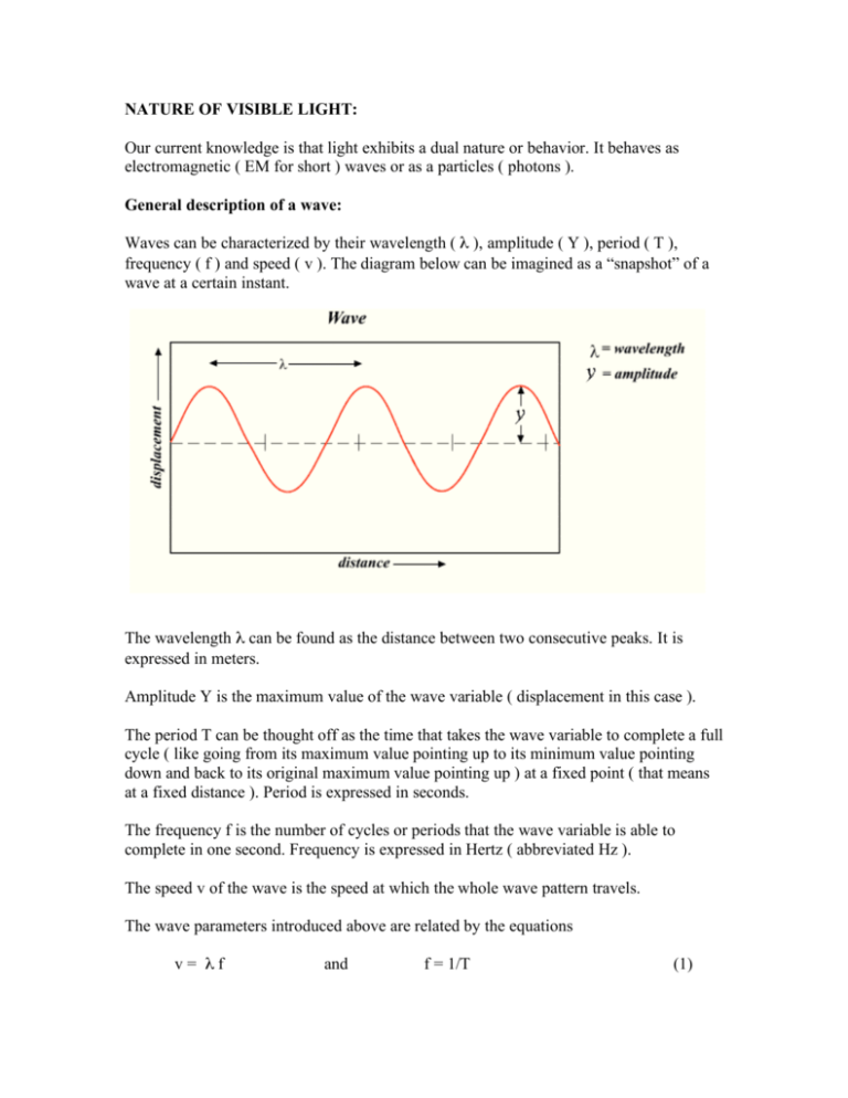
NATURE OF VISIBLE LIGHT: Our current knowledge is that light exhibits a dual nature or behavior. It behaves as electromagnetic ( EM for short ) waves or as a particles ( photons ). General description of a wave: Waves can be characterized by their wavelength ( λ ), amplitude ( Y ), period ( T ), frequency ( f ) and speed ( v ). The diagram below can be imagined as a “snapshot” of a wave at a certain instant. The wavelength λ can be found as the distance between two consecutive peaks. It is expressed in meters. Amplitude Y is the maximum value of the wave variable ( displacement in this case ). The period T can be thought off as the time that takes the wave variable to complete a full cycle ( like going from its maximum value pointing up to its minimum value pointing down and back to its original maximum value pointing up ) at a fixed point ( that means at a fixed distance ). Period is expressed in seconds. The frequency f is the number of cycles or periods that the wave variable is able to complete in one second. Frequency is expressed in Hertz ( abbreviated Hz ). The speed v of the wave is the speed at which the whole wave pattern travels. The wave parameters introduced above are related by the equations v= λf and f = 1/T (1) Electromagnetic wave nature of light: Visible light are EM waves to which the human eye is sensitive. Other EM waves like ultraviolet and infrared waves are not visible to humans. An EM wave is composed of electric ( E ) and magnetic ( M ) fields vibrating perpendicularly to each other and also perpendicularly to the direction of propagation as shown below. The speed of all EM waves in vacuum is the universal constant c = 3 x 10⁸ m/s. When EM waves travel through media, their speeds are smaller than c and depend both on the kind of media and the wavelength of the EM wave itself. To finish this section, look below at the schematic representation of the range of existing EM waves which expands from gamma rays ( from nuclear processes ) to radio waves. Note the very small range ( expanded for clarity ) that corresponds to visible light. Particle ( photon ) nature: A beam of light also behaves as a very fast ( moving at the speed c in vacuum ) stream of concentrated packets of energy ( called photons ). Each photon carries an energy E = h f where h = 6.626 x 10⁻³⁴ J.s is the Planck’s constant and f is the frequency of the EM wave associated with the photon. Photons can interact with other particles via collisions. HOW IS VISIBLE LIGHT GENERATED? Light generation via quantum energy transitions: Experiments and the quantum theory show that EM waves ( including visible light ) can be generated when a physical system ( like an atom or a solid piece of metal ) emits energy via a transition from a high to a low energy state. A first example is a sample of hydrogen gas. Electrons transitioning from higher to lower energy levels can do it following many different paths. Each transition originates an EM wave ( or photon ) of energy E = h f , where h is the Planck’s constant and f is the frequency of the EM wave ). The energy E is also the energy difference between the initial and final energy levels for the particular transition involved ( E = Ei – Ef ). There are many possible transitions and therefore many possible values of the frequencies for the EM waves emitted ranging from ultraviolet to radio waves including visible light. However, only 4 waves have frequencies in the visible part of the spectrum. The discrete nature of the energy level values is responsible for the discrete nature of the emission spectrum from the hydrogen gas sample. A second example is the hot metallic filament in an incandescent light bulb. The energy level structure of the solid metallic filament is composed of many closely spaced energy levels, so close to each other that in essence they form a continuous energy distribution. Energy transitions among these energy states produces EM waves from the ultraviolet to radio waves including visible light. In this example however, all possible frequencies in that range are included and for that reason, the spectrum is called a continuos spectrum, as opposed to a discrete spectrum for the case of the hydrogen gas previously considered. The diagram below shows a continuous energy spectrum like the one arising from a very hot ( like 3000 K ) incandescent light bulb. It can be concluded that the energy level structure of the system determines the kinds of EM waves generated. Above, two specific examples were given but every light source has its unique energy level structure and correspondingly its unique emission spectrum. For very instructive illustrations of light sources and their spectra visit: http://ioannis.virtualcomposer2000.com/spectroscope/amici Light generation via accelerated motion of charged particles: Experiments and the classical electromagnetic theory show that a charged particle generates EM waves when it is forced into accelerated motion. The stronger the acceleration, the higher the frequency of the wave. Waves ranging from X-rays to radio waves ( including visible light ) can be generated via this mechanism. COLOR TEMPERATURE: Color temperature is useful in photography, filmmaking, television, the printing and lighting industries, and astronomy because it allows a simple numerical specification of the color for many common light sources. Before defining color temperature, it is necessary to describe the radiation emitted by a perfect thermal radiator ( called a black body ). The Sun is considered a perfect radiator. A common incandescent light bulb approximates quite well a perfect radiator. As the temperature of the perfect radiator increases, its color changes as follows: At a temperature of 800 K ( K stands for Kelvin ) it glows dull red, at 2000 K it looks bright orange, at 3000 K it looks unsaturated yellow, at 6000 K it looks white and at 10,000 K it looks blue. It is possible to observe these colors ( except blue ) in an incandescent lamp by using a dimmer switch to allow more or less electricity to flow through it. The diagram on the right shows approximately the colors of the light emitted by a black body at different temperatures. The colors displayed on this diagram look slightly different from the real observed colors due to limitations in printing and displaying technologies. DO NOT confuse this diagram with a spectrum, IT IS NOT. This diagram shows how the color of an object like an incandescent light bulb changes as the temperature of the object changes. A spectrum on the other hand shows the component colors contained in the light emitted by a light source at a fixed temperature. Color temperature is a temperature value assigned to a light source. It represents the temperature of a perfect radiator glowing with the color that best matches the color exhibited by the light source under consideration. The value does not represent the temperature of the light source. Some examples are sunlight at noon on a clear day ( color temperature of 4870 K ) and skylight on an overcast day ( color temperature of 6770 K ). THE PROPAGATION OF LIGHT This section considers how light travels in transparent, homogeneous media as well as what happens to light when it reaches the interface between two different media ( like airglass ). The involved phenomena are reflection, refraction and total internal reflection. They play a fundamental role in still or video cameras, projectors, illumination equipment, binoculars, human vision and optical fibers. A light ray propagates in straight line in a homogeneous medium ( like air or glass ). However, when it reaches the interface between two homogeneous media, its direction in general changes abruptly. These changes in direction are described by the laws of reflection and refraction. Law of specular reflection: When light initially travelling through a medium (1) reaches the smooth interface between this medium and a second medium (2), reflected light ALWAYS “bounced back” to the initial medium. The incident and reflected rays stay in a plane oriented perpendicular to the reflecting surface. Also, the angle of reflection is equal to the angle of incidence as shown in the diagram below. Diffuse reflection: If the reflecting surface is rough rather than smooth, incident parallel light will reflect in all directions. However, the law of specular reflection still holds for each reflected ray. The multiple directions of reflection are due to the multiple microscopic orientations found at different locations of the rough surface. Most of the objects that we see are not light sources. We see these objects because they reflect the light generated by light sources and the reflected light reaches our eyes. If it were not for diffuse reflection, we would not see any light coming from an ordinary object ( and therefore would not be able to see it at all ) unless we were located exactly in the direction of the reflected rays as specified by the law of specular reflection. Law of refraction ( Snell’s Law ): When light initially travelling through a medium (1) reaches the smooth interface between this medium and a second medium (2), SOMETIME light passes to the second medium. This phenomenon is called refraction of light. The refracted ray follows a direction different from the direction of the incident ray ( except if the incident ray is perpendicular to the surface, in which case, the direction of the refracted ray is the same as the direction of the incident ray ). The incident and refracted rays stay in a plane oriented perpendicular to the reflecting surface. These facts are reflected in the diagram that follows. The change in the direction of travel of light as it passes from the first to the second medium has its origin in the fact that light travels at different speeds in different media. A quantity called the index of refraction of a medium has been introduced in order to reflect this fact. The index or refraction ( n ) of a medium is defined as: n=c/v (2) where c and v are the speeds of light in vacuum and the medium of concern respectively. Values of n for some media using yellow light of λ = 589 nm are: n=1 for vacuum n=1.0003 for air n=1.33 for pure water n=1.50 for glass n=2.42 for diamond The law of refraction also predicts the direction of the refracted ray from the following expression: n1 sin ( θ1 ) = n2 sin ( θ2 ) (3) where n1 and n2 are the indexes of refraction of media (1) and (2); θ1 and θ1 are the angles that the incident and refracted rays make with the perpendicular to the surface ( simply called angles of incidence and refraction ); and sin is the abbreviation for the mathematical function sine,( a function studied in trigonometry ). If n1, n2, and θ1 are known, then the equation can be solved for θ2, which specifies the direction of the refracted ray with respect to the perpendicular to the surface. Total internal reflection: While discussing what happens when light reaches the interface between two media, it was stressed out that reflection ALWAYS takes place but refraction takes place only SOMETIMES. Ignoring absorption of light by the media, all the energy of the incident light has to be distributed between the reflected and refracted rays. If refraction does not take place ( a scenario suggested by the expression SOMETIMES ), then all the energy of the incident light will go to the reflected ray. This scenario of having only reflection but not refraction is called total internal reflection. It is possible to know when this situation will happen. For it to happen, two conditions must be present simultaneously as follows: First condition: n1 > n2 For example: Light initially travelling through a glass medium ( medium (1) with n1 = 1.5 ) and attempting to pass to air ( medium (2) with n2 = 1.0003 ). Second condition: θ1 > θc For example: Angle of incidence θ1 = 60o for the media pair glass-air of θc = 41.8o. θc = sin-1( n1/n2 ) is called the critical angle for total internal reflection for the specific pair of media (1) and (2). The function sin-1 is the inverse sine function. Total internal reflection is the mechanism by which light signals travel inside the optical fibers used in computer networks that support the internet. Dispersion of light: The section deals with the discussion of the fascinating topic of dispersion of light, which has important applications and also explains how spectra are produced and rainbows are formed. A beam of light from sunlight or from an incandescent light bulb contains visible light waves of many different wavelengths as described before while explaining how light is generated. Visible light waves of different wavelengths are perceived by humans as different colors. However, the light from a very hot glowing incandescent light bulb is perceived basically white rather than colored. The reason is that all the light waves composing that light are superimposed ( added ) on each other and the perception produced is what we describe as white. This section is not about color perception but the topic was touched briefly to understand why we do not see directly the components of white light. Dispersion is a simple mechanism that allows the separation of white or other composed light into its different component waves. The light refraction phenomenon studied before is responsible for it: Assume that you start with a narrow parallel beam of white light. The task is to change the direction of each component wave by different amounts so that they appear separated from each other in a divergent beam. Lets restate some facts mentioned in previous sections: Light refraction changes the direction of a ray as it passes to a second medium. The amount of deviation ( related to the angle of refraction θ2 ) depends on the index of refraction of the second medium ( n2 ). The index of refraction of the second medium depends on the speed of the light wave in that medium. Finally, the speed of the light in a medium depends on the wavelength of the light wave. Therefore, reasoning backwards we have that light waves of different wavelengths travel at different speeds in a medium; as a result, the index of refraction of the medium for each wave is different and each wave will be deviated by a different amount. The final result is achieving the spatial separation of the light components looked for. A glass prism has been used for this purpose for many years as shown below. In the illustration, a ray of white light travelling through air reaches one side of the prism and each component of it passes into the prism travelling in different directions. All these components then reach the other side of the prism and refract again undergoing even more spatial separation. The light emerging from the prism forms a divergent beam of light where component are separated from each other. Looking at the emerging light directly or after it shines on a screen allows us to perceive the familiar colors of the white light spectrum. The same mechanism produces a rainbow, the only difference is that instead of light refraction taking place in glass prisms it takes place in small water drops. IMAGE FORMATION FROM LENSES: Characteristics of lenses: The most common type of lens is made of glass with a circular shape and bounded by spherical surfaces in the front and the back as shown below. When a beam of parallel light passes through a lens, the beam can emerge from it as a converging or diverging beam. If the beam converges, the lens is called converging, otherwise it is called diverging. The discussion here is limited to converging lenses, which are the most common. The point at which a beam of parallel light is made to converge by a converging lens is called the focal point of the lens and the distance from the center of the lens to the focal point is called the focal distance ( f ) of the lens as shown below. Do not confuse the focal distance f of a lens with the frequency f of a wave; they are completely different quantities using the same symbol. On a sunny day you can burn a whole in a piece of paper by converging the light from the Sun on the paper using a big magnifying glass. The focal length of a thin lens in air can be calculated using the lensmaker’s formula: f = ( R1 R2 ) / [(n – 1)(R2 – R1 )] (4) In this formula, n is the index of refraction of the glass while r1 and r2 are the radii of curvature of the spherical surfaces that bound the lens on the front and the back. For example, a double convex lens with n = 1.5, R1 = 20 cm and R2 = -10 cm ( explaining the sign conventions involved is beyond our interests ) will have f = 13.3 cm. Image formation by a thin lens: The formation of the real image of an object by a lens takes place in the following way: Light emanating from a point in the object spreads in all directions. This light reaches the whole exposed surface of the lens and after emerging from the lens converges into a single point ( the point of convergence can not be the focal point of the lens unless the object was located at an infinite distance from the lens ). This point of convergence is the location of the image of the point of the object. Through the same mechanism, each point of the object will have a corresponding image point on the other side of the lens and the combination of all these image points will form the full image of the object. If a screen is situated on the location where the image is formed, the image of the object will be clearly seen on the screen. The diagram below illustrates how the image of an object is formed by a converging lens. The illustration also shows the object distance do ( labeled ad S1 ) and the image distance di ( labeled as S2 ). The characteristics of the image produced by a thin lens can be determined through graphical or analytical methods. The analytical method will now be described. The lens equation allows the calculation of the image distance ( di ) which locates the position of the image as follows: di = ( do f ) / ( do – f ) (5) The lateral magnification m which specifies how large is the image with respect to the object is found by the equation: m = hi / ho = - di / do hi and ho are the heights of the image and the object (6) If the absolute value of the lateral magnification is larger than 1, the image is larger than the object. If it is 1, the image and the object have the same size. If it is smaller than 1, the image is smaller than the object. Also, if m is positive, the image has the same orientation than the object but if it is negative, the image is inverted (looks upside down with respect to the object). As an example, analyze the image of a flower located 100 cm from a camera lens with focal lens of 5 cm. From the lens equation: di = ( 100 cm x 5 cm ) / ( 100 cm – 5 cm ) = 5.26 cm. From the lateral magnification equation: m = - 5.26 cm / 100 cm = -0.0526 Therefore, the image is located 5.26 cm from the lens, it is upside down with respect to the object ( negative m ) and about 20 times smaller than the object ( 0.0526 is approximately 1/20 ). This situation is illustrated below. To finish this section it is important to mention that the location of the real image means the location where the image appears sharp ( a sharp image is commonly called an image in focus ). If a screen is located away ( but not extremely far ) from that perfect location, an image is still visible but it will look blurred ( a blurred image is commonly called out of focus ). The larger the separation from the screen to the perfect location of the image, the more out of focus it will look. If the separation is excessively large, no image will be discerned at all. Image formation by the human eye: The human eye is a remarkable natural optical device. It is schematically shown in the diagram below. The lens and the cornea ( in fact another lens ) are the image forming elements of the eye and form an inverted image of the object on the surface of the retina. Most of the bending of the light entering the eye is done via refraction in the cornea. The lens is used for fine adjustments to produce a focussed image on the surface of the retina. The iris controls how much light enters the eye by making the entrance hole ( the pupil ) larger or smaller. If the ambient light intensity is high, the iris decreases the size of the pupil. If the light intensity is low, the iris increases the size of the pupil. The normal human eye can see objects in focus from as close as 10 cm ( young children ) to as far as we desire ( for example, we can see the moon in focus ). How can the eye produce an image in focus from objects located over such a large range of distances from the eye? For the eye, the image distance di is fixed because the distance from the surface of the eye to the retina is fixed anatomically. The lens equation ( equation 5 ) indicates that a lens of fixed focal length f can not produce images with the same image distance di if the objects are located at different distances from the lens. The only possibility left is that the focal length of the optical system of the eye is adjusted as needed in order to produce the same image distance and therefore, a sharp image on the retina. This is in fact what the eye does. The way the eye achieves this is by changing the shape of the lens by the action of the ciliary muscle. When there is the need to focus a near object, the ciliary muscle presses on the lens and makes it thicker and rounder. The closer the object, the stronger the action of the muscle on the lens. To understand why this shape change produces a change in the focal distance of the lens we can go back to the lensmaker’s formula ( equation 4 ) and observe that changing the shape of the lens changes the radii of curvature R1 and R2 and therefore the focal lens. An interesting point to mention is the fact that as people get older, the elasticity of the eye lens is reduced and the ciliary muscle is not able to change enough the shape of the lens. As a result, people beyond 40 years of age normally have trouble reading without the help of reading glasses because they are not able to focus near objects. Image formation by a camera: This section deals with the formation of the image from a simple camera ( still or video ) just to illustrate the optics involved. A camera forms a real image of an object in the same way that the eye does. It can achieve this with a single converging lens called the objective. In order to be able to produce images in focus for objects that are at a range of distances from the camera, the camera lens can not be reshaped as the eye lens because the camera lens is made of rigid glass. Instead, the lens is allowed to change its distance from the image plane ( the plane where the photographic film or imaging chip is located ). Moving the lens closer to the image plane allows focusing far objects. Moving the lens farther away from the image plane allows focusing near objects. A numerical example is given below to consolidate this important point. Example: Consider a camera having an objective lens of 50 mm focal length. We want to know how far from the image plane this lens must be in order to focus an object located a) 10 meters away and b) 1 meter away. a) di = ( do f ) / ( do – f ) = ( 10,000 mm x 50 mm ) / ( 10,000 mm – 50 mm ) di = 50.3 mm b) b) di = ( do f ) / ( do – f ) = ( 1,000 mm x 50 mm ) / ( 1,000 mm – 50 mm ) di = 52.6 mm This example shows that in fact, this lens does not have to move too much in order to focus correctly objects over a substantial distance range. Beyond 10 meters, objects can be considered to be at an infinite distance using this particular lens because an object at infinite will form an image at an image distance equal to the focal length of the lens ( 50 mm in this case ). The next point that is important to consider is related to the magnification of the image. The question is what has to be done in order to obtain a larger image of an object if the camera can not be brought closer to the object? The answer is to use a lens with a larger focal length. A lens like this is called a telephoto lens. Again, to illustrate this important point, a numerical example will be given. Example: Consider a child 1.2 meters tall and 10 meters away from a camera. We want to know the size of the image of the child on the image plane of the camera using lenses of focal lengths a) 50 mm and b) 200 mm. To simplify the numerical calculations, we first combine the lens and magnification equations to obtain the following equation for the magnification: m = - f / ( do – f ). Now this equation can be used for the calculations as follows: a) m = - f / ( do – f ) = - 50 mm / ( 10,000 mm – 50 mm ) = - 0.005 From the magnification equation we get hi = m ho = -0.005 x 1200 mm = -6 mm b) m = - f / ( do – f ) = - 200 mm / ( 10,000 mm – 200 mm ) = - 0.020 From the magnification equation we get hi = m ho = -0.020 x 1200 mm = -24 mm Therefore, the size of the image of the child is 6 mm with the 50 mm lens but 24 mm with the 200 mm focal lens ( the negative sign just means that the image is inverted ). This example shows that quadrupling the focal lens of the objective quadruples the magnification of the image. In general, the magnification of the image will increase by the same factor as the focal length is increased. Cameras equipped with a zoom lens are able to have variable magnification by varying the focal length of the lens system. The term lens system is used because it is composed of more than one lens. The distances among the lenses in the system is varied to achieve the change in the focal length of the whole system. An explanation of composed lens systems goes beyond the scope of this introductory material. To finish the discussion of the camera it is important to mention that the intensity of light received at the image plane is controlled with the diaphragm, which has the same function that the iris has in the eye.


