Oxidation of the carcinogenic non-aminoazo dye 1-phenylazo
advertisement
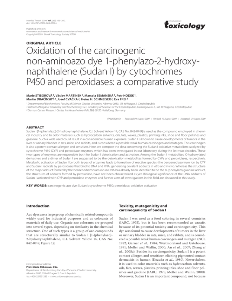
Interdisc Toxicol. 2009; Vol. 2(3): 195–200. doi: 10.2478/v10102-009-0017-z interdisciplinary Published online in: www.setox.eu/intertox & www.versita.com/science/medicine/it/ Copyright©2009 Slovak Toxicology Society SETOX ORIGINAL ARTICLE Oxidation of the carcinogenic non-aminoazo dye 1-phenylazo-2-hydroxynaphthalene (Sudan I) by cytochromes P450 and peroxidases: a comparative study Marie STIBOROVÁ 1, Václav MARTÍNEK 1, Marcela SEMANSKÁ 1, Petr HODEK 1, Martin DRAČÍNSKÝ 2, Josef CVAČKA 2, Heinz H. SCHMEISER 2, Eva FREI 3 1 Department of Biochemistry, Faculty of Science, Charles University, Albertov 2030, 128 40 Prague 2, Czech Republic 2 Institute of Organic Chemistry and Biochemistry, v.v.i., Academy of Sciences of the Czech Republic, Flemingovo n. 6, 166 10 Prague 6, Czech Republic 3 German Cancer Research Center, Im Neuenheimer Feld 280, 69120 Heidelberg, Germany ITX020309A04 • Received: 04 August 2009 • Revised: 10 August 2009 • Accepted: 12 August 2009 ABSTRACT Sudan I [1-(phenylazo)-2-hydroxynaphthalene, C.I. Solvent Yellow 14, CAS No: 842-07-9] is used as the compound employed in chemical industry and to color materials such as hydrocarbon solvents, oils, fats, waxes, plastics, printing inks, shoe and floor polishes and gasoline. Such a wide used could result in a considerable human exposure. Sudan I is known to cause developments of tumors in the liver or urinary bladder in rats, mice, and rabbits, and is considered a possible weak human carcinogen and mutagen. This carcinogen is also a potent contact allergen and sensitizer. Here, we compare the data concerning the Sudan I oxidative metabolism catalyzed by cytochrome P450 (CYP) and peroxidase enzymes, which has been investigated in our laboratory during the last two decades. These two types of enzymes are responsible both for Sudan I detoxication and activation. Among the Sudan I metabolites, C-hydroxylated derivatives and a dimer of Sudan I are suggested to be the detoxication metabolites formed by CYPs and peroxidases, respectively. Metabolic activation of Sudan I by both types of enzymes leads to formation of reactive species (the benzenediazonium ion by CYP and Sudan I radicals by peroxidase) that bind to DNA and RNA, generating covalent adducts in vitro and in vivo. Whereas the structure of the major adduct formed by the benzenediazonium ion in DNA has already been identified to be the 8-(phenylazo)guanine adduct, the structures of adducts formed by peroxidase, have not been characterized as yet. Biological significance of the DNA adducts of Sudan I activated with CYP and peroxidase enzymes and further aims of investigations in this field are discussed in this study. KEY WORDS: carcinogenic azo dye; Sudan I; cytochrome P450; peroxidase; oxidative activation Introduction Azo dyes are a large group of chemically related compounds widely used for industrial purposes and as colorants of materials of daily use. Organic azo colorants are grouped into several types, depending on similarity in the chemical structure. One of such types is a group of azo compounds that are structurally similar to Sudan I [1-(phenylazo)2-hydroxynaphthalene, C.I. Solvent Yellow 14, CAS No: 842-07-9, Figure 1)]. Correspondence address: Prof. Marie Stiborová, DSc. Department of Biochemistry, Faculty of Science, Charles University, Albertov 2030, 128 40 Prague 2, Czech Republic TEL: +420-221951285 • E-MAIL: stiborov@natur.cuni.cz Toxicity, mutagenicity and carcinogenicity of Sudan I Sudan I was used as a food coloring in several countries (IARC, 1975), but it has been recommended as unsafe, because of its potential toxicity and carcinogenicity. This dye was found to cause developments of tumors in the liver or urinary bladder in rats, mice, and rabbits, and is considered a possible weak human carcinogen and mutagen (NCI, 1982; Garner et al., 1984; Westmoreland and Gatehouse, 1991; Moller and Wallin, 2000; An et al., 2007; Zhang et al., 2008a). Besides its carcinogenicity, Sudan I is a potent contact allergen and sensitizer, eliciting pigmented contact dermatitis in human (Kozuka et al., 1980). Nevertheless, it is used to color materials such as hydrocarbon solvents, oils, fats, waxes, plastics, printing inks, shoe and floor polishes and gasoline (IARC, 1975; Moller and Wallin, 2000). Moreover, Sudan I is an important compound, not because 196 Oxidation of the carcinogenic Sudan I by cytochromes P450 and peroxidases M. Stiborová, V. Martínek, M. Semanská, P. Hodek, M. Dračínský, J. Cvačka, H.H. Schmeiser, E. Frei H O N H O N N N Figure.1. Sudan I (keto and enol forms). it is still used to color these materials, but because it is the simplest in a series of dyes and pigments including Sudan III and Sudan IV that are used in great quantities and occur everywhere in red and orange colored consumer products, foods and printed matter. Besides Sudan I, Sudan III and Sudan IV have also been found to be weak carcinogens, being classified as category 3 carcinogens by International Agency for Research on Cancer (IARC, 1975; Moller and Wallin, 2000; http://msds.chem.ox.ac.uk/SU/sudan_I.html). Even though the amounts of these azo dyes used in industry and to color the above materials have not yet been exactly evaluated, their use causes long-term harm in the environment and could result in a considerable human exposure (IARC, 1975; Moller and Wallin, 2000; http://msds.chem.ox.ac.uk/ SU/sudan_I.html). Recently, an increased attention has been paid to this dye, because it has been found as a contaminant of several European foodstuffs, being detected in chilli powder and in chilli-containing food products such as in Pixian douban, Golden Mark guilin chilli sauce, Golden Mark satay sauce, Italian pasta, chilli-snack and vegetable sauce (Mazzetti et al., 2004; Liu et al., 2007; Uematsu et al., 2007; Wang et al., 2007). More recently, levels of these dyes were evaluated; analysis of a few market samples of turmeric, chilli, and curry powders showed the presence of Sudan I (4.8–12.1 mg/g), Sudan IV (0.9–2.0 mg/g) and metanil yellow (1.5–4.6 mg/g) in loose turmeric and chilli samples (Xu et al., 2007; Dixit et al., 2008; Wang et al., 2009). In addition, there is evidence that ingestion of food products contaminated with Sudan I could lead to exposure in the human gastrointestinal tract to metabolites generated by the intestinal bacteria (Xu et al., 2007). Therefore, the use of Sudan I as an additive in food products has been prohibited in the European Union and many other countries (Federal Institute for Risk Assessment, 2003). Nevertheless, the question whether the recent detection of Sudan I and other Sudan I-derived dyes in various food commodities and additional materials is actually serious problems requires further toxicological evaluations by regulatory agencies. Such evaluations should determine the real impact of this Sudan I dye on human health (Federal Institute for Risk Assessment, 2003; Mazzetti et al., 2004; Liu et al., 2007; Uematsu et al., 2007; Wang et al., 2007; Xu et al., 2007; Dixit et al., 2008; Wang et al., 2009). Sudan I gives positive results in Salmonella typhimurium mutagenicity tests with S-9 activation (Cameron et al., 1987; Zeiger et al., 1988) and is mutagenic to mouse lymphoma L5178Y TK+/– cells in vitro, with S-9 activation (Zeiger et al., ISSN: 1337-6853 (print version) | 1337-9569 (electronic version) 1988). It is a clastogenic compound, inducing micronuclei in the bone marrow of rats (Westmoreland and Gatehouse, 1991). There is also evidence that this compound exhibits genotoxic effects, after its metabolic activation by hepatic cytochrome P450 (CYP) and peroxidase enzymes in vitro, and in the rat liver and urinary bladder in vivo (Stiborová et al., 1988b;c; 1990a;b; 1992; 1993; 1995a;b; 1999a;b; 2002; 2006; Dixit et al., 2008; Zhang et al., 2008b,), and in a human hepatoma cell line, HepG2 (An et al., 2007; Zhang et al., 2008b). Activation and detoxication metabolism of Sudan I catalyzed by cytochromes P450 While the metabolism of Sudan I is not understood in humans, its metabolism has been characterized in rabbits (Childs and Clayson, 1966), where it is metabolized primarily in the liver by oxidative or reductive reactions (Childs and Clayson, 1966). Azo-reduction of Sudan I seems to be responsible mainly for its detoxification, producing aniline and 1-amino-2-naphthol (Moller and Wallin, 2000). C-Hydroxylated metabolites 1-(4-hydroxyphenylazo)-2naphthol (4’-OH-Sudan I), 1-(phenylazo)-naphthalene-2,6diol (6-OH-Sudan I) and 1-(4-phenylazo)-naphthalene2,6-diol (4’,6-diOH-Sudan I) were found to be the major products of Sudan I oxidation in vivo and excreted in urine (Childs and Clayson, 1966; IARC, 1975), and also of its oxidation by rat hepatic microsomes in vitro (Stiborová et al., 1988a) (Figure 2). Glucuronides of the same three compounds have been detected in bile and urine of rabbits fed Sudan I (Childs and Clayson, 1966). Another C-hydroxylated metabolite, 1-(3,4-dihydroxyphenylazo)2-naphthol (3’,4’-diOH-Sudan I), was found to be formed as a minor product by Sudan I oxidation with rat hepatic microsomes in vitro (Stiborová et al., 1994) (Figure 2). Besides the C-hydroxylated metabolites, which are considered detoxication products, the benzenediazonium ion (BDI), formed by microsome-catalyzed enzymatic splitting of the azo group of Sudan I, was found to react with DNA in vitro (Stiborová et al., 1988a;b; 1995b). The major DNA adduct formed in this reaction has been characterized and identified as the 8-(phenylazo)guanine adduct (Stiborová et al., 1995b) (Figure 2). This adduct was also found in liver DNA of rats exposed to Sudan I (Stiborová et al., 2006) (Figure 3). Oxidation of Sudan I with formation of the same C-hydroxylated metabolites and Sudan I-derived DNA adducts was also demonstrated with human CYP enzymes (Stiborová et al., 2002; 2005). CYP1A1 is the major enzyme oxidizing Sudan I in human tissues rich in this enzyme, while CYP3A4 is also active in human liver (Stiborová et al., 2002; 2005) (Figure 2). Interestingly, even though levels of CYP1A1 expression in human livers are low, <0.7% of total hepatic CYP, the CYP1A1 contribution to oxidation of carcinogenic Sudan I in the set of human liver microsomes of Caucasian donors tested in our former study ranges from 12 to 30% (Stiborová et al., 2005). Moreover, the CYP1A1 enzyme is strongly induced by Sudan I itself in rats and human cells in culture, due to activating the cytosolic aryl hydrocarbon receptor (Lubet et al., 1983). Hence, long-term occupational exposure of humans to Sudan I might be an Interdisciplinary Toxicology. 2009; Vol. 2(3): 195–200 Also available online on setox.eu/intertox & versita.com/science/medicine/it H Peroxidase DNA, RNA and protein adducts 4' CYP1A1 N (CYP3A) + N 6 (CYP3A) H HO + N CYP1A1 Benzendiazonium ion O H N HO (CYP3A) Sudan I CYP1A1 Peroxidase O N HO Naphthalene-1,2-diol O N N N O O OH 6-OH-Sudan I CYP CYP + DNA 4'-OH-Sudan I CYP 1,2-Naphthoquinone O H HO HO O H N HO N N N HN O N N H 2N N N N + DNA OH 4',6-diOH-Sudan I 3',4'-diOH-Sudan I unknown adducts 8-Phenylazoguanine in DNA Figure 2. Scheme of Sudan I metabolism. important risk factor for individuals, improving Sudan I metabolism and binding to DNA, thereby increasing its toxicological relevance. Activation and detoxication metabolism of Sudan I by peroxidases While microsomal CYPs were found to be responsible for the activation of Sudan I in human or animal liver (Stiborová et al., 1988b; 1995b; 2002; 2005; Martínek and Stiborová 2002), they play a marginal role in the in vivo metabolic activation of Sudan I in the urinary bladder, because this organ has little or no detectable CYP enzymes. But relatively high levels of peroxidases are expressed in this tissue (Wise et al., 1984). We have found that in addition to microsomal CYP enzymes, Sudan I and its C-hydroxylated metabolites are also oxidized by peroxidases such as a model plant peroxidase from horseradish as well as the mammalian enzyme, prostaglandin H synthase (cyclooxygenase), as a consequence DNA, RNA and protein adducts are formed (Stiborová et al., 1988c; 1990a;b; 1991; 1992; 1993; 1995a; 1999b) (Figure 2). In bladder, therefore, peroxidase-catalyzed activation of Sudan I has been suggested, similar to other carcinogens such as carcinogenic aromatic amines (Wise et al., 1984; Yamazoe et al., 1985; Eling et al., 1990; Chen et al., 1996). We have suggested a CYP- or peroxidase-mediated activation of Sudan I or a combination of both mechanisms as an explanation for the organ specificity of this carcinogen for liver and urinary bladder in animals (Stiborová et al., 1988b; 1990a;b; 1992; 1995b; 2002; 2005; Martínek and Stiborová, 2002). Indeed, the 8-(phenylazo)guanine DNA adduct generated from the BDI, the product of CYP activation, was found in the liver of rats treated with Sudan I (Stiborová et al., 2006) (Figure 3), whereas the physico-chemical properties of DNA adducts found in the urinary bladder are identical to those formed by the peroxidase-mediated Sudan I activation that contain the whole molecule of Sudan I (Stiborová et al., 1999a). Unfortunately, neither the structures of DNA- or RNAadduct(s), nor those of the ultimate carcinogen(s) formed by peroxidase from Sudan I are known as yet. This knowledge is, however, crucial for the comparative study with the in vivo products (Stiborová et al., 1999a, 2006). In our former studies with peroxidase activation of Sudan I, we have identified BDI and C-hydroxy derivatives of Sudan I [6-OH-Sudan I and 4’,6-di(OH)-Sudan I] as only minor metabolites, while products of a suggested polymerization of the primarily formed Sudan I radicals were more abundant (Stiborová et al., 1988c; 1991; 1996). In the meantime, we have confirmed this for the two major metabolites (Semanská et al., 2008), one is a Sudan I dimer (Figure 4A) generated by a Sudan I one electron oxidation, while the other is the product of secondary, enzyme independent reactions of this Sudan I dimer, spiro-bezoxadiazine derivative of Sudan I (Semanská et al., 2008) (Figure 4B). Plant horseradish peroxidase (HRP) was found in these studies to be an acceptable model for Sudan I oxidation by mammalian enzymes such as non-specific urinary bladder peroxidases and/or cyclooxygenases (Stiborová et al., 1990a;b; 1996; 2000; 2002; 2006). Even though mammalian and plant peroxidases are Copyright © 2009 Slovak Toxicology Society SETOX 197 198 Oxidation of the carcinogenic Sudan I by cytochromes P450 and peroxidases M. Stiborová, V. Martínek, M. Semanská, P. Hodek, M. Dračínský, J. Cvačka, H.H. Schmeiser, E. Frei A O B 8-Phenylazoguanine N HN N N H 2N C N N DNA 3 1 2 Figure 3. Autoradiographs of PEI-cellulose thin layer chromatography (TLC) maps of 32P-labeled digests of calf thymus DNA reacted with Sudan I, NADPH and human recombinant CYP1A1 in Supersomes (A) and of liver DNA of rats treated with Sudan I (C). (B) Schematic figure of adducts with assigned numbers and the structure of adduct 1 (closed circle). Analysis was performed by the nuclease P1 version of the assay. Chromatographic conditions are described (Stiborová et al., 1995; 2006). Autoradiography was at -80oC for 6 (A) and 3 h (C). Origins are located in the bottom left corners (Stiborová et al., 2006). A O N O N N N O B 5 4 6 3 7´ 8´ 6´ 4a 2 1 O 7 8a 8 8a´ 5´ 4a ´ N 1´ N 1´´ 4´ 2´ 3´ O 2´´ 3´´ 4´´ Figure 4. Sudan I metabolites generated during its peroxidase mediated oxidation. (A) Sudan I dimer, (B) spiro-bezoxadiazine derivative of Sudan (Semanská et al., 2008). ISSN: 1337-6853 (print version) | 1337-9569 (electronic version) structurally different proteins, the oxidation mechanisms are similar due to analogous arrangements of their active sites (Wise et al., 1984; Yamazoe et al., 1985; Eling et al., 1990; Stiborová et al., 1991; 2000; Chen et al., 1996). It should be noted that the Sudan I metabolites formed by peroxidase (Figure 3) are much less likely to be formed physiologically than in the in vitro system (Semanská et al., 2008), because many nucleophilic molecules are present in cells to scavenge the Sudan I reactive species. Indeed, during Sudan I oxidation by peroxidase in the presence of nuclephiles such as DNA, tRNA polydeoxynucleotides, polynucleotides and or proteins, the formation of adducts runs parallel to a decrease in generation of Sudan I metabolites (Stiborová et al., 1990a;b; 1991; 1999b; and Stiborova et al., unpublished data). Hence, formation of adducts of Sudan I reactive species with these nucleophilic compounds seems to be the preferred reaction under physiological conditions. Using the 32P-postlabeling assay (Randerath et al., 1981; Gupta 1985) we have already analyzed Sudan I-DNA adducts formed by peroxidases (HRP, cyclooxygenase) (Stiborová et al., 1990a;b; 1992; 1999b). Deoxyguanosine was the major target for Sudan I-DNA binding, followed by deoxyadenosine (Stiborová et al., 1992). Likewise, guanosine was found to be the major target for peroxidaseactivated Sudan I binding in RNA (Stiborová et al., 1995a). Autoradiographs of thin layer chromatography (TLC) maps of 32P-labelled digests of tRNA and polyguanosine modified by Sudan I activated with peroxidase, shown in Figure 5, also indicate that guanosine is the target for binding of Sudan I activated with peroxidase in tRNA. It has been postulated by Eling and coworkers (Eling et al., 1990) that characterization of peroxidase-mediated adducts derived from carcinogens is exceptionally difficult due to problems in preparing the DNA (or RNA) adducts in sufficient quantities and purity for structural analysis. Indeed, our earlier studies with peroxidase activation of Interdisciplinary Toxicology. 2009; Vol. 2(3): 195–200 Also available online on setox.eu/intertox & versita.com/science/medicine/it A B H O N N 11 7 8 1’ 12 C 2 9 6 10 1 3 5 4 D 2 3 5 1 4 Figure 5. Autoradiographs of PEI-cellulose TLC maps of P-labeled digests of the following: (A) rat liver tRNA treated with Sudan I, peroxidase and hydrogen peroxide, (C) polyguanosine treated with Sudan I, peroxidase and hydrogen peroxide. (B) and (D) Schematic figures of adducts formed in tRNA (B) and polyguanosine (D) with assigned numbers. Analysis was performed by the nuclease P1 enhanced version of the assay. Chromatographic conditions are described (Stiborová et al., 1990a;b; 2002). Autoradiography was at 25 °C for 25 min (A) and for 5 min (C). Origins are located at the bottom left corners (D3 from bottom to top and D4 from left to right). Sudan I in the presence of DNA, tRNA, (deoxy)guanosine 3´-monophosphate or (deoxy)guanosine 5´-monophosphate did not yield enough adducts for their structural characterization (Stiborová et al., 1992; 1995a). Acknowledgement The research was supported in part Grant Agency of the Czech Republic (grants 303/09/0472 and 203/09/0812), the Ministry of Education of the Czech Republic (grant MSM0021620808, 1M0505 and RP MSMT 14/63). Conclusion Based on the above findings, the study which will lead to prepare and isolate deoxyguanosine- and guanosine adducts formed by the peroxidase-activated Sudan I in sufficient amounts and purity is essential for their structural characterization. Therefore, such a study is under way in our laboratory. It will contribute to establish the molecular mechanisms explaining the formation of two Sudan I metabolites formed by peroxidase (Figure 3) and contribute to determine structures of adducts that were found to be formed in DNA/RNA in vitro (Stiborová et al., 1990a;b; 1992; 1995a;b; 1999b) and in vivo (Stiborová et al., 1999a, 2006). REFERENCES An Y, Jiang L, Cao J, Geng C, Zhong L. (2007). Sudan I induces genotoxic effects and oxidative DNA damage in HepG2 cells. Mutat Res 627: 164–170. Cameron TP, Hughes TJ, Kirby PE, Fung VA, Dunkel VC. (1987). Mutagenic activity of 27 dyes and related chemicals in the Salmonella/microsome and mouse lymphoma TK+/– assays. Mutat Res 189: 223–261. Chen L, Devanesan PD, Higginbotham S, Ariese F, Jankowiak R, Small GJ, Rogan EG, Cavalieri EL. (1996). Expanded analysis of benzo[a]pyrene-DNA adducts formed in vitro and in mouse skin: their significance in tumor initiation. Chem Res Toxico 9: 897–903. Childs JJ, Clayson DS. (1966). The metabolism of 1-phenylazo-2-naphthol in the rabbit. Biochem Pharmacol 15: 1247–1258. Copyright © 2009 Slovak Toxicology Society SETOX 199 200 Oxidation of the carcinogenic Sudan I by cytochromes P450 and peroxidases M. Stiborová, V. Martínek, M. Semanská, P. Hodek, M. Dračínský, J. Cvačka, H.H. Schmeiser, E. Frei Dixit S, Khanna SK, Das M. (2008). A simple 2-directional high-performance thin-layer chromatographic method for the simultaneous determination of curcumin, metanil yellow, and sudan dyes in turmeric, chili, and curry powders. J AOAC Int 91: 1387–1396. Eling TE, Thompson DC, Foureman GL, Curtis JF, Hughes MF. (1990). Prostaglandin H synthase and xenobiotic oxidation. Annu Rev Pharmacol Toxicol 30: 1–45. Federal Institute for Risk Assessment. (2003). Dyes Sudan I to IV in food. In Opinion of 10 November 2003. Federal Institute for Risk Assessment, Berlin, Germany. Garner RC, Martin CN, Clayson DB. (1984). Carcinogenic aromatic amines and related compounds. In Chemical Carcinogens, 2nd Edn, Vol. 1, CS Monograph 182, (Searle, C. ed.), American Chemical Society, Washington, DC, pp. 175–302. Gupta RC. (1985). Enhanced sensitivity of 32P-postlabeling analysis of aromatic carcinogenic:DNA adducts. Cancer Res 45: 5656–5662. http://msds.chem.ox.ac.uk/SU/sudan_I.html – downloaded on August 4, 2009 International Agency for Research on Cancer (IARC) (1975). Sudan I. In IARC Monographs, Vol. 8, IARC, Lyon, France, pp. 225–231. Kozuka T, Tashiro M, Sano S, Fujimoto K, Nakamura Y, Hashimoto S, Nakaminami G. (1980). Pigmented contact dermatitis from azo dyes. I. Cross-sensitivity in humans. Contact Dermatitis 6: 330–336. Liu Y, Song Z, Dong F, Zhang L. (2007). Flow injection chemiluminescence determination of Sudan I in hot chilli sauce. J Agric Food Chem 55: 614–617. Lubet RA, Connolly G, Kouri RE, Nebert DW, Bigelow SW. (1983). Biological effects of Sudan I dyes. Role of the cytosolic Ah receptor. Biochem Pharmacol 32: 3053–3058. Martínek V, Stiborová M. (2002). Metabolism of carcinogenic azo dye Sudan I by rat, rabbit, minipig and human hepatic microsomes. Collect Czech Chem Commun 67: 1883–1898. Mazzetti M, Fascioli R, Mazzoncini I, Spinelli G, Morelli I, Bertoli A. (2004). Determination of 1-phenylazo-2-naphthol (Sudan I) in chilli powder and in chillicontaining food products by GPC clean-up and HPLC with LC/MS confirmation. Food Addit Contam 21: 935–941. Moller P, Wallin H. (2000). Genotoxic hazards of azo pigments and other colorants related to 1-phenylazo-2-hydroxynaphthalene. Mutat Res 462: 13–30. NCI (1982). Carcinogenesis bioassay of C.I. Solvent Yellow 14 in F344/N rats and B6C3F1 mice. In Technical Report No. 226, US National Cancer Institute, Bethesda. Randerath K, Reddy MV, Gupta RC. (1981). 32P-labeling test for DNA damage. Proc Natl Acad Sci USA 78: 6126–6129. Semanská M, Martínek V, Hudeček J, Hodek P, Dračínský M, Frei E, Stiborová M. (2008). A one-electron oxidation of carcinogenic non-aminoazo dye Sudan I by horseradish peroxidase. Neuro Endocrinol Lett 29: 712–716. Stiborová M, Asfaw B, Anzenbacher P, Lešetický L, Hodek P. (1988a). The first identification of the benzenediazonium ion formation from a non-aminoazo dye, 1-phenylazo-2-hydroxynaphthalene (Sudan I) by microsomes of rat livers. Cancer Lett 40: 319–326. Stiborová M, Asfaw B, Anzenbacher P, Hodek P. (1988b). A new way to carcinogenicity of azo dye: the benzenediazonium ion formed from non-aminoazo dye, 1-phenylazo-2-hydroxynaphthalene (Sudan I) by microsomal enzymes binds to deoxyguanosine residues of DNA. Cancer Lett 40: 327–333. Stiborová M, Asfaw B, Anzenbacher P. (1988c). Activation of carcinogens by peroxidase. Horseradish peroxidase-mediated formation of benzenediazonium ion from a non-aminoazo dye, 1-phenylazo-2-hydroxynaphthalene (Sudan I) and its binding to DNA. FEBS Lett 232: 387–390. Stiborová M, Frei E, Klokow K, Wiessler M, Šafařík L, Anzenbacher P, Hradec J. (1990a). Peroxidase-mediated reaction of the carcinogenic non-aminoazo dye 1-phenylazo-2-hydroxynaphthalene with transfer ribonucleic acid. Carcinogenesis 11: 1789–1794. Stiborová M, Frei E, Schmeiser HH, Wiessler M, Hradec J. (1990b). Mechanism of formation and 32P-postlabeling of DNA adducts derived from peroxidative activation of carcinogenic non-aminoazo dye 1-phenylazo-2-hydroxynaphthalene (Sudan I). Carcinogenesis 11: 1843–1848. Stiborová M, Frei E, Anzenbacher P. (1991). Study of oxidation and binding to macromolecules of the carcinogenic non-aminoazo dye 1-phenylazo2-hydroxynaphthalene catalyzed by horseradish (Armoracia rusticana L.) peroxidase. Biochem Physiol Pflanzen 187: 227–236. Stiborová M, Frei E, Schmeiser HH, Wiessler M. (1992). 32P-Postlabeling analysis of adducts formed from 1-phenylazo-2-hydroxynaphthalene (Sudan I, Solvent Yellow 14) with DNA and homopolydeoxyribonucleotides. Carcinogenesis 13: 1221–1225. ISSN: 1337-6853 (print version) | 1337-9569 (electronic version) Stiborová M, Frei E, Schmeiser HH, Wiessler M, Hradec J. (1993). Detoxication products of the carcinogenic azodye Sudan I (Solvent Yellow 14) bind to nucleic acids after activation by peroxidase. Cancer Lett 68: 43–47. Stiborová M, Asfaw B, Frei E, Schmeiser HH, Wiessler M. (1994). Identification of 1-(3,4-dihydroxyphenylazo)-2-hydroxynaphthalene as the product of oxidation of 1-phenylazo-2-hydroxynaphthalene (Sudan I, Solvent Yellow 14) by rat liver microsomes. Collect Czech Chem Commun 59: 2727–2733. Stiborová M, Asfaw B, Frei E. (1995a). Peroxidase-activated carcinogenic azo dye Sudan I (Solvent Yellow 14) binds to guanosine in transfer ribonucleic acid. Gen Physiol Biophys 14: 39–49. Stiborová M, Asfaw B, Frei E, Schmeiser HH, Wiessler M. (1995b). Benzenediazonium ion derived from Sudan I forms an 8-(phenylazo)guanine adduct. Chem Res Toxicol 8: 489–498. Stiborová M, Asfaw B, Frei E, Schmeiser HH. (1996). Oxidation of azo dyes by peroxidase: additional evidence of a one-electron mechanism of oxidation of dimethylaminoazobenzene and Sudan I (Solvent Yellow 14). Collect Czech Chem Commun 61: 962–972. Stiborová M, Schmeiser HH, Breuer A, Frei E. (1999a). 32P-Postlabelling analysis of DNA adducts with 1-(phenylazo)-2-naphthol (Sudan I, Solvent Yellow 14) formed in vivo in Fisher 344 rats. Collect Czech Chem Commun 64: 1335– 1347. Stiborová M, Schmeiser HH, Frei E. (1999b). Prostaglandin H synthase mediated oxidation and binding to DNA of a detoxication metabolite of carcinogenic Sudan I 1-(phenylazo) 2,6-dihydroxynaphthalene. Cancer Lett 142: 53–60. Stiborová M, Mikšanová M, Martínek V, Frei E. (2000). Heme peroxidases: structure, function, mechanism and involvement in activation of carcinogens. A review. Collect Czech Chem Commun 65: 297–325. Stiborová M, Martínek V, Rýdlová H, Hodek P, Frei E. (2002). Sudan I is a potential carcinogen for humans: evidence for its metabolic activation and detoxication by human recombinant cytochrome P450 1A1 and liver microsomes. Cancer Res 62: 5678–5684. Stiborová M, Martínek V, Rýdlová H, Koblas T, Hodek P. (2005). Expression of cytochrome P450 1A1 and its contribution to oxidation of a potential human carcinogen 1-phenylazo-2-naphthol (Sudan I) in human livers. Cancer Lett 220: 145–154. Stiborová M Martínek V Schmeiser HH, Frei E. (2006). Modulation of CYP1A1mediated oxidation of carcinogenic azo dye Sudan I and its binding to DNA by cytochrome b5. Neuro Endocrinol. Lett. 27 (Suppl. 2): 35–39. Uematsu Y, Ogimoto M, Kabashima J, Suzuki K, Ito K. (2007). Fast cleanup method for the analysis of Sudan I–IV and para red in various foods and paprika color (oleoresin) by high-performance liquid chromatography/diode array detection: focus on removal of fat and oil as fatty acid methyl esters prepared by transesterification of acylglycerols. J AOAC Int 90: 437–945. Wise RW, Zenser TV, Kadlubar FF, Davis BB. (1984). Metabolic activation of carcinogenic aromatic amines by dog bladder and kidney prostaglandin H synthase. Cancer Res 44: 1893–1897. Wang S, Xu Z, Fang G, Duan Z, Zhang Y, Chen S. (2007). Synthesis and characterization of a molecularly imprinted silica gel sorbent for the on-line determination of trace Sudan I in Chilli powder through high-performance liquid chromatography. J Agric Food Chem 55: 3869–3876. Wang Y, Wei D, Yang H, Yang Y, Xing W, Li Y, Deng A. (2009). Development of a highly sensitive and specific monoclonal antibody-based enzyme-linked immunosorbent assay (ELISA) for detection of Sudan I in food samples. Talanta 77: 1783–1789. Westmoreland C, Gatehouse DG. (1991). The differential clastogenicity of Solvent Yellow 14 and FD & C Yellow No. 6 in vivo in the rodent micronucleus test (observation of species and tissues specificity). Carcinogenesis 12: 1403– 1407. Xu H, Heinze TM, Chen S, Cerniglia CE, Chen H. (2007). Anaerobic metabolism of 1-amino-2-naphthol-based azo dyes (Sudan dyes) by human intestinal microflora. Appl Environ Microbiol 73: 7759–7762. Yamazoe Y, Miller DW, Wies CC, Dooley KL, Zenser TV, Beland FA, Kadlubar FF. (1985). DNA adducts formed by ring-oxidation of the carcinogen 2-naphthylamine with prostaglandin H synthase in vitro and in the dog urothelium in vivo. Carcinogenesis 6: 1379–1387. Zeiger E, Andersen B, Haworth S, Lawlor T, Mortelmans K. (1988). Salmonella mutagenicity tests. IV. Results from the testing of 300 chemicals. Environ. Mutagen 11, Suppl. 12: 1–158. Zhang X, Jiang L, Geng C, Hu C, Yoshimura H, Zhong L. (2008a). Inhibition of Sudan I genotoxicity in human liver-derived HepG2 cells by the antioxidant hydroxytyrosol. Free Radic Res 42: 189–195. Zhang X, Juany L, Geng C, Hu C, Yoshimura H, Zhong L. (2008b). Inhibition of Sudan I genotoxicity in human liver-derived HepG2 cells by the antioxidant hydroxytyrosol. Free Radic Res 42: 189–195.
